. 2011;6(5):e20220.
doi: 10.1371/journal.pone.0020220. Epub 2011 May 26.

Pre-microRNA and mature microRNA in human mitochondria

Affiliations

Pre-microRNA and mature microRNA in human mitochondria

Eric Barrey et al. PLoS One. 2011.

Abstract

Background: Because of the central functions of the mitochondria in providing metabolic energy and initiating apoptosis on one hand and the role that microRNA (miRNA) play in gene expression, we hypothesized that some miRNA could be present in the mitochondria for post-transcriptomic regulation by RNA interference. We intend to identify miRNA localized in the mitochondria isolated from human skeletal primary muscular cells.

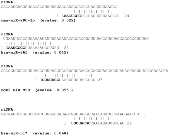
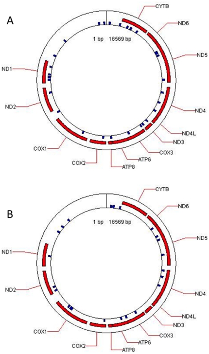
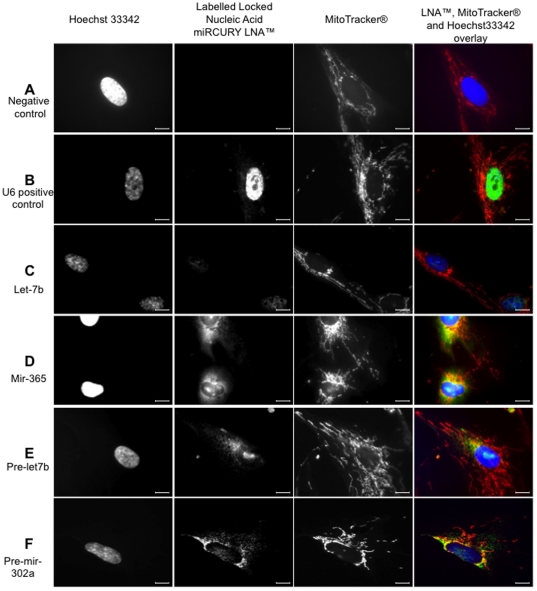
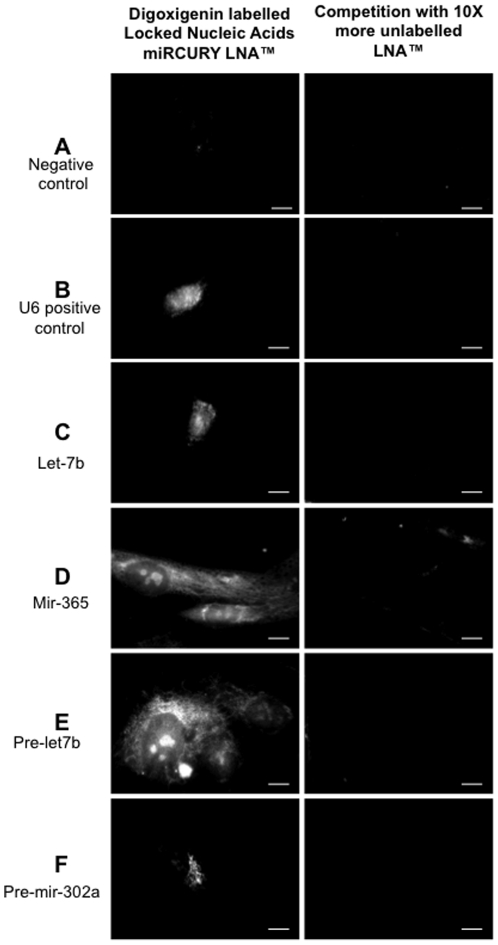
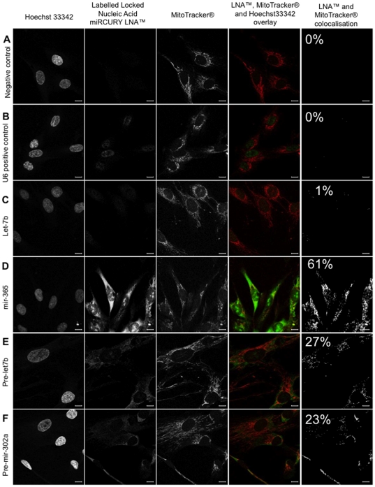
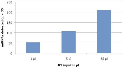
Methodology/principal findings: To investigate the potential origin of mitochondrial miRNA, we in-silico searched for microRNA candidates in the mtDNA. Twenty five human pre-miRNA and 33 miRNA aligments (E-value<0.1) were found in the reference mitochondrial sequence and some of the best candidates were chosen for a co-localization test. In situ hybridization of pre-mir-302a, pre-let-7b and mir-365, using specific labelled locked nucleic acids and confocal microscopy, demonstrated that these miRNA were localized in mitochondria of human myoblasts. Total RNA was extracted from enriched mitochondria isolated by an immunomagnetic method from a culture of human myotubes. The detection of 742 human miRNA (miRBase) were monitored by RT-qPCR at three increasing mtRNA inputs. Forty six miRNA were significantly expressed (2(nd) derivative method Cp>35) for the smallest RNA input concentration and 204 miRNA for the maximum RNA input concentration. In silico analysis predicted 80 putative miRNA target sites in the mitochondrial genome (E-value<0.05).

Conclusions/significance: The present study experimentally demonstrated for the first time the presence of pre-miRNA and miRNA in the human mitochondria isolated from skeletal muscular cells. A set of miRNA were significantly detected in mitochondria fraction. The origin of these pre-miRNA and miRNA should be further investigate to determine if they are imported from the cytosol and/or if they are partially processed in the mitochondria.


Conflict of interest statement

Competing Interests: The authors have declared that no competing interests exist.

Figures

Figure 1
Figure 1. Examples of alignment results of human mature miRNA 365 and 31.
The nucleotides in bold letters indicate the seed region of the miRNA.
Figure 2
Figure 2. Mitochondrial map of the human pre-mature (A) and mature miRNA (B) candidates identified by alignements between the reference mitochondrial sequence and miRBase data.
(A) The locations of the human pre-miRNA described in Table 3 are represented as blue squares on the mitochondrial genome. (B) The locations of the human mature miRNA described in Table 4 are represented as blue squares on the mitochondrial genome.
Figure 3
Figure 3. In situ hybridization pattern of digoxigenin-labeled Locked Nucleic Acid (LNA) for specific miR and pre-miR in human skeletal muscle myoblasts cells observed in classic optic microscopy.
Using locked nucleic acid (LNA) probes digoxigenin labelled, we determined the in situ hybridization pattern of mir and pre-mir. Hoechst 33342 staining of nuclei (lane 1), specific signal of scramble miRNA (A; negative control), U6 small nuclear RNA (B; positive control), let-7b (C), mir-365 (D), pre-let-7b (E) and pre-mir-302a (F) probes (lane 2) and MitoTracker® Red CM-H2XRos staining of respiring mitochondria (lane 3) are represented in gray scale. All these images were acquired using an Olympus BX61 straight microscope controlled with Metamorph software (Molecular Devices, Downington, PA19335) using a 100× oil-immersion objective. In the overlays (lane 4) provided by Image J software, positive in situ hybridization signals are visualized in green, respiring mitochondria signal in red and nuclei staining in blue. Yellow staining suggests co-localization of LNA probes (green fluorescence) and MitoTracker® Red CM-H2Ros (red fluorescence). Scale bars = 10 µm. Images are not scaled to the same intensity range. Positive in situ hybridization signals were normalized by scramble miR signal intensity (negative control).
Figure 4
Figure 4. Specificity of the locked nucleic acid (LNA) probes for the detection of miR and pre-miR using in situ hybridization.
Using 2.5 pmol of locked nucleic acid (LNA) probes prelabelled with digoxigenin, we determined in situ hybridization patterns of scramble miR (A; negative control), U6 small nuclear RNA (B; positive control), let-7b (C), mir-365 (D), pre-let-7b (E) and pre-mir-302a (F) probes (lane 1). In the second lane, Digoxigenin-labeled probes were competed with excessive amount (25 pmol ie 10× more) of unlabeled probes. Most of the signal observed in the panels of the first lane are absent from the panels in the second lane. Scale bars = 10 µm. Images are not scaled to the same intensity range. Positive in situ hybridization signals showed were normalized by scramble mirna signal intensity (negative control).
Figure 5
Figure 5. In situ signal of mir-365, pre-mir-let7b and pre-mir-302a co-localized with functioning mitochondria in human myoblasts observed in confocal microscopy.
Using locked nucleic acid (LNA) probes, we performed in situ hybridization to localized mir-365, mir-let-7b, pre-let-7b and pre-mir-302a within the cell. Hoechst 33342 staining of nuclei (lane 1), specific signal of scramble miR (A; negative control), U6 small nuclear RNA (B; positive control), let-7b (C), mir-365 (D), pre-let-7b (E) and pre-mir-302a (F) provided by locked nucleic acid (LNA) probes (lane 2) and MitoTracker® Red CM-H2XRos staining of functioning mitochondria (lane 3) are represented in gray scale. All these images were acquired with a Leica TSC-P2 confocal microscope using a 63× oil-immersion objective. A sequential mode for three colour of acquisition (FITC for LNA probes signals, Dapi for Hoechst staining and Cy3 for MitoTracker® Red CM-H2XRos) has been used. In the overlays (lane 4) provided by Image J software, positive in situ hybridization signal are visualized in green and respiring mitochondria signal in red. Yellow corresponded to red and green overlay. The pixels with co-localized signals (lane 5) from functioning mitochondria and specific LNA probe for mir or pre-mir are determined using the Image J MBF plugging “co-localization highlighter”. Scale bars = 10 µm. The raw images are showed in the figure. The percentage of pixels with co-localized miRNA and mitochondrial signals were determined using Isodata plugging threshold (Image J software) and indicated on the figure (% in lane 5).
Figure 6
Figure 6. Number of significant miRNA detected in three increasing mitochondrial mtRNA inputs.
The number of assays giving a significant signal (Cp<35) was 46, 106 and 204 respectively. The maximum number of assays giving a significant signal represents 27% of the total amount of miRNA assays tested. The number of miRNAs did as expected followed the amount of RNA input and demonstrated the quality of the mitochondrial mRNA extract for miRNA profiling.

References

    1. Trenker M, Malli R, Fertschai I, Levak-Frank S, Graier WF. Uncoupling proteins 2 and 3 are fundamental for mitochondrial Ca2+ uniport. Nat Cell Biol. 2007;2007 9:445–452. Erratum in: Nat Cell Biol 10: 1371. - PMC - PubMed
    1. Taanman JW. The mitochondrial genome: structure, transcription, translation and replication. Biochim Biophys Acta. 1999;1410:103–123. - PubMed
    1. Mokranjac D, Neupert W. Protein import into isolated mitochondria. Methods Mol Biol. 2007;372:277–286. - PubMed
    1. Schmidt O, Pfanner N, Meisinger C. Mitochondrial protein import: from proteomics to functional mechanisms. Nat Rev Mol Cell Biol. 2010;11:655–667. - PubMed
    1. Chinnery PF, Howell N, Andrews RM, Turnbull DM. Clinical mitochondrial genetics. J Med Genet. 1999;36:425–436. - PMC - PubMed

Publication types