. 2011 Sep;14(3):331-43.
doi: 10.1007/s10456-011-9217-1. Epub 2011 Jun 3.

EAF2 loss enhances angiogenic effects of Von Hippel-Lindau heterozygosity on the murine liver and prostate

Affiliations

EAF2 loss enhances angiogenic effects of Von Hippel-Lindau heterozygosity on the murine liver and prostate

Laura E Pascal et al. Angiogenesis. 2011 Sep.

Abstract

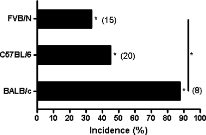
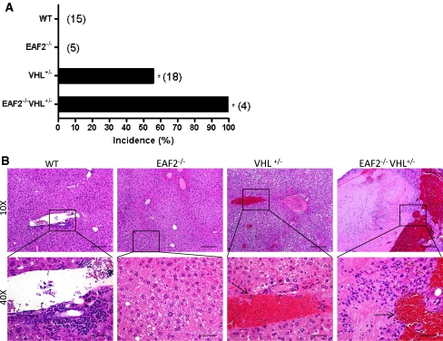
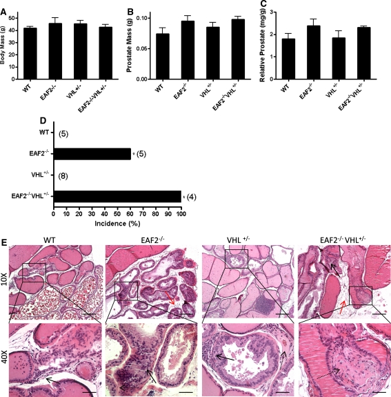
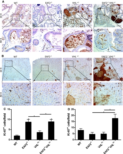
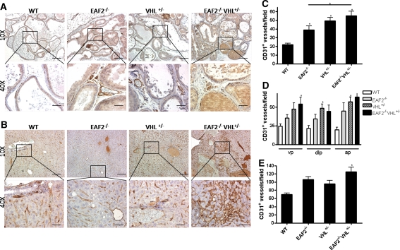
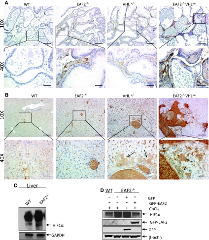
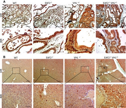
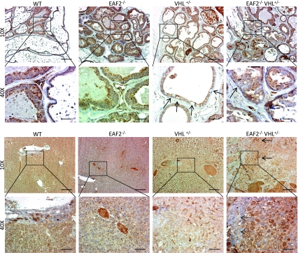
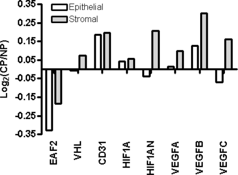
Von Hippel-Lindau (VHL) disease results from the inactivation of the VHL gene and is characterized by highly vascular tumors. A consequence of VHL loss is the stabilization of hypoxia-inducible factor (HIF) alpha subunits and increased expression of HIF target genes, which include pro-angiogenic growth factors such as vascular endothelial growth factor (VEGF). In mice, homozygous deletion of VHL is embryonic lethal due to vascular abnormalities in the placenta; and, VHL(+/-) mice develop proliferative vascular lesions in several major organs, most prominently the liver. Loss of ELL-associated factor (EAF2) in murine models has also been shown to induce increased vascular density in the liver as well as the prostate. Previously, EAF2 was determined to be a binding partner of VHL and loss of EAF2 induced a reduction in pVHL levels and an increase in hypoxia induced factor 1α (HIF1α) levels in vitro. Here we characterized the cooperative effects of VHL- and EAF2-deficiency on angiogenesis in the liver and prostate of male mice. VHL deficiency consistently increased the incidence of hepatic vascular lesions across three mouse strains. These vascular lesions where characterized by an increase in microvessel density, and staining intensity of VHL target proteins HIF1α and VEGF. EAF2(-/-)VHL(+/-) mice had increased incidence of proliferative hepatic vascular lesions (4/4) compared to VHL(+/-) (10/18) and EAF2(-/-) (0/5) mice. Prostates of EAF2(-/-)VHL(+/-) mice also displayed an increase in microvessel density, as well as stromal inflammation and prostatic intraepithelial neoplasia. These results suggest that cooperation of VHL and EAF2 may be critical for angiogenic regulation of the liver and prostate, and concurrent loss of these two tumor suppressors may result in a pro-angiogenic phenotype.


Figures

Fig. 1
Fig. 1
Strain-specific incidence rate of hepatic vascular lesions in VHL+/− mice at ages 12–15 mos. No hepatic vascular lesions were found in any wild-type control mice from each strain (*P < 0.05)
Fig. 2
Fig. 2
Effect of combined EAF2- and VHL-deficiency on the incidence rate of hepatic vascular lesions at age 20–24 mos. (*P < 0.05). a Incidence rate of hepatic vascular lesions. b H&E stain of transverse sections of liver from wild-type control (WT), EAF2−/−, VHL+/− and EAF2−/−VHL+/− mice at age 20–24 mos. VHL+/− and EAF2−/−VHL+/− livers displayed angiectasis (black arrows)
Fig. 3
Fig. 3
Effect of combined EAF2- and VHL-deficiency on the prostate at age 20–24 mos. a Body mass. b Absolute prostate mass. c Relative prostate mass. Data represent average of 4–8 mice per group (P > 0.05). d Incidence rate of prostatic intraepithelial neoplasia (*P < 0.05). e H&E stain of transverse sections of prostate ventral lobes from wild-type control (WT), EAF2−/−, VHL+/− and EAF2−/−VHL+/− mice at age 20–24 mos. All groups displayed increased lymphocytic infiltration (black arrow, WT inset). EAF2−/− mice displayed prostatic intraepithelial neoplasia (black arrow), stromal fibrosis and fibroplasia (dashed arrow) and stromal edema (red arrow). VHL+/− mice displayed lymphocytic infiltration (black arrow), stromal fibrosis and fibroplasia (dashed arrow), but not prostatic intraepithelial neoplasia. EAF2−/−VHL+/− mice displayed prostatic intraepithelial neoplasia (black arrow), stromal fibrosis and fibroplasia (dashed arrow) and stromal edema (red arrow)
Fig. 4
Fig. 4
Effects of EAF2- and VHL-deficiency on cellular proliferation in the prostate and liver. a Ki-67 immunostaining in transverse sections of prostate ventral lobes from wild-type control (WT), EAF2−/−, VHL+/− and EAF2−/−VHL+/− mice at age 20–24 mos. b Ki-67 immunostaining in transverse sections of liver from WT, EAF2−/−, VHL+/− and EAF2−/−VHL+/− mice at age 20–24 mos. Original magnification × 10, inset × 40. Scale bars indicate 200 mm in × 10, 50 mm in × 40. c Quantification of Ki-67+ epithelial cells in ventral prostate. d Quantification of Ki-67+ cells in liver. Data represent average of 4–8 mice per group (*P < 0.05)
Fig. 5
Fig. 5
Cooperative effects of EAF2- and VHL-deficiency on CD31+ blood vessel formation in the prostate and liver. a CD31 immunostaining of vessels in transverse sections of prostate ventral lobes from wild-type control (WT), EAF2−/−, VHL+/− and EAF2−/−VHL+/− mice at age 20–24 mos. b CD31 immunostaining in transverse sections of liver from WT, EAF2−/−, VHL+/− and EAF2−/−VHL+/− mice at age 20–24 mos. Original magnification × 10, inset × 40. Scale bars indicate 200 mm in × 10, 50 mm in × 40. c Quantification of CD31+ vessels in prostate. d Quantification of CD31+ vessels in the ventral (vp), dorsal-lateral (dlp) and anterior (ap) prostate. e Quantification of CD31+ vessels in the liver. Data represent average of 5–8 mice per group (*P < 0.05)
Fig. 6
Fig. 6
Expression of HIF1α in prostate and liver. a HIF1α immunostaining of vessels in transverse sections of prostate ventral lobes from wild-type control (WT), EAF2−/−, VHL+/− and EAF2−/−VHL+/− mice at age 20–24 mos. b HIF1α immunostaining of vessels in transverse sections of liver from wild-type control (WT), EAF2−/−, VHL+/− and EAF2−/−VHL+/− mice at age 20–24 mos. VHL+/− mice displayed cytoplasmic staining of hepatocytes and sinusoidal lining cells proximal to portal areas (black arrow). EAF2−/−VHL+/− mice displayed cytoplasmic staining of hepatocytes and sinusoidal lining cells (black arrows) in areas of increased oval cell proliferation (dashed arrow). c Western blot analysis of HIF1α expression in liver extracts of wild-type or EAF2−/− mice at age 12 mos. Blots were reprobed with GAPDH antibody to confirm equal protein loading. d Modulation of HIF1α expression by EAF2 in MEF cells. EAF2−/− MEF cells transfected with GFP-EAF2 had reduced HIF1α expression. Blots were reprobed with β-actin antibody to confirm equal protein loading. Western blot images are representative of at least 3 experiments
Fig. 7
Fig. 7
Expression of VEGF in prostate and liver. a VEGF immunostaining in transverse sections of prostate ventral lobes from wild-type control (WT), EAF2−/−, VHL+/− and EAF2−/−VHL+/− mice at age 20–24 mos. b VEGF immunostaining of in transverse sections of liver from wild-type control (WT), EAF2−/−, VHL+/− and EAF2−/−VHL+/− mice at age 20–24 mos. EAF2−/−VHL+/− mice displayed areas of intense cytoplasmic staining of hepatocytes (black arrow)
Fig. 8
Fig. 8
Expression of VHL in prostate and liver. a VHL immunostaining in transverse sections of prostate ventral lobes from wild-type control (WT), EAF2−/−, VHL+/− and EAF2−/−VHL+/− mice at age 20–24 mos. VHL+/− mice displayed sporadic negative VHL immunoreactivity (black arrows), and EAF2−/−VHL+/− mice displayed negative VHL immunoreactivity in PIN lesions (black arrow). b VHL immunostaining of in transverse sections of liver from wild-type control (WT), EAF2−/−, VHL+/− and EAF2−/−VHL+/− mice at age 20–24 mos. Proliferating oval cells near areas of angiectasis in VHL+/− animals were negative (black arrow) and surrounding hepatocytes had reduced VHL immunoreactivity. EAF2−/−VHL+/− mice displayed areas of intense cytoplasmic staining of hepatocytes (black arrows) and negative VHL immunoreactivity in proliferating oval cells and hepatocytes (inset, dashed arrow) near areas of angiectasis
Fig. 9
Fig. 9
Expression profile of EAF2 and VHL pathway genes in prostate cancer and cancer-associated fibroblasts compared to normal prostate epithelial and stromal cells as determined by analysis of sorted CD26+ (cancer and normal epithelial) cells, CD90+ (cancer-associated fibroblast) cells and CD49a+ (normal stromal) from human tissue specimens. Positive log2 (Cancer/Normal) on the y-axis indicates increased cancer expression while negative log2 indicates decreased cancer expression

Similar articles

Cited by

References

    1. Maxwell PH, Wiesener MS, Chang GW, Clifford SC, Vaux EC, Cockman ME, Wykoff CC, Pugh CW, Maher ER, Ratcliffe PJ. The tumour suppressor protein VHL targets hypoxia-inducible factors for oxygen-dependent proteolysis. Nature. 1999;399:271–275. doi: 10.1038/20459. - DOI - PubMed
    1. Ohh M, Park CW, Ivan M, Hoffman MA, Kim TY, Huang LE, Pavletich N, Chau V, Kaelin WG. Ubiquitination of hypoxia-inducible factor requires direct binding to the beta-domain of the von Hippel-Lindau protein. Nat Cell Biol. 2000;2:423–427. doi: 10.1038/35017054. - DOI - PubMed
    1. Rathmell WK, Simon MC. VHL: oxygen sensing and vasculogenesis. J Thromb Haemost. 2005;3:2627–2632. doi: 10.1111/j.1538-7836.2005.01441.x. - DOI - PubMed
    1. Liu W, Xin H, Eckert DT, Brown JA, Gnarra JR. Hypoxia and cell cycle regulation of the von Hippel-Lindau tumor suppressor. Oncogene. 2010;30:21–31. doi: 10.1038/onc.2010.395. - DOI - PMC - PubMed
    1. Hu CJ, Wang LY, Chodosh LA, Keith B, Simon MC. Differential roles of hypoxia-inducible factor 1alpha (HIF-1alpha) and HIF-2alpha in hypoxic gene regulation. Mol Cell Biol. 2003;23:9361–9374. doi: 10.1128/MCB.23.24.9361-9374.2003. - DOI - PMC - PubMed

Publication types

MeSH terms

Substances