Review
. 2011:12:301-25.
doi: 10.1146/annurev-genom-082410-101440.

Recent advances in the genetics of Parkinson's disease

Affiliations
Review

Recent advances in the genetics of Parkinson's disease

Ian Martin et al. Annu Rev Genomics Hum Genet. 2011.

Abstract

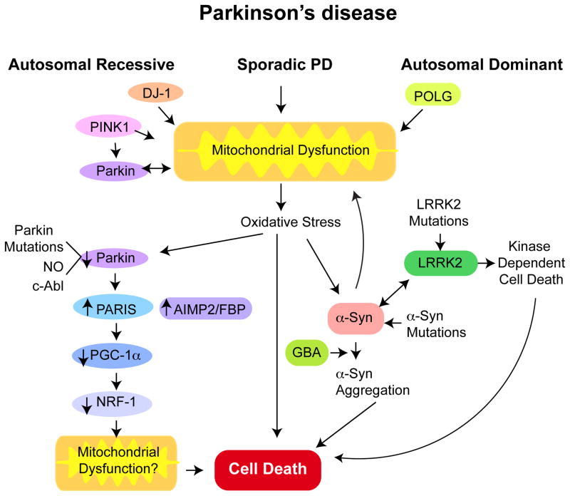
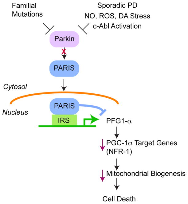
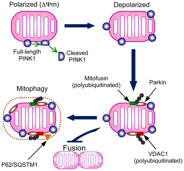
Genetic studies have provided valuable insight into the pathological mechanisms underlying Parkinson's disease (PD). The elucidation of genetic components to what was once largely considered a nongenetic disease has given rise to a multitude of cell and animal models enabling the dissection of molecular pathways involved in disease etiology. Here, we review advances obtained from models of dominant mutations in α-synuclein and LRRK2 as well as recessive PINK1, parkin and DJ-1 mutations. Recent genome-wide association studies have implicated genetic variability at two of these loci, α-synuclein and LRRK2, as significant risk factors for developing sporadic PD. This, coupled with the established role of mitochondrial impairment in both familial and sporadic PD, highlights the likelihood of common mechanisms fundamental to the etiology of both.


Figures

Figure 1
Figure 1. PD gene product domains and pathogenic mutations
Domains are arranged from N- to C- termini. For LRRK2: Ank (Ankyrin-like repeats); LRR (leucine-rich repeats); ROC (Ras of complex GTPase domain); COR (C-terminal of ROC); Kinase and WD40. PINK1: MTS (mitochondrial targeting sequence); TM (putative transmembrane domain); and serine/threonine kinase. For Parkin: UBL (ubiquitin-like) and two RING domains separated by an IBR (in-between RING) domain. α-synuclein has a number of imperfect KTKEGV repeat sequences (white stripes) in the N-terminal region and central NAC (non-amyloid component) region. DJ-1 is a single domain protein. Numbers under the protein indicate domain boundaries. Mutations that segregate with PD are annotated at their approximate position along the protein’s length.
Figure 2
Figure 2. Potential pathogenic pathways linking the genetic and sporadic causes of PD
A key feature of both sporadic and genetic causes of PD is mitochondrial dysfunction. Mutations in the autosomal recessive genes PINK1, Parkin and DJ-1 may directly cause mitochondrial dysfunction. PINK1 may act upstream of Parkin. PINK1 and Parkin may regulate mitochondria mitophagy. Parkin may directly regulate mitochondrial biogenesis through PARIS, which is a transcriptional repressor of the PGC-1α, the master regulator of mitochondrial biogenesis. Dominant mutations in POLG, the catalytic subunit of mitochondrial DNA polymerase causes Parkinsonism in some families. Dominant mutations in LRRK2 and α-synuclein (α-syn) cause PD. Oxidative stress including nitrosative stress and c-Abl phosphorylation of Parkin leads to its inactivation in sporadic PD and subsequent accumulation of substrates that are degraded by the ubiquitin proteasome system, such as AIMP2, FBP1 and PARIS. PARIS may be the key pathogenic Parkin substrate as knockdown of PARIS in an adult conditional knockout of Parkin completely rescues neurodegeneration of DA neurons. α-Synuclein aggregation is a key step in DA neuron degeneration in PD. Oxidative and nitrosative (NO) stress can accelerate α-synuclein and aggregated α-synuclein can damage mitochondria setting in motion a feed forward mechanism. GBA mutations also seem to accelerate α-synuclein aggregation and LRRK2 and α-synuclein interact at some level in the pathogenesis of PD. LRRK2 mutations lead to DA neurodegeneration that is kinase dependent.
Figure 3
Figure 3. α-Synuclein aggregation and pathophysiology
α-Synuclein natively unfolded monomers assemble to form β-sheet-rich soluble oligomers, which further aggregate to form mature fibrils. Aggregates at both stages are thought to act as templates that seed further α-synuclein fibrillogenesis in a feed-forward cycle. Fibrils can deposit into Lewy bodies or might break down into small transmissible aggregates via incomplete degradation that are able to transmit between cells, facilitating the spread of α-synuclein aggregation and toxicity in a prion-like manner. The mechanism of transmission from one neuron to another is unknown. Prevention of oligomer and fibril formation and the disaggregation of mature fibrils to non-toxic breakdown products are therapeutic goals and several compounds, including EGCG, baicalein, N-methylated peptides and catechol-based compounds have been put forward based on their effectiveness in vitro.
Figure 4
Figure 4. Parkin controls the expression of PGC-1α through PARIS
PARIS is a key pathogenic substrate of Parkin that accumulates in models of Parkin inactivation and in patients with Parkin mutations and PD patients due to nitrosative (NO), reactive oxygen species (ROS) and dopamine (DA) stress as well as c-Abl phosphorylation. PARIS is a transcriptional repressor that selectively down regulates PGC1-α by binding to insulin response sequences (IRS) in the PGC1-α promoter leading to downregulation of NRF-1, which transcriptionally controls mitochondrial biogenesis leading to DA neuron degeneration.
Figure 5
Figure 5. Interaction of PINK1 and Parkin in regulating mitochondrial turnover
The model proposed is based on recent evidence supporting PINK1-dependent recruitment of Parkin to mitochondria and subsequent Parkin substrate polyubiquitination to promote autophagy. On polarized mitochondria (mitochondrial membrane potential intact), PINK1 is cleaved into a short ~52 kDa fragment, which is released into the cytosol. This cleavage is voltage-dependent and impaired by membrane depolarization in dysfunctional mitochondria. Retention of PINK1 at the mitochondrial membrane leads to recruitment of Parkin by unknown mechanisms. Once localized to mitochondria, Parkin polyubiquitylates mitofusin and VDAC1 (voltage-dependent anion channel) and VDAC1 is required for Parkin-mediated mitochondrial clearance. Polyubiqutination of mitofusin may inactivate it and prevent fusion of the dysfunctional mitochondrion with the pool of healthy mitochondria. The autophagic adaptor protein P62/SQSTM1 is recruited to mitochondria and is required for mitochondrial clearance.

References

    1. Alegre-Abarrategui J, Ansorge O, Esiri M, Wade-Martins R. LRRK2 is a component of granular alpha-synuclein pathology in the brainstem of Parkinson’s disease. Neuropathol Appl Neurobiol. 2008;34:272–83. - PMC - PubMed
    1. Aleyasin H, Rousseaux MW, Marcogliese PC, Hewitt SJ, Irrcher I, et al. DJ-1 protects the nigrostriatal axis from the neurotoxin MPTP by modulation of the AKT pathway. Proc Natl Acad Sci U S A. 2010;107:3186–91. - PMC - PubMed
    1. Andres-Mateos E, Mejias R, Sasaki M, Li X, Lin BM, et al. Unexpected lack of hypersensitivity in LRRK2 knock-out mice to MPTP (1-methyl-4-phenyl-1,2,3,6-tetrahydropyridine) J Neurosci. 2009;29:15846–50. - PMC - PubMed
    1. Andres-Mateos E, Perier C, Zhang L, Blanchard-Fillion B, Greco TM, et al. DJ-1 gene deletion reveals that DJ-1 is an atypical peroxiredoxin-like peroxidase. Proc Natl Acad Sci U S A. 2007;104:14807–12. - PMC - PubMed
    1. Autere J, Moilanen JS, Finnila S, Soininen H, Mannermaa A, et al. Mitochondrial DNA polymorphisms as risk factors for Parkinson’s disease and Parkinson’s disease dementia. Hum Genet. 2004;115:29–35. - PubMed

Publication types