. 2011 Aug 1;317(13):1860-71.
doi: 10.1016/j.yexcr.2011.05.006. Epub 2011 May 18.

Decreased cell adhesion promotes angiogenesis in a Pyk2-dependent manner

Affiliations

Decreased cell adhesion promotes angiogenesis in a Pyk2-dependent manner

Colette J Shen et al. Exp Cell Res. .

Abstract

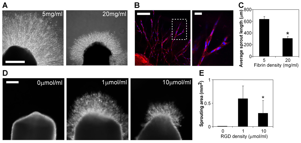
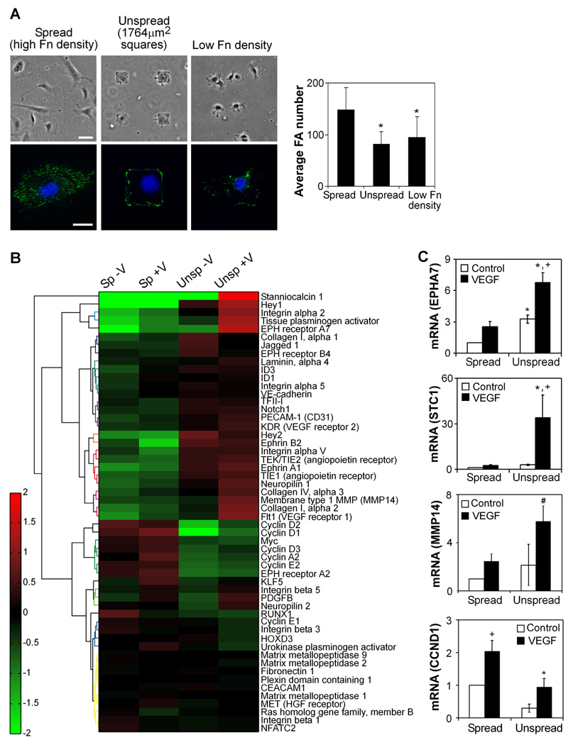
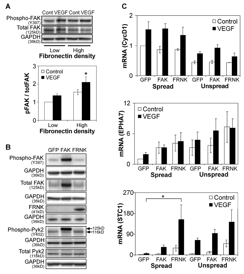
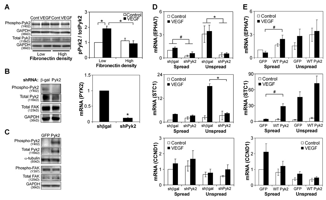
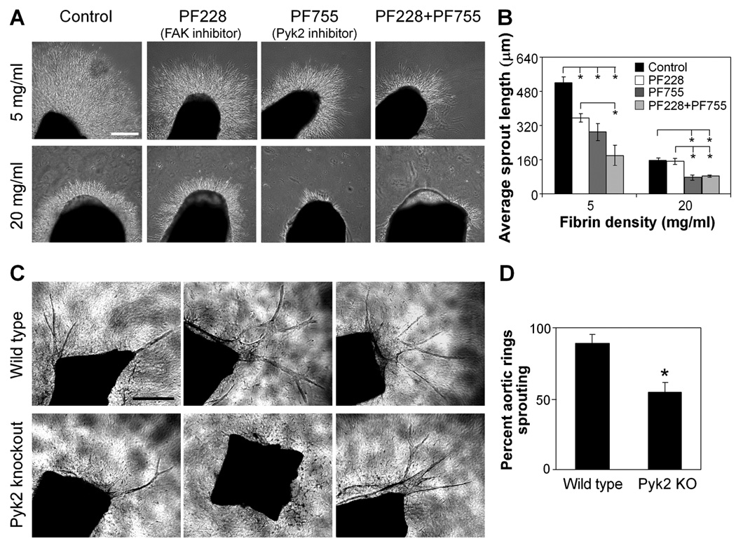
Angiogenesis is regulated by both soluble growth factors and cellular interactions with the extracellular matrix (ECM). While cell adhesion via integrins has been shown to be required for angiogenesis, the effects of quantitative changes in cell adhesion and spreading against the ECM remain less clear. Here, we show that angiogenic sprouting in natural and engineered three-dimensional matrices exhibited a biphasic response, with peak sprouting when adhesion to the matrix was limited to intermediate levels. Examining changes in global gene expression to determine a genetic basis for this response, we demonstrate a vascular endothelial growth factor (VEGF)-induced upregulation of genes associated with vascular invasion and remodeling when cell adhesion was limited, whereas cells on highly adhesive surfaces upregulated genes associated with proliferation. To explore a mechanistic basis for this effect, we turned to focal adhesion kinase (FAK), a central player in adhesion signaling previously implicated in angiogenesis, and its homologue, proline-rich tyrosine kinase 2 (Pyk2). While FAK signaling had some impact, our results suggested that Pyk2 can regulate both gene expression and endothelial sprouting through its enhanced activation by VEGF in limited adhesion contexts. We also demonstrate decreased sprouting of tissue explants from Pyk2-null mice as compared to wild type mice as further confirmation of the role of Pyk2 in angiogenic sprouting. These results suggest a surprising finding that limited cell adhesion can enhance endothelial responsiveness to VEGF and demonstrate a novel role for Pyk2 in the adhesive regulation of angiogenesis.


Figures

Figure 1
Figure 1. Decreased adhesive ligand density enhances angiogenic sprouting
(A) Sprouting of chick aortic rings embedded in 5 mg/ml (left) versus 20 mg/ml (right) fibrin in EGM-2 medium at 48 hours, bar=500 µm. (B) Endothelial cells in sprouts labeled with lectin (red) and Hoechst 33342 (blue), with region outlined by dotted line magnified on the right. Bar (left image), 100 µm; bar (right image), 20 µm. (C) Quantification of average sprout length for chick aortic rings in 5 mg/ml versus 20 mg/ml fibrin. Graph represents means±SEM (n=4), with each experiment averaged over at least 3 aortic rings. (D) Sprouting of chick aortic rings embedded in PEGDAAm gels with 0 µmol/ml adhesive RGDS + 10 µmol/ml non-adhesive RGES (left), 1 umol/ml RGDS + 9 umol/ml RGES (middle), and 10 umol/ml RGDS + 0 umol/ml RGES (right) in EGM-2 medium at 48 hours, bar=200 µm. (E) Quantification of sprouting area for chick aortic rings in PEGDAAm gels of varying RGDS density. Graph represents means±SD (n=8). *, p<0.05, compared to 1 umol/ml RGDS, as calculated by one-way ANOVA and post-hoc Tukey’s HSD test.
Figure 2
Figure 2. Cell adhesion and spreading regulate the expression of genes associated with angiogenesis
(A) Cell adhesion is controlled by constraining cells to micropatterned islands of fibronectin (“unspread,” 1764 µm2 area, high density 20 µg/ml fibronectin) or reducing fibronectin density (5 µg/ml), as compared to fully spread cells on high density fibronectin (“spread”). Phase images in top row, bar=50 µm. Immunofluorescent images in bottom row with vinculin (green) and Hoechst 33342 (blue), bar=20 µm. The average focal adhesion number per cell is quantified for each condition (means±SD, n=15 cells per condition). *, p<0.05 versus high fibronectin density, as calculated by one-way ANOVA and post-hoc Tukey’s HSD test. (B) Heatmap of expression of genes associated with angiogenesis in four conditions (spread no VEGF, spread with VEGF, unspread no VEGF, unspread with VEGF) after 18 hours of culture. Heatmap values represent log-transformed ratios of expression in one condition to the average expression value over all conditions for a given gene, averaged over three replicates. Genes are clustered based on similarities in expression patterns using Ward’s hierarchical clustering method. (C) HUVECs were cultured as spread or unspread, in starvation medium with or without 25 ng/ml VEGF for 16–18 hours and analyzed for expression of select genes by quantitative real-time PCR analysis (EPHA7=Eph Receptor A7, MMP14=Membrane type 1 MMP, STC1=Stanniocalcin 1, CCND1=Cyclin D1). Data represent means±SEM (n≥3). *, p<0.05 compared to the spread condition, and +, p<0.05 compared to no VEGF control, as calculated by two-way ANOVA and post-hoc Tukey’s HSD test. #, p<0.05 compared to the spread condition, as calculated by Student’s t-test (not significant by ANOVA).
Figure 3
Figure 3. FAK is not a major regulator of limited adhesion-induced angiogenic gene expression
(A) Western blot of FAK phosphorylation in HUVECs cultured on high (20 µg/ml) and low (5 µg/ml) density fibronectin-coated surfaces with or without VEGF stimulation for 30 minutes. Data represent means±SEM (n=3). *, p<0.05 compared to low density fibronectin, as calculated by two-way ANOVA and post-hoc Tukey’s HSD test. (B) Western blot of FAK and Pyk2 phosphorylation in GFP-, FAK-, and FRNK-overexpressing HUVECs cultured on high density fibronectin without VEGF stimulation. Note that for phospho-Pyk2, the antibody interacts non-specifically with FAK and thus results in a higher molecular weight band (125kD) when FAK is overexpressed; the Pyk2 Y402 band is the lower molecular weight band at 116kD. (C) Gene expression of GFP-, FRNK-, and FAK-overexpressing HUVECs after 16–18 hours of culture in spread or unspread conditions with or without VEGF stimulation. Data represent means±SEM (n=3). *, p<0.05 compared to GFP, as calculated by three-way ANOVA and post-hoc Tukey’s HSD test.
Figure 4
Figure 4. Pyk2 regulates the expression of genes associated with angiogenesis
(A) Western blot of Pyk2 phosphorylation in HUVECs cultured on high (20 µg/ml) and low (5 µg/ml) density fibronectin-coated surfaces with or without VEGF stimulation for 30 minutes. Data represent means±SEM (n=4) and *, p<0.05, as calculated by two-way ANOVA and post-hoc Tukey’s HSD test. (B) Pyk2 and FAK protein expression by Western blot (left) and Pyk2 mRNA (right) in HUVECs transfected with control (β-gal) versus Pyk2 shRNA cultured on high density fibronectin without VEGF stimulation. mRNA graph data represent means±SEM (n=4). *, p<0.05, as calculated by Student’s t-test. (C) Western blot of Pyk2 and FAK phosphorylation in GFP- and Pyk2-overexpressing HUVECs cultured on high density fibronectin without VEGF stimulation. (D) Gene expression of HUVECs transfected with control versus Pyk2 shRNA after 16–18 hours of culture in spread or unspread conditions with or without VEGF stimulation. Data represent means±SEM (n=3). *, p<0.05, as calculated by three-way ANOVA and post-hoc Tukey’s HSD test. (E) Gene expression of GFP- and Pyk2-overexpressing HUVECs after 16–18 hours of culture in spread or unspread conditions with or without VEGF stimulation. Data represent means±SEM (n=3). #, p<0.05, as calculated by Student’s t-test (not significant by ANOVA).
Figure 5
Figure 5. Pyk2 regulates angiogenic sprouting
(A) Sprouting of chick aortic rings in the presence of DMSO control, 500 nM PF228, 500 nM PF755, or both 500 nM PF228 and 500 nM PF755 in low (5 mg/ml) versus high (20 mg/ml) density fibrin in EGM-2 medium at 48 hours, bar=300 µm. (B) Quantification of average sprout length for aortic rings in (A). Data represent means±SEM (n=3), with each experiment averaged over at least 2 aortic rings. *, p<0.05, as calculated by one-way ANOVA and post-hoc Tukey’s HSD test. (C) Sprouting of aortic rings isolated from Pyk2 knockout versus wild type mice in 2.5mg/ml collagen I gels in VEGF-containing medium at 8 days, bar=500 µm. (D) Quantification of percent of aortic rings with sprouting in the conditions in (C). Data represent means±SEM (n=4), with each experiment averaged over at least 8 aortic rings. *, p<0.05, as calculated by Student’s t-test.

References

    1. Adams RH, Alitalo K. Molecular regulation of angiogenesis and lymphangiogenesis. Nat Rev Mol Cell Biol. 2007;8:464–478. - PubMed
    1. Ferrara N, Gerber HP, LeCouter J. The biology of VEGF and its receptors. Nat Med. 2003;9:669–676. - PubMed
    1. Carmeliet P, Ferreira V, Breier G, Pollefeyt S, Kieckens L, Gertsenstein M, Fahrig M, Vandenhoeck A, Harpal K, Eberhardt C, Declercq C, Pawling J, Moons L, Collen D, Risau W, Nagy A. Abnormal blood vessel development and lethality in embryos lacking a single VEGF allele. Nature. 1996;380:435–439. - PubMed
    1. Ferrara N, Carver-Moore K, Chen H, Dowd M, Lu L, O'Shea KS, Powell- Braxton L, Hillan KJ, Moore MW. Heterozygous embryonic lethality induced by targeted inactivation of the VEGF gene. Nature. 1996;380:439–442. - PubMed
    1. Miquerol L, Langille BL, Nagy A. Embryonic development is disrupted by modest increases in vascular endothelial growth factor gene expression. Development. 2000;127:3941–3946. - PubMed

Substances