Review
. 2011 Jul;41(4):182-99.
doi: 10.1016/j.jchemneu.2011.05.004. Epub 2011 May 27.

The serotonergic anatomy of the developing human medulla oblongata: implications for pediatric disorders of homeostasis

Affiliations
Review

The serotonergic anatomy of the developing human medulla oblongata: implications for pediatric disorders of homeostasis

Hannah C Kinney et al. J Chem Neuroanat. 2011 Jul.

Abstract

The caudal serotonergic (5-HT) system is a critical component of a medullary "homeostatic network" that regulates protective responses to metabolic stressors such as hypoxia, hypercapnia, and hyperthermia. We define anatomically the caudal 5-HT system in the human medulla as 5-HT neuronal cell bodies located in the raphé (raphé obscurus, raphé magnus, and raphé pallidus), extra-raphé (gigantocellularis, paragigantocellularis lateralis, intermediate reticular zone, lateral reticular nucleus, and nucleus subtrigeminalis), and ventral surface (arcuate nucleus). These 5-HT neurons are adjacent to all of the respiratory- and autonomic-related nuclei in the medulla where they are positioned to modulate directly the responses of these effector nuclei. In the following review, we highlight the topography and development of the caudal 5-HT system in the human fetus and infant, and its inter-relationships with nicotinic, GABAergic, and cytokine receptors. We also summarize pediatric disorders in early life which we term "developmental serotonopathies" of the caudal (as well as rostral) 5-HT domain and which are associated with homeostatic imbalances. The delineation of the development and organization of the human caudal 5-HT system provides the critical foundation for the neuropathologic elucidation of its disorders directly in the human brain.


Figures

Figure 1
Figure 1
Schematic diagram of the caudal 5-HT system and its relationship to homeostatic regulation. This system: 1) receives sensory input about the internal milieu via the nucleus of the solitary tract (visceral sensory) in the autonomic nervous system, as well as its own chemo- and glucose receptors in close relationship to arteries and/or cerebrospinal fluid; 2) modulates adjustments to homeostatic stresses via its projections to the major medullary effector nuclei (HG, hypoglossal nucleus; NTS, nucleus of the solitary tract; DMX, dorsal motor nucleus of the vagus; preBöt, preBötzinger complex, Phr Nucl, phrenic nucleus in the cervical spinal cord; and IML, intermediolateral column in the thoracic spinal cord); 3) receives modulatory input itself from the hypothalamus other limbic forebrain sites relevant to sleep/wake cycle regulation and cardiorespiratory effects via receptor-mediated interactions with diverse neurotransmitters and neuromodulators; and 4) interfaces with the cytokine system which is critical to homeostasis in its mediation of “protective” sickness behaviors and cellular defenses against tissue damage. Abbreviations: O2, oxygen, CO2, carbon dioxide; temp, temperature; glc, glucose.
Figure 2
Figure 2
Schematic diagram of the topography of the caudal 5-HT system in the human infant medulla. The 5-HT neurons are adjacent to effector nuclei of cardiorespiratory function (solid gray). The source 5-HT neurons are located in the raphé (blue) and extra-raphé (green), and along the ventral medullary surface in the arcuate nucleus (red). Each circle represents a 5-HT neuron in the infant medulla. Abbreviations: GC, gigantocellularis; PGCL, paragigantocellularis lateralis; nAm, nucleus ambiguus; preBöt, putative locus of the human preBötzinger complex; PIO, principal inferior olive; NTS, nucleus of the solitary tract; HG, hypoglossal nucleus; DMX, dorsal motor nucleus of the vagus. The numbers stand for the cranial nerves.
Figure 3
Figure 3
Comparative neuroanatomy of the ventral surface of the medulla in the cat (left) versus human infant (right). The red neuronal clusters represent cytological and positional homologous neuronal sites between the human arcuate nucleus (hARC) and the cat (arcuate-like) sites (cARC) that are respiratory chemosensitive fields based upon physiological testing (Filiano et al., 1990). Other homologous cell populations are the cCT (cat conterminalis-like) and hCT (human conterminalis), as well as thickened marginal glia (TMG). Abbreviations: RP, raphé pallidus; PYR, pyramid; HN, hypoglossal nucleus; NTS, nucleus of the solitary tract; hCT, human nucleus conterminalis; CN 12, cranial nerve 12. Scale bar=5 milllimeters.
Figure 4
Figure 4
Immunostained 5-HT neurons extend ventrally from the region of the raphé pallidus (midline) (A) along the ventral surface of the pyramids (B) in the human infant medulla (Kinney et al., 2007). Abbreviations: pyr, pyramid; VMS, ventral medullary surface. A. ×4, scale bar=200 microns. B. ×10, scale bar=80 microns.
Figure 5
Figure 5
Three-dimensional view of the ventral surface of the medulla in the cat (A), rat (B), and human infant (C). The classical distribution of the ventrolateral respiratory chemosensitive zones (M, S, and L regions) are illustrated in the cat diagram (A). The relationship of 5-HT neurons (green) to the major arterial system of the medulla (red) is shown in the rat (B) (Bradley et al., 2002). The topographic distribution of immunostained 5-HT neurons in the human infant medulla are shown in the raphé (blue) and extra-raphé (paragigantocellularis lateralis) (green), and ventral surface (arcuate nucleus) (red) (Kinney et al., 2007). Similar 5-HT neuronal “columns” in the rostrocaudal axis are noted in the rat (B) and human infant (C) and are in a homologous lateral positions to the M, S, and L regions in the cat (A). See text.
Figure 6
Figure 6
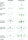
Computer-based graphic plots of the topography of 5-HT neurons in the developing human medulla (Kinney et al., 2007). The time period spans from 7–8 gestational weeks to 6 postnatal months, i.e., a critical period in homeostatic maturation in early human life. Each circle represents a 5-HT neuron in the raphé (blue), extra-raphé, and ventral surface (red). The medullary sections (cross-section) are to scale. By 15–22 gestational weeks, the basic topography is adult-like.
Figure 7
Figure 7
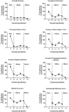
The developmental profile of 5-HT receptor binding with the broad radioligand 3H-LSD based upon tissue receptor autoradiography in 8 medullary nuclei (Paterson et al., 2004). In all sites sampled, there is a marked reduction in binding over the second half of gestation; binding is relatively uniform through infancy. Scale bar= 1 centimeter.
Figure 8
Figure 8
Montage of computer-based images of autoradiograms for 5-HT receptors (5-HT1A and 5-HT2A) and transporter (5-HTT) in the human infant medulla at the mid- and rostral levels in the infant medulla at 2–3 postnatal months. The tisse sections are from normative infants without neurological disorders or neuropatoalogic findings at autopsy. Computer-generated plots of 5-HT neurons at this level are provided for reference. The receptor binding patterns for inter-relating systems are also shown, i.e., cholinergic nicotinic and GABAA receptors. The binding levels of each computer-generated mosaic of the autoradiograms are scaled to the individual ligands and not across ligands. See text for description and references to methods. Abbreviations: Arc, arcuate nucleus; GC, gigantocellularis; PGCL, paragigantocellularis lateralis; nAm, nucleus ambiguus; preBöt, preBötzinger complex; ROb, raphé obscurus; NTS, nucleus of the solitary tract; HG, hypoglossal nucleus; DMX, dorsal motor nucleus of the vagus; PIO, principal inferior olive; LSD, lysergic acid diethylamide; Nic, nicotine; Epi, epibatidine; Cyt, cytisine; Bgt, α-bungarotoxin. The autoradiographic images were obtained from the laboratory's archives. Scale bar=5 millimeters.
Figure 9
Figure 9
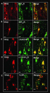
Expression of different receptors of key neurotransmitters (5-HT, GABAA and acetylcholine [nicotinic receptors]) and cytokine (IL-6R) that interface with 5-HT neurons, as demonstrated with double-label immunocytochemistry in the raphé obscurus in the normative human infant medulla at 2–3 postnatal months. Scale bar=100 microns. See text.
Figure 10
Figure 10
Schematic diagram of 5-HT synthetic and degradative pathways, receptors, and transporter. The sites of the different defects in developmental serotonopathies involving the caudal 5-HT domain are indicated. See text.
Figure 11
Figure 11
Decreased 5-HT receptor binding with 3H-LSD in the arcuate nucleus (encircled in red oval) in a SIDS case who died at 2 postnatal weeks compared to two age-related controls (Control A and Control B) (Kinney et al., 2005). This infant was studied prospectively with different parameters of central cardiorespiratory control at 2 days of life. The bottom three panels show the histograms and fitted normal curves for respiratory rate in active sleep, difference of heart rate between active and quiet sleep, and sustained change in beat-to-beat heart rate variability. The y-axis in each of the three graphs is the proportion of the subjects with a particular parameter. This infant had values for each of these parameters (red dot) that were outside the 10th percentile of values. Abbreviations: NTS, nucleus of the solitary tract; ION, inferior olivary nucleus; Arc, arcuate nucleus; br/min, breaths per minute; AS, active sleep; HR, heart rate; QS, quiet sleep; HRV, heart rate variability; %, percent.

References

    1. Abdala AP, Dutschmann M, Bissonnette JM, Paton JF. Correction of respiratory disorders in a mouse model of Rett syndrome. Proc Natl Acad Sci U S A. 2010;107:18208–18213. - PMC - PubMed
    1. Adamsen D, Meili D, Blau N, Thony B, Ramaekers V. Autism associated with low 5-hydroxyindolacetic acid in CSF and the heterozygous SLC6A4 gene Gly56Ala plus 5-HTTLPR L/L promoter variants. Mol Genet Metab. 2011;102:368–373. - PubMed
    1. Akefeldt A, Ekman R, Gillberg C, Mansson JE. Cerebrospinal fluid monoamines in Prader-Willi syndrome. Biol Psychiatry. 1998;44:1321–1328. - PubMed
    1. Akefeldt A, Mansson JE. Is monoamine oxidase activity elevated in Prader-Willi syndrome? Eur Child Adolesc Psychiatry. 1998;7:163–165. - PubMed
    1. Amaral DG, Schumann CM, Nordahl CW. Neuroanatomy of autism. Trends Neurosci. 2008;31:137–145. - PubMed

Publication types

MeSH terms

LinkOut - more resources