. 2011 Jun 10;145(6):863-74.
doi: 10.1016/j.cell.2011.05.020.

Kynurenine 3-monooxygenase inhibition in blood ameliorates neurodegeneration

Affiliations

Kynurenine 3-monooxygenase inhibition in blood ameliorates neurodegeneration

Daniel Zwilling et al. Cell. .

Abstract

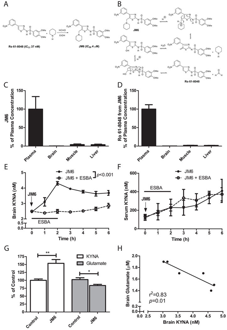
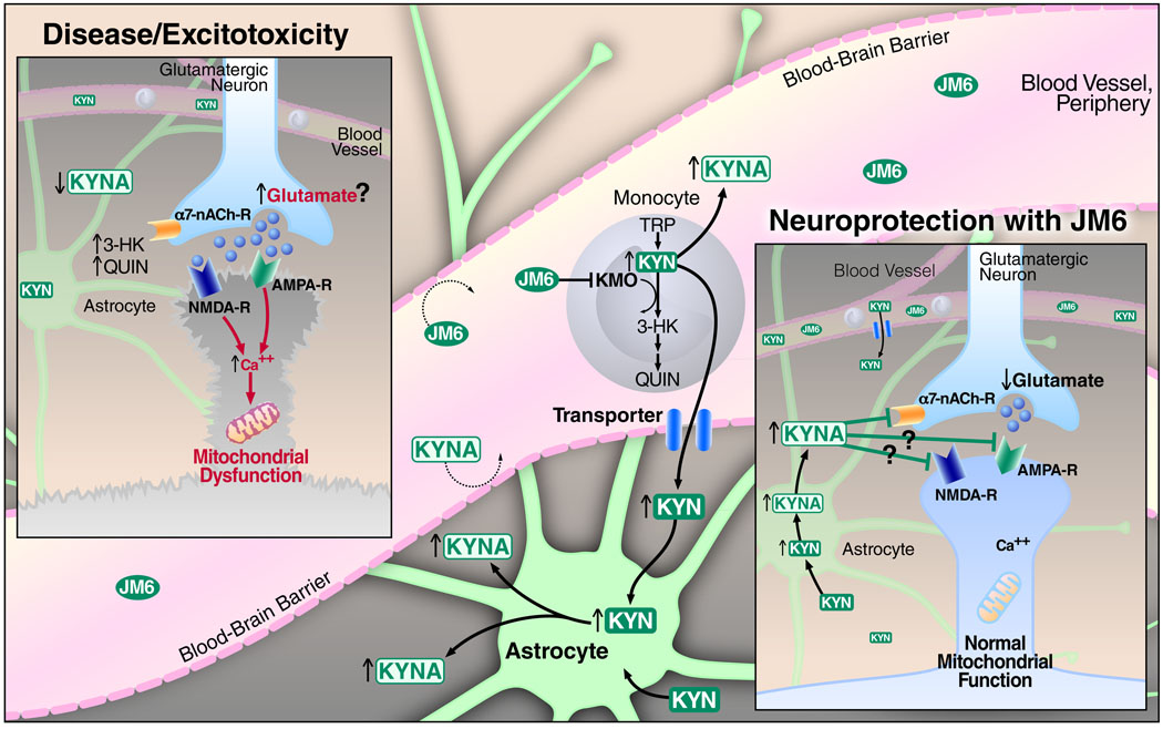
Metabolites in the kynurenine pathway, generated by tryptophan degradation, are thought to play an important role in neurodegenerative disorders, including Alzheimer's and Huntington's diseases. In these disorders, glutamate receptor-mediated excitotoxicity and free radical formation have been correlated with decreased levels of the neuroprotective metabolite kynurenic acid. Here, we describe the synthesis and characterization of JM6, a small-molecule prodrug inhibitor of kynurenine 3-monooxygenase (KMO). Chronic oral administration of JM6 inhibits KMO in the blood, increasing kynurenic acid levels and reducing extracellular glutamate in the brain. In a transgenic mouse model of Alzheimer's disease, JM6 prevents spatial memory deficits, anxiety-related behavior, and synaptic loss. JM6 also extends life span, prevents synaptic loss, and decreases microglial activation in a mouse model of Huntington's disease. These findings support a critical link between tryptophan metabolism in the blood and neurodegeneration, and they provide a foundation for treatment of neurodegenerative diseases.


Figures

Figure 1
Figure 1
The kynurenine pathway (KP) of tryptophan degradation in mammals. KMO functions at a key branching point in the KP. Inhibition of KMO causes the accumulation of its substrate kynurenine and a shunt in the KP, leading to increased production of the neuroprotective molecule KYNA.
Figure 2
Figure 2
JM6 is a novel pro-drug inhibitor of KMO that increases brain KYNA by blocking KMO in the blood. (A) Chemical synthesis of JM6. JM6 is a pro-drug of the KMO inhibitor Ro 61-8048. (B) Hypothetical mechanism for the acid-induced release of Ro 61-8048 from JM6. JM6 (C) and Ro 61-8048 released from JM6 (D) accumulate at high levels in plasma but not in brain or other tissues 5 h after JM6 administration (300 mg/kg p.o.) in mice. Absolute concentrations of JM6 were 40 µM (plasma), 119 nM (brain), 1.6 µM (muscle) and 5 µM (liver) (n = 5). Absolute concentrations of Ro 61-8048 were 7 µM (plasma), 18 nM (brain), 149 nM (muscle) and 132 nM (liver) (n = 5). (E) Extracellular KYNA levels in the striatum of rats measured by microdialysis following acute JM6 treatment (100 mg/kg, p.o.; arrow). In addition, in one group of animals, the KAT II inhibitor ESBA (1 mM) was applied by reverse dialysis for the first 2 h, and prevented the JM6-induced increase in KYNA levels (p<0.001, repeated-measures 2-way ANOVA; n = 5 per treatment group). (F) In the same rats, serum KYNA levels rose equally over time in the two groups, i.e. intracerebral ESBA administration did not influence circulating KYNA levels in JM6-treated rats. (G) Extracellular KYNA and glutamate were determined by in vivo microdialysis in the prefrontal cortex of rats treated for 7 days with JM6 (75 mg/kg/day p.o.). Baseline values for control rats were 2.5 ± 0.1 nM (KYNA) and 2.0 ± 0.2 µM (glutamate). (H) Extracellular KYNA correlates negatively with extracellular glutamate in these animals. *p<0.05, **p<0.01 (t-test). (n = 6 per group). Values in (C-G) are means ± s.e.m.
Figure 3
Figure 3
JM6 prevents spatial memory loss and anxiety deficits in a mouse model of AD. (A) JM6 rescues spatial memory deficits in APPtg mice in the Morris water maze (MWM). Mice were trained to find a hidden platform in the MWM using spatial cues. Spatial memory was assessed 24 h after training by removing the hidden platform and quantifying how much time mice spent in the target quadrant where the platform had been placed previously. APPtg mice that received JM6 (75 mg/kg/day p.o. for 120 days) spend significantly more time in the target quadrant than vehicle-treated controls. Solid bar, target quadrant; white bar, average of three other quadrants. (B) JM6 reduces anxiolytic behavior in APPtg mice in the elevated plus maze (EPM). APPtg mice are disinhibited and spend an equal amount of time in the open and closed arms in the EPM. However, APPtg mice that receive JM6 spend significantly more time in the closed arm than in the open arm and are not significantly different from WT mice. Values are means ± s.e.m. *p<0.05, **p<0.01, ***p<0.001. For (A,B), t tests were used to calculate differences between target vs. other quadrants, and time spent in open vs. closed arm, respectively (n = 8–14 mice per group). One-way ANOVA was used to calculate differences among the four experimental groups. JM6 reduces the distance traveled by APPtg mice in the open arm of the elevated plus maze. (C,D) APPtg mice that receive JM6 (75 mg/kg/day p.o. for ~120 days starting at day 30) travel significantly less distance in the open arm of the elevated plus maze than vehicle-treated controls, but the total travel distance in both arms of the maze is not different. Values are means ± s.e.m. ***p<0.001, t-test, open vs. closed arm. **p<0.01, ***p<0.001, one-way ANOVA comparing open arms between groups (n = 8–14 per group). ns: not significant.
Figure 4
Figure 4
JM6 prevents synaptic loss in a mouse model of AD. (A-D) JM6 (75 mg/kg/day p.o. for 120 days) prevents synaptic loss in the hippocampus and cortex of 7–8-month-old APPtg mice. (A,C) Representative images (630X) of serial sections of the hippocampus (A) and frontal cortex (C) of WT or APPtg mice immunostained with an antibody for synaptophysin. (B,D) Quantification of synaptophysin levels in the hippocampus (B) and frontal cortex (D) of APPtg mice treated with JM6. Synaptophysin levels in APPtg mice treated with JM6 are not significantly (n.s.) different from those found in WT mice. Values are means ± s.e.m. **p<0.01, ***p<0.001, one-way ANOVA (n = 5–9 per group).
Figure 5
Figure 5
JM6 increases brain KYNA levels in a mouse model of AD. (A) Compared to WT mice, cortical KYNA levels are significantly reduced in APPtg mice, and the deficit is normalized by JM6 treatment (75 mg/kg/day p.o. for 120 days); (B) Plasma KYNA measurements in the same three groups show a similar pattern as in the brain. (C-F) No group differences are seen in cortical KMO activity (C), 3-HK levels (D) and QUIN levels (E), as well as in plasma QUIN levels (F). Values are means ± s.e.m. [n = 5–15 per group, except APPtg controls in (B) (n = 3)]. *p<0.05, **p<0.01 (t-test); ns: not significant.
Figure 6
Figure 6
JM6 prevents neurodegeneration in a mouse model of HD. (A) Kaplan-Meier survival analysis shows that chronic JM6 administration (7.5 or 25 mg/kg/day p.o., starting at 4 weeks of age) increases survival in R6/2 mice in the absence of behavioral enrichment (log rank: p = 0.017; n = 8–13 per group). (B) Kaplan-Meier survival analysis shows that chronic JM6 administration (25 mg/kg/day p.o. starting at 4 weeks of age) increases survival in an independent cohort of R6/2 that received behavioral enrichment (log rank: p = 0.005; n = 14–15 per group). (C,D) JM6 (7.5 or 25 mg/kg/day p.o., starting at 4 weeks of age) prevents synaptic loss in R6/2 mice. (C) Representative images (630X) of serial sections of striatum from WT or R6/2 mice immunostained with an antibody for synaptophysin. (D) Quantification of synaptophysin levels in R6/2 mice treated with JM6. (E,F) JM6 (7.5 or 25 mg/kg/day p.o.) prevents the loss of Fos, a marker for neuronal activity, in R6/2 mice. (E) Representative images (440X) of serial sections of the striatum from WT or R6/2 mice immunostained with an antibody for Fos. (F) Quantification of Fos levels in R6/2 mice treated with JM6. (G,H) JM6 decreases CNS inflammation in R6/2 mice. (G) Representative images (440X) of serial sections of the striatum from WT or R6/2 mice immunostained with an antibody against Iba1 that labels microglia. (H) Iba1 levels in R6/2 mice treated with JM6 (7.5 or 25 mg/kg/day p.o., starting at 4 weeks of age). Levels of synaptophysin, Fos and Iba1 in R6/2 mice treated with JM6 are not significantly different from those in WT mice. (I) Brain sections from the cortex of 97–139-day-old R6/2 mice that received JM6 (25 mg/kg/day p.o., starting at 4 weeks of age) were immunostained with an anti-huntingtin antibody (EM48). Inclusion bodies increase in size and abundance between 97 and 139 days of age. (J) In the same animals JM6 does not significantly decrease the abundance of inclusion bodies, as determined by quantification of EM48 immunostaining. Values are means ± s.e.m. **p<0.01, ***p<0.001, one-way ANOVA (n = 5–13 per group). ns: not significant.
Figure 7
Figure 7
A model illustrating the mechanism by which KMO inhibition in blood cells leads to elevated brain KYNA levels and neuroprotection. In neurodegenerative diseases like HD and AD, increased levels of the toxic kynurenine pathway metabolites 3-HK and QUIN and decreased levels of the neuroprotective pathway metabolite KYNA might contribute to increased glutamatergic neurotransmission, elevation of intracellular calcium levels, mitochondrial dysfunction, and ultimately neuronal dysfunction and cell death (inset labeled “Disease/Excitotoxicity”). We hypothesize that the biotransformation of JM6 to Ro 61-8048 in the gut (not shown) results in KMO inhibition in peripheral monocytes, causing the accumulation of both kynurenine (KYN) and KYNA in blood. Unlike KYNA, KYN is then actively transported into the brain, where it is rapidly converted by astrocytes to KYNA. KYNA released from astrocytes mediates neuroprotection, at least in part, by decreasing glutamate levels via antagonism of pre-synaptic α7 nicotinic acetylcholine receptors (inset labeled “Neuroprotection with JM6”). However, at high local concentrations, KYNA might also directly block glutamate receptors to reduce excitotoxicity. Neuroprotection by KYNA might also involve a decrease in inflammation and modulation of mitochondrial function in immune cells (not shown).

Comment in

References

    1. Andiné P, Lehmann A, Ellren K, Wennberg E, Kjellmer I, Nielsen T, Hagberg H. The excitatory amino acid antagonist kynurenic acid administered after hypoxic-ischemia in neonatal rats offers neuroprotection. Neurosci Lett. 1988;90:208–212. - PubMed
    1. Beal MF, Matson WR, Storey E, Milbury P, Ryan EA, Ogawa T, Bird ED. Kynurenic acid concentrations are reduced in Huntington’s disease cerebral cortex. J Neurol Sci. 1992;108:80–87. - PubMed
    1. Björkqvist M, Wild EJ, Thiele J, Silvestroni A, Andre R, Lahiri N, Raibon E, Lee RV, Benn CL, Soulet D, et al. A novel pathogenic pathway of immune activation detectable before clinical onset in Huntington's disease. J Exp Med. 2008;205:1869–1877. - PMC - PubMed
    1. Boegman RJ, el-Defrawy SR, Jhamandas K, Beninger RJ, Ludwin SK. Quinolinic acid neurotoxicity in the nucleus basalis antagonized by kynurenic acid. Neurobiol Aging. 1985;6:331–336. - PubMed
    1. Campesan S, Green EW, Breda C, Sathyasaikumar KV, Muchowski PJ, Schwarcz R, Kyriacou CP, Giorgini F. The kynurenine pathway modulates neurodegeneration in a Drosophila model of Huntington’s disease. Curr Biol. 2011 in press. - PMC - PubMed

Publication types

Substances