. 2011 Oct;91(2):485-9.
doi: 10.1016/j.yexmp.2011.05.003. Epub 2011 May 27.

Delta-aminolevulinic dehydratase is a proteasome interacting protein

Affiliations

Delta-aminolevulinic dehydratase is a proteasome interacting protein

Fawzia Bardag-Gorce et al. Exp Mol Pathol. 2011 Oct.

Abstract

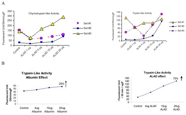
The proteasome interacts with a large number of proteins which regulate specific cellular functions. The focus of this study is to examine the proteasome interaction with Delta-aminolevulinate dehydratase (ALAD). ALAD is involved in the heme biosynthesis pathway and was co-isolated, with the 20S proteasome using several chromatographic purification steps. The MALDI-TOF mass spectrometry analysis identified this proteasome co-isolated protein as ALAD. When the proteasome was isolated using density-gradient centrifugation, ALAD was also found in the 26S proteasome fractions. It co-isolated with the 20S more than with the 26S proteasome. Furthermore, immunoprecipitated ALAD stained positive with antibodies to proteasome subunits. These results indicate that ALAD might interact with the proteasome. It is possible that ALAD is involved in modulating proteasome activity. When purified proteasomes were incubated with ALAD it was found that ALAD changes proteasome activity in a dose dependent manner. This indicates that ALAD may play a significant role in regulating proteasome activity. The data supports the hypothesis that ALAD, an important enzyme for heme synthesis, is also important as a proteasome interacting protein.


Figures

Fig. 1
Fig. 1
Panel A shows: (A) Chromatogram of 20S proteasome purified from rat liver. The spectra represents the protein measurement at 280nm using the optical unit linked to the FPLC system used for the proteasome purification. The columns used for this purification are described in MM. The red line is the KCl gradient applied for the 20S isolation. Note the peak preceding the 20S peak was an unknown protein Px, which co-isolated with the 20S, and, therefore, was isolated along with the 20S proteasome in the purification process. (B) and (C): SDS-Page electrophoresis and Coomassie Blue stained gels of Px Peak fractions (B) and 20S peak fraction (C). Note that the 44 kDa Px protein was highly concentrated and purified with the 20S. The band was cut out of the gel and analyzed by mass spectrometry.
Fig. 2
Fig. 2
The 20S co-isolated protein Px was analyzed by matrix-assisted laser desorption/ ionization-time of flight (MALDI-TOF) mass spectrometry. The trypsin digested peptides showed a high score of identification with the Delta aminolevulinic acid dehydratase sequences (a). The protein was identified in the database as Heme2 mouse Delta-aminolevulinic acid dehydratase (b)
Fig. 3
Fig. 3
ALAD and proteasome interaction. (a) Immunoprecipitation of ALAD and Western blot of the eluates using antibodies to the proteasome subunits Rpt4, a 19S ATPase subunit and to the 20S alpha 2 subunit. (b) Western blot analysis of the Px fraction collected from the 20S isolation using ALAD antibody. (c) 26S proteasome isolation. (d) Western blot of 26S proteasome fraction analyzed with ALAD antibody. (a) The “higher MW” corresponds to the unspecific binding of the 2nd antibody against the mouse monoclonal specific to Rpt4 or to α2. Possibly this binding is due to the protein-G that co-isolate with the immunoprecipitated complex. The protein-G has high affinity for the mouse monoclonal antibody. (b) The lower MW is certainly antigens detected by the antibody used to detect ALAD. However these lowers MW are not at the right size of ALAD that was detected efficiently with this antibody. The Markers are purchased from Bio-Rad and they are Blue when transferred to the membrane in the Western blot analysis. The detection method was done using Alkaline phosphatase Kit (colorimetric method). Therefore there is no need to visualize the markers.
Fig. 4
Fig. 4
ALAD modulated proteasome activity. (a) Chymotrypsin-like and trypsin-like activities were measured after incubating the proteasomes with ALAD for 10 min at 37°C. The three traces are measurements using three different proteasome preparation. (b) Control for ALAD-proteasome interaction effect. Bovin albumin was used as a nonspecific protein control.

References

    1. Bardag-Gorce F, Venkatesh R, Li J, French BA, French SW. Hyperphosphorylation of rat liver proteasome subunits: the effects of ethanol and okadaic acid are compared. Life Sci. 2004;75(5):585–597. - PubMed
    1. Bercovich B, Stancovski I, Mayer A, Blumenfeld N, Laszlo A, Schwartz AL, Ciechanover A. Ubiquitin-dependent degradation of certain protein substrates in vitro requires the molecular chaperone Hsc70. J. Biol. Chem. 1997;272(14):9002–9010. - PubMed
    1. Bose S, Stratford FL, Broadfoot KI, Mason GG, Rivett AJ. Phosphorylation of 20S proteasome alpha subunit C8 (alpha7) stabilizes the 26S proteasome and plays a role in the regulation of proteasome complexes by gamma-interferon. Biochem. J. 2004;378(Pt 1):177–184. - PMC - PubMed
    1. Bousquet-Dubouch MP, Nguen S, Bouyssié D, Burlet-Schiltz O, French SW, Monsarrat B, Bardag-Gorce F. Chronic ethanol feeding affects proteasome-interacting proteins. Proteomics. 2009 Jul;9(13):3609–22. [PMid: 19609968 doi:10.1002/pmic.200800959. - PMC - PubMed
    1. Bradford MM. A rapid and sensitive method for the quantitation of microgram quantities of protein utilizing the principle of protein-dye binding. Anal. Biochem. 1976;7:72, 248–54. - PubMed

Publication types

Substances