Comparative Study
. 2011 Jul 20;499(2):109-13.
doi: 10.1016/j.neulet.2011.05.044. Epub 2011 May 26.

Differential expression of miRNA-146a-regulated inflammatory genes in human primary neural, astroglial and microglial cells

Affiliations
Comparative Study

Differential expression of miRNA-146a-regulated inflammatory genes in human primary neural, astroglial and microglial cells

Yuan Yuan Li et al. Neurosci Lett. .

Erratum in

  • Neurosci Lett. 2012 Jun 27;520(1):126

Abstract

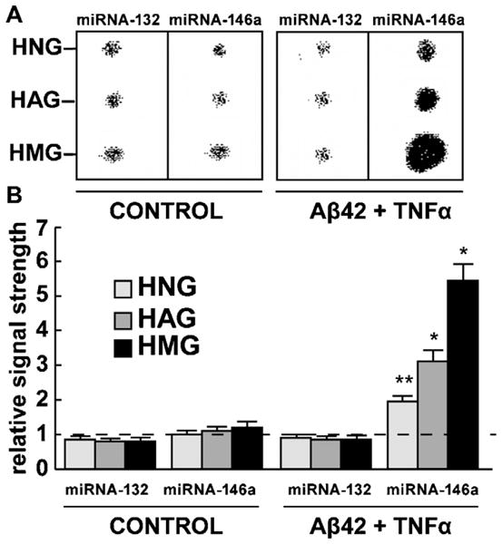
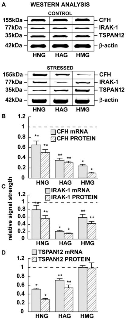
MicroRNA-146a (miRNA-146a) is an inducible, 22 nucleotide, small RNA over-expressed in Alzheimer's disease (AD) brain. Up-regulated miRNA-146a targets several inflammation-related and membrane-associated messenger RNAs (mRNAs), including those encoding complement factor-H (CFH) and the interleukin-1 receptor associated kinase-1 (IRAK-1), resulting in significant decreases in their expression (p<0.05, ANOVA). In this study we assayed miRNA-146a, CFH, IRAK-1 and tetraspanin-12 (TSPAN12), abundances in primary human neuronal-glial (HNG) co-cultures, in human astroglial (HAG) and microglial (HMG) cells stressed with Aβ42 peptide and tumor necrosis factor alpha (TNFα). The results indicate a consistent inverse relationship between miRNA-146a and CFH, IRAK-1 and TSPAN12 expression levels, and indicate that HNG, HAG and HMG cell types each respond differently to Aβ42-peptide+TNFα-triggered stress. While the strongest miRNA-146a-IRAK-1 response was found in HAG cells, the largest miRNA-146a-TSPAN12 response was found in HNG cells, and the most significant miRNA-146a-CFH changes were found in HMG cells, the 'resident scavenging macrophages' of the brain.


Figures

Fig. 1
Fig. 1
Primary cultures of human neuronal-glial [HNG] cells (A), human astroglia [HAG] cells (B), and human microglial [HMG] cells (C) are currently used to study human brain aging- and AD-relevant disease mechanisms [15,33,22,6,27,20,13,18,1,11,24,5,38]. HNG and HAG cells are stained using antibody to glial fibrillary acidic protein (GFAP), a glial-specific marker (green fluorescence; λmax = 556 nm); HMG cells are stained with fluorescent antibody to a human microglial-specific Iba1 (green fluorescence; λmax = 504 nm; sc-32725, Santa Cruz); in addition to GFAP, HNG cells are stained with βTUBIII, a neuron-specific marker (red; λmax = 702 nm) and Hoescht 33258 to highlight apoptotic features of cell nuclei (blue; λmax = 461 nm) (C). Brain cell types and numbers can be electronically quantified according to differential nuclear and cytosol staining and λmax [1,21,38]. Using the λmax index after 2 weeks in culture HNG cells Exhibit 50/50 neuronal/astroglial cells, HAG cultures exhibit at least 95% of cells staining with a λmax = 556 nm and HMG cultures exhibit at least 95% of cells staining with a λmax = 504 nm [,; unpublished]. Interestingly, highly purified monocultures of HMG cells begin expressing GFAP mRNA and react with antibody to GFAP after 2 or more weeks in culture [37]; each cell type shown here are 2 weeks in culture; HNG and HAG cells are about 50% confluent; HMG cells are about 25% confluent; all magnifications 20× [21,38].
Fig. 2
Fig. 2
Analysis of a control miRNA-132 and an inducible miRNA-146a in HNG, HAG and HMG cells both in untreated controls and in Aβ42 peptide + TNFα-stressed cells. Northern dot blot analysis (A) and signal quantitation using qPCR analysis (B); similar results were obtained using either miRNA array, Northern dot blot or qPCR analysis. While the levels of miRNA-132 were found to not change under any treatment condition, miRNA-146a was induced 2.1-, 3.2- and 5.4-fold over controls in HNG, HAG and HMG cells, respectively after the stress treatments indicated. Signals were quantified against internal 5SRNA levels; for ease of comparison a dashed horizontal line at 1.0 indicates miRNA-146a levels in control HNG cells; N = 5 for each cell type determination; significance over controls *p < 0.01; **p < 0.05 (ANOVA).
Fig. 3
Fig. 3
Levels of complement factor H (CFH), interleukin-1 receptor associated kinase 1 (IRAK-1) and TSPAN12 protein in control and in stressed (Aβ42 peptide + TNFα) HNG, HAG and HMG cells compared to β-actin levels within the same sample (A). Relative mRNA and protein levels (as determined by qPCR or Western analysis, respectively) are quantified for CFH (B), IRAK-1 (C) or TSPAN12 (D). For ease of comparison a dashed horizontal line in (B–D) at 1.0 indicates relative control levels. Interestingly, TSPAN12 mRNA and protein signals remained unchanged in HMG cells after any treatment condition studied here (D); N = 8 for each cell type; significance compared to control levels *p < 0.01; **p < 0.05 (ANOVA).

References

    1. Alexandrov PN, Zhao Y, Pogue AI, Tarr MA, Kruck TPA, Percy ME, Lukiw WJ. Synergistic effects of iron and aluminum on stress-related gene expression in primary human neural cells. J Alzheimers Dis. 2005;8:117–127. - PubMed
    1. Aronica E, Fluiter K, Iyer A, Zurolo E, Vreijling J, van Vliet EA, Baayen JC, Gorter JA. Expression pattern of miRNA-146a, an inflammation-associated miRNA, in experimental and human temporal lobe epilepsy. Eur J Neurosci. 2010;31:1100–1107. - PubMed
    1. Baltimore D, Boldin MP, O’Connell RM, Rao DS, Taganov KD. MiRNAs:, new regulators of immune cell development and function. Nat Immunol. 2008;9:839–845. - PubMed
    1. Colangelo V, Schurr J, Ball MJ, Pelaez RP, Bazan NG, Lukiw WJ. Gene expression profiling of 12633 genes in Alzheimer hippocampal CA1: transcription and neurotrophic factor down-regulation and up-regulation of apoptotic and pro-inflammatory signaling. J Neurosci Res. 2002;70:462–473. - PubMed
    1. Cui JG, Kuroda H, Chandrasekharan NV, Pelaez RP, Simmons DL, Bazan NG, Lukiw WJ. Cyclooxygenase-3 gene expression in Alzheimer hippocampus and in stressed human neural cells. Neurochem Res. 2004;29:1731–1737. - PubMed

Publication types

MeSH terms

LinkOut - more resources