

Pathogenesis of Influenza A/H5N1 virus infection in ferrets differs between intranasal and intratracheal routes of inoculation
- PMID: 21640972
- PMCID: PMC3123863
- DOI: 10.1016/j.ajpath.2011.03.026
Pathogenesis of Influenza A/H5N1 virus infection in ferrets differs between intranasal and intratracheal routes of inoculation
Abstract
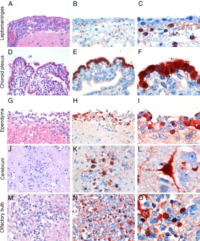
Most patients infected with highly pathogenic avian influenza A/H5N1 virus develop severe pneumonia resulting in acute respiratory distress syndrome, with extrarespiratory disease as an uncommon complication. Intranasal inoculation of ferrets with influenza A/H5N1 virus causes lesions in both the respiratory tract and extrarespiratory organs (primarily brain). However, the route of spread to extrarespiratory organs and the relative contribution of extrarespiratory disease to pathogenicity are largely unknown. In the present study, we characterized lesions in the respiratory tract and central nervous system (CNS) of ferrets (n = 8) inoculated intranasally with influenza virus A/Indonesia/5/2005 (H5N1). By 7 days after inoculation, only 3 of 8 ferrets had a mild or moderate bronchointerstitial pneumonia. In contrast, all 8 ferrets had moderate or severe CNS lesions, characterized by meningoencephalitis, choroiditis, and ependymitis, and centered on tissues adjoining the cerebrospinal fluid. These findings indicate that influenza A/H5N1 virus spread directly from nasal cavity to brain, and that CNS lesions contributed more than pulmonary lesions to the pathogenicity of influenza A/H5N1 virus infection in ferrets. In comparison, intratracheal inoculation of ferrets with the same virus reproducibly caused severe bronchointerstitial pneumonia. The method of virus inoculation requires careful consideration in the design of ferret experiments as a model for influenza A/H5N1 in humans.
Copyright © 2011 American Society for Investigative Pathology. Published by Elsevier Inc. All rights reserved.
Figures

References
-
- Abdel-Ghafar A.N., Chotpitayasunondh T., Gao Z., Hayden F.G., Nguyen D.H., de Jong M.D., Naghdaliyev A., Peiris J.S., Shindo N., Soeroso S., Uyeki T.M., Writing Committee of the Second World Health Organization Consultation on Clinical Aspects of Human Infection with Avian Influenza A Virus Update on avian influenza A (H5N1) virus infection in humans. N Engl J Med. 2008;358:261–273. - PubMed
-
- Gambotto A., Barratt-Boyes S.M., de Jong M.D., Neumann G., Kawaoka Y. Human infection with highly pathogenic H5N1 influenza virus. Lancet. 2008;371:1464–1475. - PubMed
-
- Tanaka H., Park C.H., Ninomiya A., Ozaki H., Takada A., Umemura T., Kida H. Neurotropism of the 1997 Hong Kong H5N1 influenza virus in mice. Vet Microbiol. 2003;95:1–13. - PubMed
-
- Rimmelzwaan G.F., van Riel D., Baars M., Bestebroer T.M., van Amerongen G., Fouchier R.A., Osterhaus A.D., Kuiken T. Influenza A virus (H5N1) infection in cats causes systemic disease with potential novel routes of virus spread within and between hosts. Am J Pathol. 2006;168:176–183. quiz 364. - PMC - PubMed
Publication types
MeSH terms
LinkOut - more resources
Full Text Sources
Medical
