. 2011 Jun;178(6):2461-9.
doi: 10.1016/j.ajpath.2011.01.058.

Resident tissue-specific mesenchymal progenitor cells contribute to fibrogenesis in human lung allografts

Affiliations

Resident tissue-specific mesenchymal progenitor cells contribute to fibrogenesis in human lung allografts

Natalie Walker et al. Am J Pathol. 2011 Jun.

Abstract

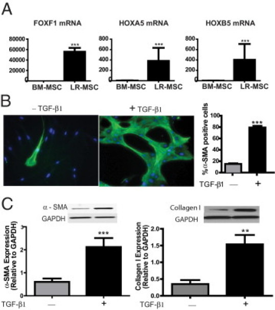
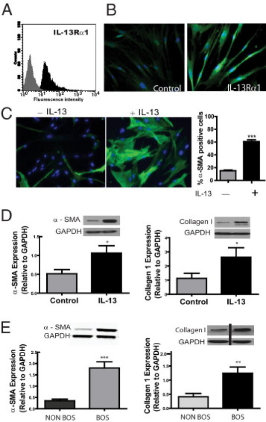
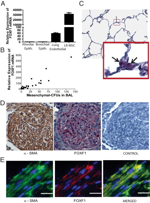
Fibrotic obliteration of the small airways leading to progressive airflow obstruction, termed bronchiolitis obliterans syndrome (BOS), is the major cause of poor outcomes after lung transplantation. We recently demonstrated that a donor-derived population of multipotent mesenchymal stem cells (MSCs) can be isolated from the bronchoalveolar lavage (BAL) fluid of human lung transplant recipients. Herein, we study the organ specificity of these cells and investigate the role of local mesenchymal progenitors in fibrogenesis after lung transplantation. We demonstrate that human lung allograft-derived MSCs uniquely express embryonic lung mesenchyme-associated transcription factors with a 35,000-fold higher expression of forkhead/winged helix transcription factor forkhead box (FOXF1) noted in lung compared with bone marrow MSCs. Fibrotic differentiation of MSCs isolated from normal lung allografts was noted in the presence of profibrotic mediators associated with BOS, including transforming growth factor-β and IL-13. MSCs isolated from patients with BOS demonstrated increased expression of α-SMA and collagen I when compared with non-BOS controls, consistent with a stable in vivo fibrotic phenotype. FOXF1 mRNA expression in the BAL cell pellet correlated with the number of MSCs in the BAL fluid, and myofibroblasts present in the fibrotic lesions expressed FOXF1 by in situ hybridization. These data suggest a key role for local tissue-specific, organ-resident, mesenchymal precursors in the fibrogenic processes in human adult lungs.


Figures

Figure 1
Figure 1
A: Increased expression of embryonic lung mesenchyme-associated transcription factors in lung-derived MSCs. mRNA expression of FOXF1, HOXA5, and HOXB5 in LR-MSCs isolated from BAL fluid of lung allografts (n = 10 LR-MSC lines derived from individual patients) was compared with that in BM-MSCs (n = 3) by real-time PCR. ***P < 0.0001. B and C: LR-MSCs demonstrate myofibroblast differentiation potential in response to the profibrotic mediator TGF-β. LR-MSCs isolated from normal lung allografts, without evidence of acute or chronic rejection, were treated with or without TGF-β1 (2 ng/mL) for 24 hour. In B, immunofluorescence staining of LR-MSCs demonstrated an increase in α-SMA–positive stress fibers in response to TGF-β1. Right: A quantitative analysis of α-SMA–positive cells across 10 high-power fields in three normal cell lines is shown. ***P < 0.0001. C: Effect of TGF-β on α-SMA and collagen I protein expression, analyzed by Western blot analysis. Immunoblots shown are from a representative experiment, with graphical data representing the densitometric ratio of the protein of interest to loading control proteins. Data represent the mean ± SEM of experiments with LR-MSCs derived from 10 lung transplant recipients. ***P = 0.0002 and **P = 0.006. GAPDH indicates glyceraldehyde-3-phosphate dehydrogenase.
Figure 2
Figure 2
A and B: LR-MSCs express IL-13 receptor α1 (IL-13Rα1). A: Immunophenotyping by flow cytometric analysis demonstrates positive IL-13Rα1 expression on LR-MSCs isolated from human lung allografts. The histogram shows IL-13Rα1 staining in black and isotype control staining in gray (n = 5). B: Immunofluorescent staining of LR-MSCs demonstrates IL-13Rα1–positive staining (compared with control unstained). C and D: LR-MSCs demonstrate profibrotic differentiation in response to IL-13. LR-MSCs isolated from normal lung allografts, without evidence of acute or chronic rejection, were treated with or without IL-13 (10 ng/mL) for 24 hours. C: Expression of α-SMA in LR-MSCs with or without IL-13 is shown using immunofluorescent staining. A quantitative analysis of α-SMA–positive cells across 10 high-power fields in three normal cell lines is shown (right). ***P < 0.0001. D: Effect of IL-13 on α-SMA and collagen I protein expression, analyzed by Western blot analysis. Data represent the mean ± SEM of experiments with LR-MSCs derived from 10 lung transplant recipients. *P < 0.05. E: LR-MSCs isolated from patients with BOS demonstrate a profibrotic phenotype. α-SMA and collagen I protein expression in LR-MSCs isolated from patients with and without BOS was compared by Western blot analysis. Data represent the mean ± SEM of experiments with LR-MSCs derived from 10 lung transplant recipients in each group. ***P < 0.0001 and **P = 0.003. GAPDH indicates glyceraldehyde-3-phosphate dehydrogenase.
Figure 3
Figure 3
A: FOXF1 expression in other endogenous lung cellular populations. FOXF1 mRNA expression in human lung allograft-derived MSCs (LR-MSCs), human alveolar epithelial (Epith.) cells (A549), human lung primary airway epithelial cells, and human pulmonary artery endothelial cells by real-time qPCR is shown. B: FOXF1 mRNA expression in BAL fluid correlates with the number of LR-MSCs in human lung transplant recipients. FOXF1 expression in 1 × 10⁶ nucleated BAL fluid cells was studied by real-time PCR. The numbers of LR-MSCs in those BAL samples were quantitated by measuring colony forming unit-fibroblast. A significant correlation was noted between number of LR-MSCs and FOXF1 mRNA in the BAL fluid (Pearson r = 0.92; 95% CI, 0. 86 to 0.95; P < 0.001). n = 50 BAL fluid samples. C: FOXF1 expression in normal adult lungs. The expression of FOXF1 in normal human lung was assessed by in situ hybridization using a digoxigenin-labeled RNA probe, followed by hematoxylin counterstaining. The enlarged box indicates the lesion marked by the box within the figure. The black arrows show cells positive for FOXF1. Original magnification: ×100 (top); ×600 (bottom). D: FOXF1 expression in fibrotic lesions in human lung allografts. Representative sections of a transbronchial lung biopsy specimen demonstrated organizing pneumonia in a lung transplant recipient, stained for α-SMA (by IHC staining) and FOXF1 (by in situ hybridization). Left: A discrete area of organizing pneumonia with intense α-SMA staining (brown) signifying infiltration by myofibroblasts. Center: FOXF1 mRNA expression was detected by in situ hybridization in the fibrotic area. Discrete spindle-shaped cells demonstrating red staining, consistent with FOXF1 expression, are noted in the area of organizing pneumonia. Right:In situ hybridization using digoxigenin-labeled control mRNA. Control for α-SMA staining is shown in Supplemental Figure S2 (available at http://ajp.amjpathol.org). Original magnification, ×400. E: Coexpression of FOXF1 and α-SMA in fibrotic lesions. A section from a human lung allograft biopsy specimen demonstrating fibrotic lesions was examined for the expression of FOXF1 and α-SMA using double-immunofluorescence microscopy. Rhodamine and fluorescein tyramide signal amplifications were used to detect the signal for FOXF1 and α-SMA, respectively. Colocalization of FOXF1 and α-SMA in spindle-shaped cells demonstrated FOXF1 expression in myofibroblasts. Original magnification, ×600 (oil). Scale bar = 20 μm.

References

    1. Estenne M., Hertz M.I. Bronchiolitis obliterans after human lung transplantation. Am J Respir Crit Care Med. 2002;166:440–444. - PubMed
    1. Boehler A., Estenne M. Obliterative bronchiolitis after lung transplantation. Curr Opin Pulm Med. 2000;6:133–139. - PubMed
    1. Hinz B., Phan S.H., Thannickal V.J., Galli A., Bochaton-Piallat M.L., Gabbiani G. The myofibroblast: one function, multiple origins. Am J Pathol. 2007;170:1807–1816. - PMC - PubMed
    1. Lama V.N., Harada H., Badri L.N., Flint A., Hogaboam C.M., McKenzie A., Martinez F.J., Toews G.B., Moore B.B., Pinsky D.J. Obligatory role for interleukin-13 in obstructive lesion development in airway allografts. Am J Pathol. 2006;169:47–60. - PMC - PubMed
    1. Estenne M., Maurer J.R., Boehler A., Egan J.J., Frost A., Hertz M., Mallory G.B., Snell G.I., Yousem S. Bronchiolitis obliterans syndrome 2001: an update of the diagnostic criteria. J Heart Lung Transplant. 2002;21:297–310. - PubMed

Publication types

MeSH terms