. 2011 Jun;178(6):2536-46.
doi: 10.1016/j.ajpath.2011.02.045.

Spontaneous lung dysfunction and fibrosis in mice lacking connexin 40 and endothelial cell connexin 43

Affiliations

Spontaneous lung dysfunction and fibrosis in mice lacking connexin 40 and endothelial cell connexin 43

Michael Koval et al. Am J Pathol. 2011 Jun.

Abstract

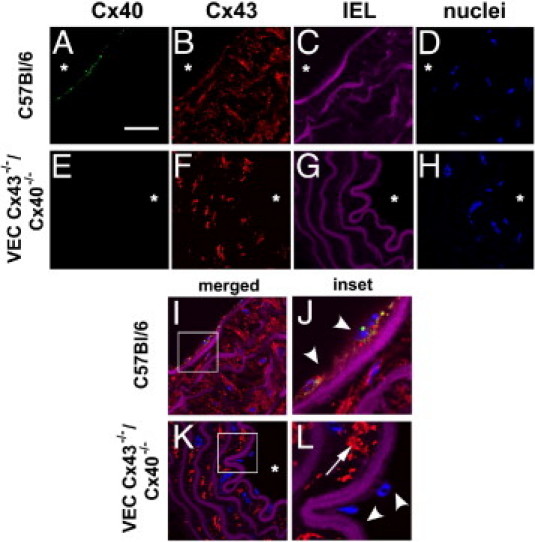
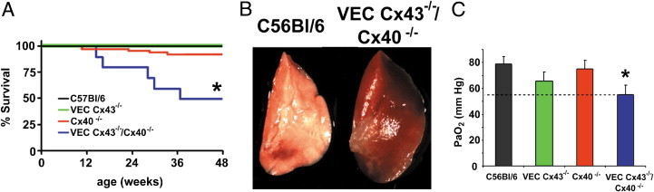
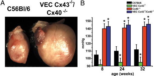
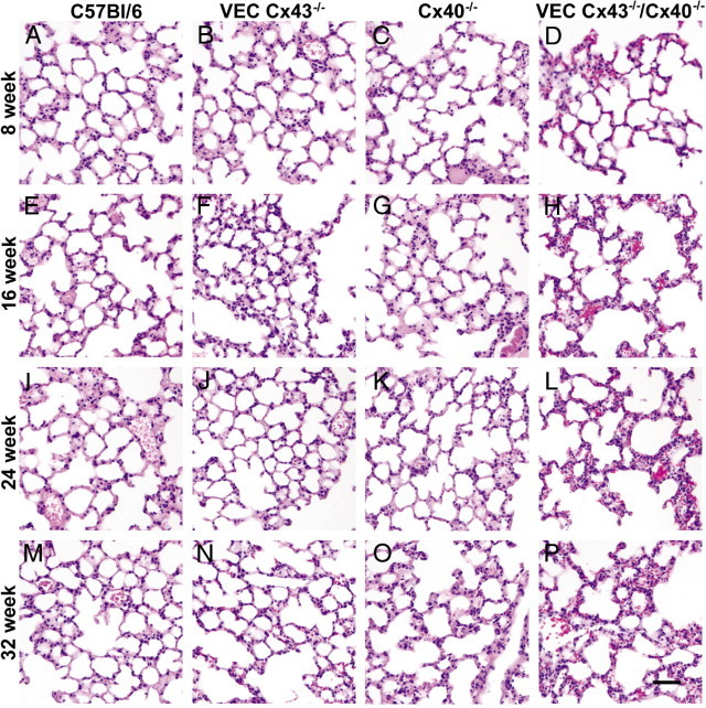
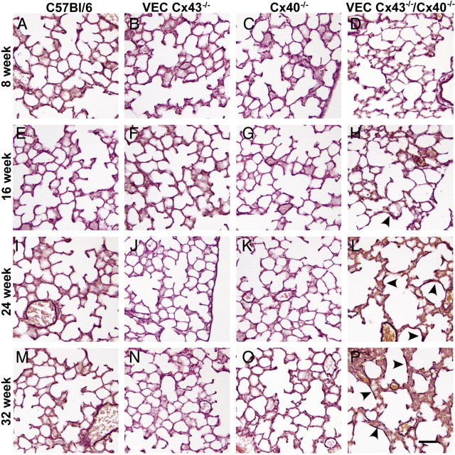
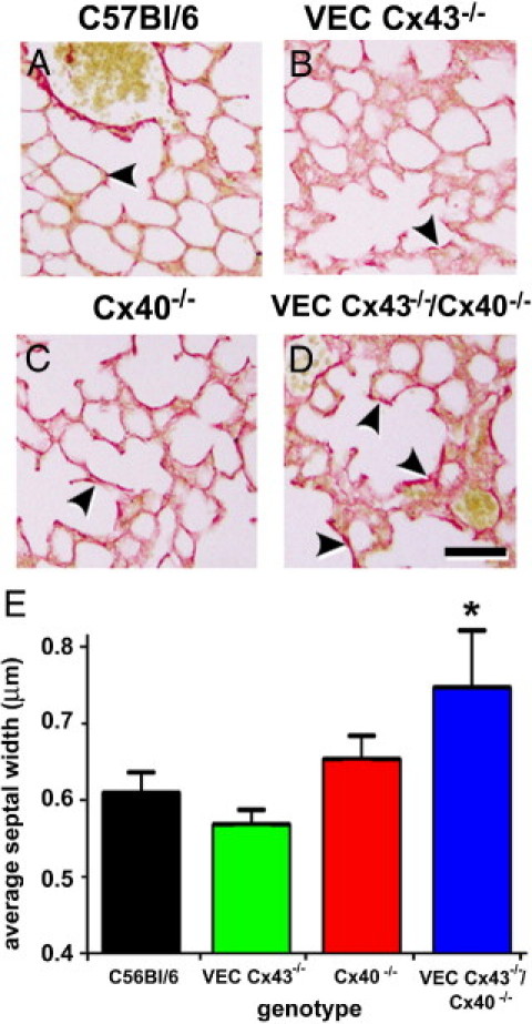
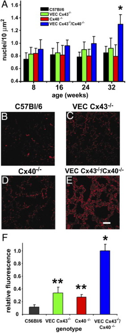
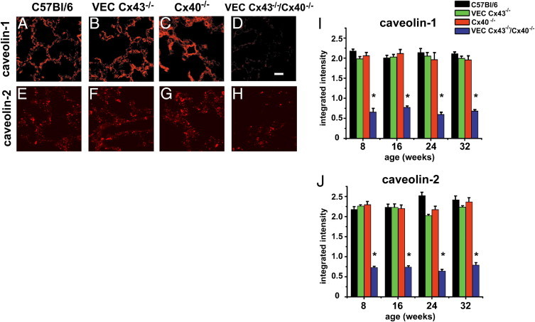
Gap junction proteins (connexins) facilitate intercellular communication and serve several roles in regulation of tissue function and remodeling. To examine the physiologic effects of depleting two prominent endothelial connexins, Cx40 and Cx43, transgenic mice were generated by breeding Cx40-deficient mice (Cx40(-/-)) with a vascular endothelial cell (VEC)-specific Cx43-deficient mouse strain (VEC Cx43(-/-)) to produce double-connexin knockout mice (VEC Cx43(-/-)/Cx40(-/-)). The life span in VEC Cx43(-/-)/Cx40(-/-) mice was dramatically shortened, which correlated with severe spontaneous lung abnormalities as the mice aged including increased fibrosis, aberrant alveolar remodeling, and increased lung fibroblast content. Moreover, VEC Cx43(-/-)/Cx40(-/-) mice exhibited cardiac hypertrophy and hypertension. Because VEC Cx43(-/-)/Cx40(-/-) mice demonstrated phenotypic hallmarks that were remarkably similar to those in mice deficient in caveolin-1, pulmonary caveolin expression was examined. Lungs from VEC Cx43(-/-)/Cx40(-/-) mice demonstrated significantly decreased expression of caveolin-1 and caveolin-2. This suggests that expression of caveolin-1 may be linked to expression of Cx40 and endothelial Cx43. Moreover, the phenotype of caveolin-1(-/-) mice and VEC Cx43(-/-)/Cx40(-/-) mice may arise via a common mechanism.


Figures

Figure 1
Figure 1
Cx40 and Cx43 expression in mouse aorta of control and VEC Cx43−/−/Cx40 −/− mice. Sections from C57Bl/6 mice (A–D, I, and J) and VEC Cx43−/−/Cx40 −/− mice (E–H, K, and L) were stained with anti-Cx40 [A and E (green)] and anti-Cx43 [B and F (red)]. C, G, and I–L: Violet areas represent autofluorescence of internal elastic lamina (IEL). D, H, and I–L: Nuclei were stained with Sytox Blue. I–L: Merged images. Note lack of Cx40 labeling in aortas from VEC Cx43−/−/Cx40 −/− mice; Cx43 was depleted from the endothelial layer in VEC Cx43−/−/Cx40 −/− mice (arrowheads) but remained present in the smooth muscle layer (arrows). J and L: Insets show threefold magnification of the regions of interest indicated in I and K. Scale bars = 100 μm.
Figure 2
Figure 2
VEC Cx43−/−/Cx40−/− mice have a shortened life span and demonstrate lung injury and decreased blood oxygenation. A: C57Bl/6, Cx40−/−, and VEC Cx43−/− mice all had normal viability; however, nearly half of the VEC Cx43−/−/Cx40−/− mice died by age 32 weeks. *Significantly different from control and single-knockout mice based on efficient score method.B: Gross anatomy of the left lobe of lungs from viable 32-week-old VEC Cx43−/−/Cx40−/− mice demonstrates a characteristic hemorrhagic appearance indicative of lung injury, as compared with healthy age-matched C57Bl/6 mice. C: Carotid arterial paO2 for C57Bl/6, VEC Cx43−/−, Cx40−/−, and VEC Cx43−/−/Cx40−/− mice measured at age 32 weeks (mean ± SE; n = 4). VEC Cx43−/−/Cx40−/− mice demonstrated significantly lower pO2 than did C57Bl/6 mice. *P < 0.05.
Figure 3
Figure 3
Compared with VEC Cx43−/−/Cx40−/− mice, Cx40−/− mice demonstrated increased blood pressure. A: Gross anatomy of hearts from 32-week-old VEC Cx43−/−/Cx40 −/− mice shows hypertrophy. B: Mean blood pressure measurements from C57Bl/6, VEC Cx43−/−, Cx40−/−, and VEC Cx43−/−/Cx40−/− mice measured at age 8, 24, or 32 weeks (mean ± SE; n = 4). Blood pressure in VEC Cx43−/− mice was significantly lower than C57Bl/6 mice at 24 and 32 weeks. *P < 0.05. Cx40−/− and VEC Cx43−/−/Cx40−/− mice had comparable blood pressure, which was significantly higher than C57Bl/6 mice. *P < 0.05.
Figure 4
Figure 4
Altered architecture of lungs from VEC Cx43−/−/Cx40−/− mice. Paraffin-embedded distal lung sections from C57Bl/6 (A, E, I, and M), VEC Cx43−/− (B, F, J, and N), Cx40−/− (C, G, K, and O), and VEC Cx43−/−/Cx40−/− (D, H, L, and P) mice at age 8 weeks (A–D), 16 weeks (E–H), 24 weeks (I–L), or 32 weeks (M–P) were stained with H&E and imaged. Compared with the other strains examined, lungs from VEC Cx43−/−/Cx40−/− mice exhibited disrupted alveolar architecture and alveolar wall thickening as they aged. Scale bars = 60 μm.
Figure 5
Figure 5
Increased elastin deposition in lungs from VEC Cx43−/−/Cx40−/− mice. Paraffin-embedded distal lung sections from C57Bl/6 (A, E, I, and M), VEC Cx43−/− (B, F, J, and N), Cx40−/− (C, G, K, and O), and VEC Cx43−/−/Cx40−/− (D, H, L, and P) mice at age 8 weeks (A–D), 16 weeks (E–H), 24 weeks (I–L), or 32 weeks (M–P) were labeled using Verhoeff to stain elastin fibers dark brown, and were imaged. Compared with the other strains examined, lungs from VEC Cx43−/−/Cx40−/− mice showed increased elastin labeling (arrowheads). Scale bars = 60 μm.
Figure 6
Figure 6
Collagen deposition in lungs from VEC Cx43−/−/Cx40−/− mice. Paraffin-embedded distal lung sections from C57Bl/6 (A), VEC Cx43−/− (B), Cx40−/− (C), and VEC Cx43−/−/Cx40−/− (D) mice at age 32 weeks were labeled using Picro-sirius Red to stain collagen, and were imaged. Compared with the other strains of mice examined, lungs from VEC Cx43−/−/Cx40−/− mice showed enhanced collagen staining (arrowheads). Scale bars = 60 μm. E: Paraffin-embedded distal lung sections from C57Bl/6, VEC Cx43−/−, Cx40−/−, and VEC Cx43−/−/Cx40−/− mice at age 32 weeks were analyzed using morphometric analysis. Compared with the other strains examined, VEC Cx43−/−/Cx40−/− mice demonstrated significantly thicker alveolar septal width between the pulmonary endothelium and the alveolar epithelium. *P < 0.05.
Figure 7
Figure 7
Lungs from VEC Cx43−/−/Cx40−/− mice are enriched in fibroblasts. A: Lung sections from mice at age 8, 16, 24, and 32 weeks were stained with Sytox Blue, and the number of nuclei per 10 μm2 were scored. At age 32 weeks, VEC Cx43−/−/Cx40−/− mice demonstrated significantly more nuclei than did the other strains examined, indicating enhanced cell proliferation (P < 0.05). B–E: Immunofluorescence analysis of paraffin-embedded lung sections from 32-week-old C57Bl/6 (B), VEC Cx43−/− (C), Cx40−/− (D), and VEC Cx43−/−/Cx40−/− (E) mice were immunostained for the fibroblast marker prolyl 4-hydroxylase. Scale bars = 60 μm. F: Quantification of fibroblast immunofluorescence from stained lung sections. Compared with the other strains examined, lungs from VEC Cx43−/−/Cx40−/− mice demonstrated significantly more fibroblasts at 32 weeks. *P < 0.01. The fibroblast content of lungs from single-knockout VEC Cx43−/− and Cx40−/− mice also had significantly more fibroblasts than did wild-type lungs at age 32 weeks. **P < 0.05.
Figure 8
Figure 8
Decreased caveolin-1 and caveolin-2 expression by lungs from VEC Cx43−/−/Cx40−/− mice. Paraffin-embedded distal lung sections from C57Bl/6 (A and E), VEC Cx43−/− (B and F), Cx40−/− (C and G), and VEC Cx43−/−/Cx40−/− (D and H) mice at age 32 weeks were immunostained for caveolin-1 (A–D) or caveolin-2 (E–H) and imaged. Scale bars = 60 μm. Caveolin-1 (I) and Caveolin-2 (J) were quantified by immunofluorescence analysis of paraffin-embedded lung sections from C57Bl/6, VEC Cx43−/−, Cx40−/−, and VEC Cx43−/−/Cx40−/− mice at age 8, 16, 24, and 32 weeks (Figures S2 and S3). Compared with the other strains of mice examined, VEC Cx43−/−/Cx40−/− mice demonstrate decreased caveolin-1 and caveolin-2, as early as age 8 weeks. *P < 0.05.

Similar articles

Cited by

References

    1. Koval M. Pathways and control of connexin oligomerization. Trends Cell Biol. 2006;16:159–166. - PMC - PubMed
    1. Laird D.W. Life cycle of connexins in health and disease. Biochem J. 2006;394:527–543. - PMC - PubMed
    1. Sohl G., Willecke K. Gap junctions and the connexin protein family. Cardiovasc Res. 2004;62:228–232. - PubMed
    1. Koval M. Sharing signals: connecting lung epithelial cells with gap junction channels. Am J Physiol Lung Cell Mol Physiol. 2002;283:L875–L893. - PubMed
    1. Abraham V., Chou M.L., George P., Pooler P., Zaman A., Savani R.C., Koval M. Heterocellular gap junctional communication between alveolar epithelial cells. Am J Physiol Lung Cell Mol Physiol. 2001;280:L1085–L1093. - PubMed

Publication types

MeSH terms