. 2011 Jun;178(6):2641-53.
doi: 10.1016/j.ajpath.2011.02.044.

Apoptosis-like cell death induction and aberrant fibroblast properties in human incisional hernia fascia

Affiliations

Apoptosis-like cell death induction and aberrant fibroblast properties in human incisional hernia fascia

Ramon Diaz et al. Am J Pathol. 2011 Jun.

Abstract

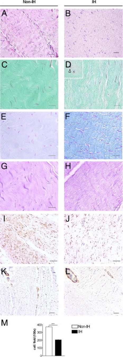
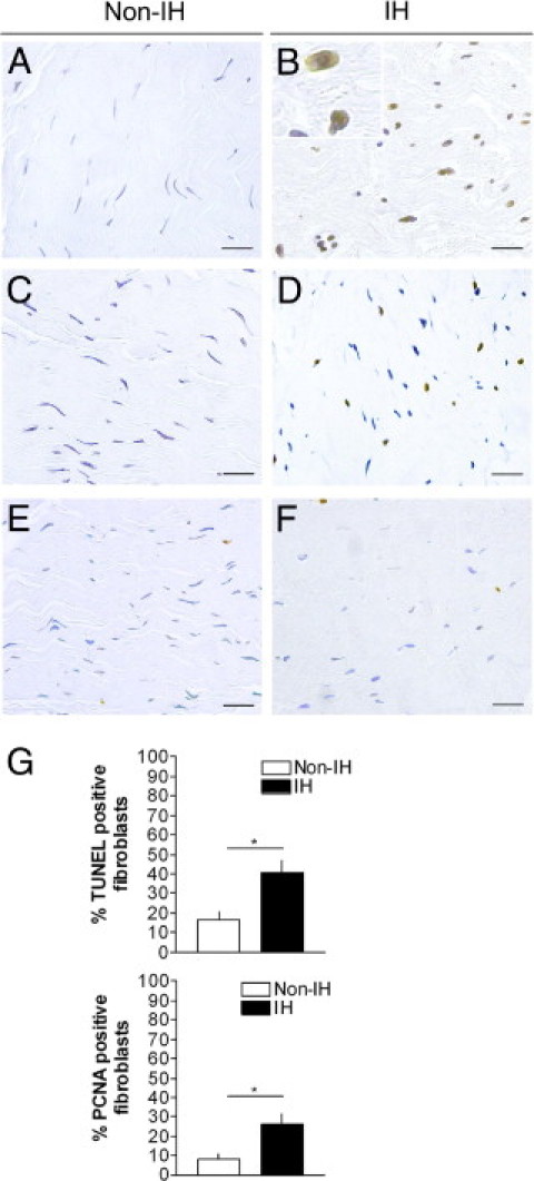
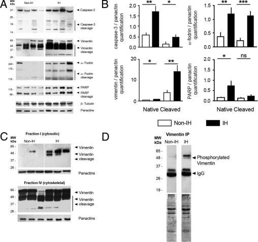
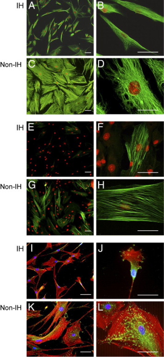
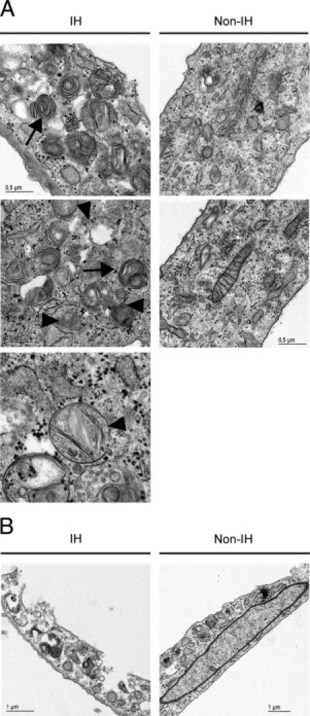
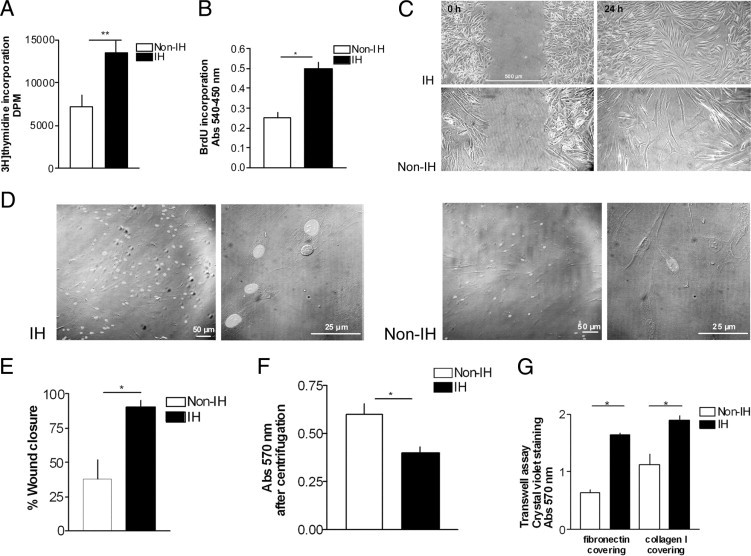
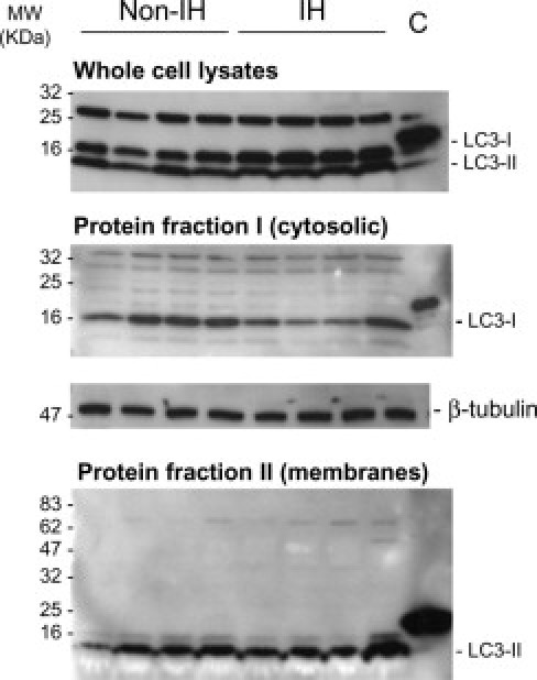
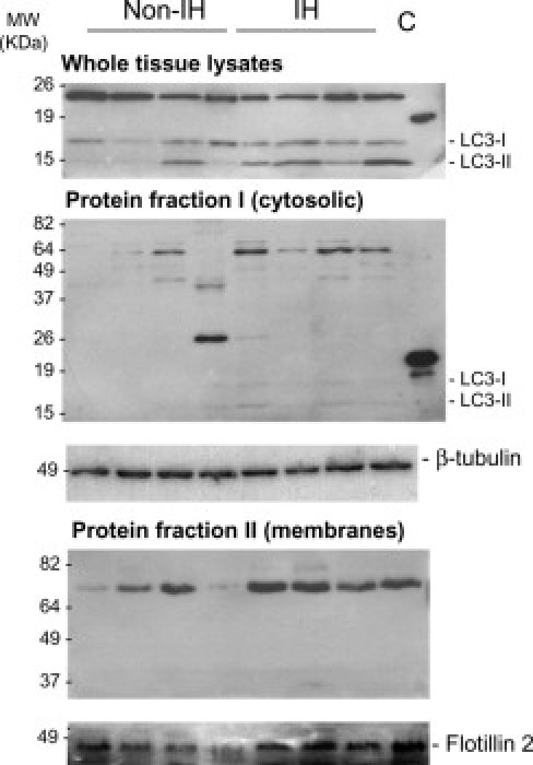
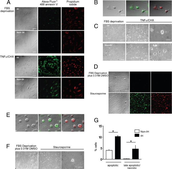
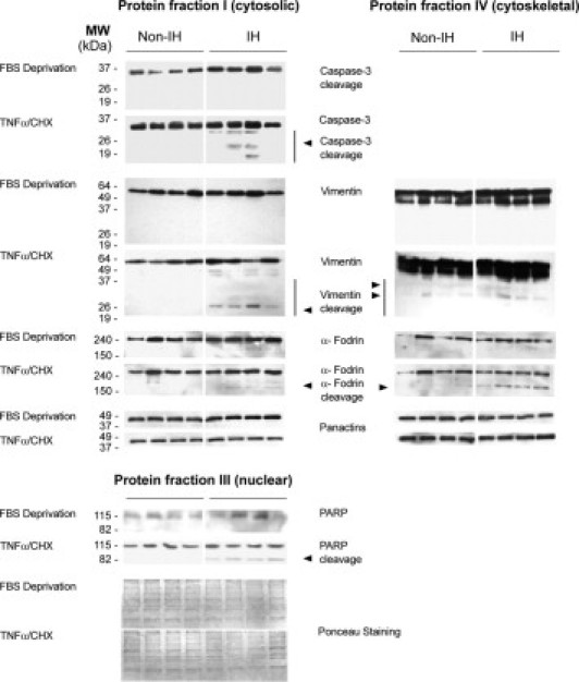
Incisional hernia often occurs following laparotomy and can be a source of serious problems. Although there is evidence that a biological cause may underlie its development, the mechanistic link between the local tissue microenvironment and tissue rupture is lacking. In this study, we used matched tissue-based and in vitro primary cell culture systems to examine the possible involvement of fascia fibroblasts in incisional hernia pathogenesis. Fascia biopsies were collected at surgery from incisional hernia patients and non-incisional hernia controls. Tissue samples were analyzed by histology and immunoblotting methods. Fascia primary fibroblast cultures were assessed at morphological, ultrastructural, and functional levels. We document tissue and fibroblast loss coupled to caspase-3 activation and induction of apoptosis-like cell-death mechanisms in incisional hernia fascia. Alterations in cytoskeleton organization and solubility were also observed. Incisional hernia fibroblasts showed a consistent phenotype throughout early passages in vitro, which was characterized by significantly enhanced cell proliferation and migration, reduced adhesion, and altered cytoskeleton properties, as compared to non-incisional hernia fibroblasts. Moreover, incisional hernia fibroblasts displayed morphological and ultrastructural alterations compatible with autophagic processes or lysosomal dysfunction, together with enhanced sensitivity to proapoptotic challenges. Overall, these data suggest an ongoing complex interplay of cell death induction, aberrant fibroblast function, and tissue loss in incisional hernia fascia, which may significantly contribute to altered matrix maintenance and tissue rupture in vivo.


Figures

Figure 1
Figure 1
Fascia histological features. Sections of fascia from non-incisional hernia (non-IH) and IH patients were stained with H&E (A and B), Masson's trichrome (C and D), Alcian Blue (E and F), PAS (G and H), and vimentin (I and J). In IH fascia, fibroblasts tend to be round or oblong (arrowheads), and some are surrounded by an open space (D, inset); K and L: α-SMA immunostaining. Magnification: H&E, vimentin, and α-SMA, ×100, scale bar = 100 μm; Masson's trichrome and Alcian Blue, ×400, scale bar = 50 μm. M: Mean fibroblast cells per field from non-IH (n = 20) and IH (n = 21), as measured in H&E-stained fascia sections. ***P < 0.001, Student's t-test.
Figure 2
Figure 2
Apoptosis and cell proliferation markers in fascia tissue. Terminal deoxynucleotidyl transferase-mediated dNTP-biotin nick end labeling (TUNEL) (A and B), proliferating cell nuclear antigen (PCNA) (C and D), and Ki-67 (E and F) immunostaining of non-IH and IH fascia tissue (magnification, ×200; scale bars: 50 μm). G: Quantitative analyses of TUNEL-positive and PCNA-positive fibroblast nuclei, in non-IH (n = 20) and IH (n = 21) fascia; *P < 0.05 (Student's t-test).
Figure 3
Figure 3
Proteolytic cleavage of caspase-3 and cytoskeletal substrates in fascia tissue. A: Representative immunoblot analyses from non-IH and IH fascia. Pan-actin was used as a loading control for normalization purposes. MW, molecular weight. B: Densitometric analysis of native and cleaved protein expression, in non-IH (n = 8) and IH (n = 8) samples (ns, nonsignificant; *P < 0.05, **P < 0.01, ***P < 0.001; Student's t-test). C: Representative immunoblots for the cytosol- (soluble) and cytoskeletal-enriched fractions from non-IH and IH fascia homogenates. D: Representative immunoblots of cytosol-enriched fractions, after immunoprecipitation with a specific polyclonal anti-phospho-Ser/Thr antibody (details in the Materials and Methods section).
Figure 4
Figure 4
Fascia fibroblast characterization. Representative immunofluorescent staining of primary fibroblasts from non-IH and IH fascia, for vimentin (green, A–D), α-SMA (green, E–H), and vinculin/phalloidin (green/red, I–L). Magnification: ×100 (A, C, E, and G), ×200 (I and K), ×400 (B, D, F, H, J, and L); Scale bar = 50 μm.
Figure 5
Figure 5
Ultrastructural appearance. A: Representative ultrastructural pictures of primary fascia fibroblasts. Outstandingly, IH fibroblasts showed autophagic vacuoles, autophagolysome-like structures, multilayered lamellar and fingerprint profiles (arrows), and mitochondrial swelling (arrowheads). B: Evidence of IH fibroblast fragility at preparation for transmission electron microscopy analysis.
Figure 6
Figure 6
Analysis of primary fibroblast behavior. Proliferation assays: (A) [3H]-thymidine and (B) 5-bromo-2′-deoxy-uridine (BrdU) incorporation into DNA, in non-IHFs (n = 5) and IHFs (n = 5). Abs, absorption. Migration and adhesion assay: (C and E) representative images of the in vitro scratch assay captured at 0 hours to 24 hours postwounding, in non-IHFs and IHFs, and comparison of wound closure percentage between non-IHFs (n = 5) and IHFs (n = 5) (details in the Materials and Methods section). D: BrdU in situ immunostaining. Magnification: ×100, ×600. F: Cell adhesion of non-IHFs (n = 5) and IHFs (n = 5), quantified by inverted centrifugation. G: Migration of non-IHFs (n = 5) and IHFs (n = 5) across fibronectin- and type I collagen-coated inserts in transwell assays. *P < 0.05, **P < 0.01
Figure 7
Figure 7
LC3 immunoblots in fascia fibroblasts. Representative immunoblots of LC3-I/LC3-II in whole-cell lysates (1 × 10⁶ cells; RIPA lysis buffer), and cytosolic- and membrane-enriched fractions (ProteoExtract Subcellular Proteome Extraction Kit) from cultured non-IH fibroblasts (non-IHFs) and IHFs. Proteins were resolved on 4% to 20% polyacrylamide gradient SDS gels. Cytosolic and membrane protein fractions were concentrated to about 10-fold on Amicon Ultra 0.5-mL centrifugal filters (Millipore, Billerica, MA). Positive controls for anti-LC3 antibody were used (MBL International Corp.). β-Tubulin detection in cytosolic-enriched protein extracts was used as a loading control (C) for normalization purposes.
Figure 8
Figure 8
LC3 immunoblots in fascia tissue. Representative immunoblots of LC3-I/LC3-II in whole lysates (RIPA lysis buffer), cytosolic- and membrane-enriched fractions (ProteoExtract Subcellular Proteome Extraction Kit) from non-IH and IH fascia tissues. SDS-PAGE was performed using 12.5% polyacrylamide gels and positive controls for anti-LC3 antibody (MBL). The cytosolic and membrane protein fractions were further concentrated to about sevenfold on Amicon Ultra 0.5-mL centrifugal filters (Millipore) and resolved in 11% and 8% SDS polyacrylamide gels, respectively. β-Tubulin detection in cytosolic protein extracts was used as a loading control for normalization purposes. Flotillin-2 was used as a membrane marker.
Figure 9
Figure 9
Fibroblast susceptibility to proapoptotic stimuli. Apoptotic response of non-IHFs (n = 5) and IHFs (n = 5), as measured by Vybrant Apoptosis Assay kit II (details in the Materials and Methods section). A–C, and G: TNF-α/CHX-treatment (72 hours): (A) Bright-field and immunofluorescence images, ×100; (B) merged images, ×600; (C) phase contrast images: left and center, ×100; right, ×600; (G) percentage of apoptotic cells (*P < 0.05, Mann-Whitney U-test). D–F: Staurosporine treatment (16 hours): (D) bright-field and immunofluorescence images, ×100; (E) merged images, ×600; (F) phase contrast images: left and center, ×100; right, ×600. Scale bars: 100 μm (×100); 50 μm (×600).
Figure 10
Figure 10
Immunoblots of proteins involved in apoptosis. Representative immunoblot analyses, obtained from cytosolic-, nuclear-, and cytoskeletal-enriched fractions from non-IHFs and IHFs, after TNFα/CHX treatment. Proteins were extracted by the ProteoExtractTM Subcellular Proteome Extraction Kit (details in Materials and Methods).

References

    1. Muysoms F.E., Miserez M., Berrevoet F., Campanelli G., Champault G.G., Chelala E., Dietz U.A., Eker H.H., El Nakadi I., Hauters P., Hidalgo Pascual M., Hoeferlin A., Klinge U., Montgomery A., Simmermacher R.K., Simons M.P., Smietański M., Sommeling C., Tollens T., Vierendeels T., Kingsnorth A., Muysoms F.E., Miserez M., Berrevoet F. Classification of primary and incisional abdominal wall hernias. Hernia. 2009;13:407–414. - PMC - PubMed
    1. Flum D.R., Horvath K., Koepsell T. Have outcomes of incisional hernia repair improved with time?: A population-based analysis. Ann Surg. 2003;237:129–135. - PMC - PubMed
    1. Arbos M.A., Ferrando J.M., Quiles M.T., Vidal J., López-Cano M., Gil J., Manero J.M., Peña J., Huguet P., Schwartz-Riera S., Reventós J., Armengol M. Improved surgical mesh integration into the rat abdominal wall with arginine administration. Biomaterials. 2006;27:758–768. - PubMed
    1. den Hartog D., Dur A.H., Tuinebreijer W.E., Kreis R.W. Open surgical procedures for incisional hernias. Cochrane Database Syst Rev. 2008;(3) CD006438. - PMC - PubMed
    1. Yahchouchy-Chouillard E., Aura T., Picone O., Etienne J.C., Fingerhut A. Incisional hernias: I. Related risk factors. Dig Surg. 2003;20:3–9. - PubMed

Publication types

MeSH terms