. 2011 Jun;178(6):2716-25.
doi: 10.1016/j.ajpath.2011.02.015.

Autoreactive cytotoxic T lymphocytes acquire higher expression of cytotoxic effector markers in the islets of NOD mice after priming in pancreatic lymph nodes

Affiliations

Autoreactive cytotoxic T lymphocytes acquire higher expression of cytotoxic effector markers in the islets of NOD mice after priming in pancreatic lymph nodes

Kate L Graham et al. Am J Pathol. 2011 Jun.

Abstract

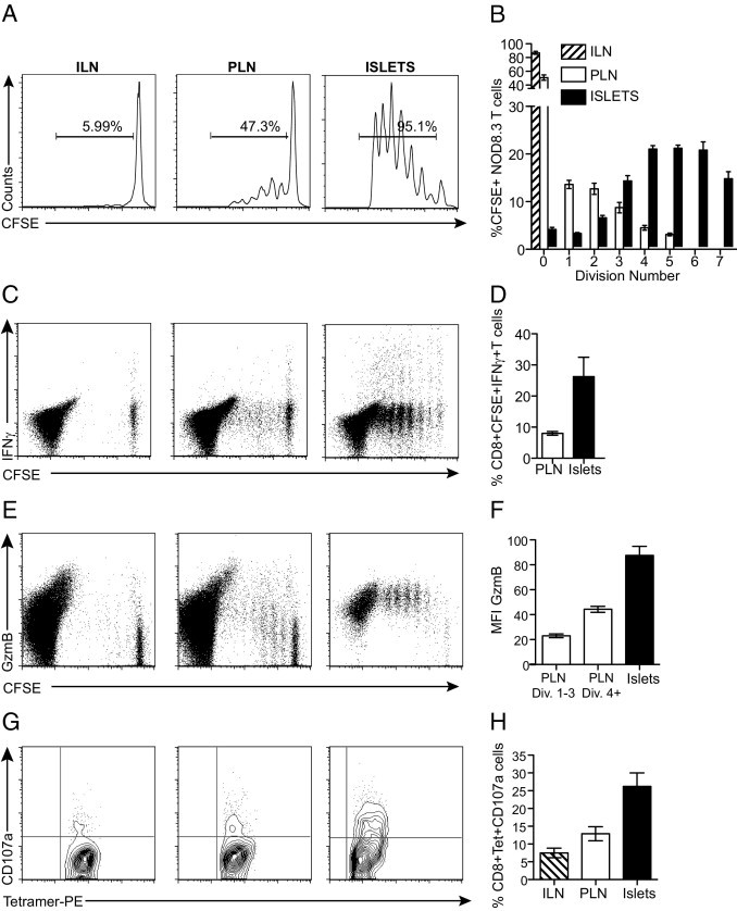
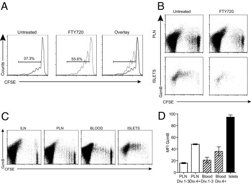
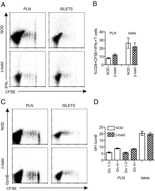
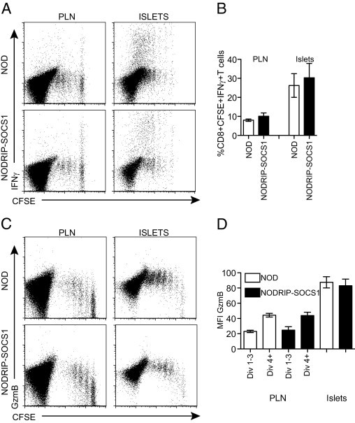
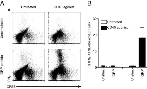
Cytotoxic T lymphocytes (CTLs) that cause type 1 diabetes are activated in draining lymph nodes and become concentrated as fully active CTLs in inflamed pancreatic islets. It is unclear whether CTL function is driven by signals received in the lymph node or also in the inflamed tissue. We studied whether the development of cytotoxicity requires further activation in islets. Autoreactive CTLs found in the islets of diabetes-prone NOD mice had acquired much higher expression of the cytotoxic effector markers granzyme B, interferon γ, and CD107a than had those in the pancreatic lymph node (PLN). Increased expression seemed to result from stimulation in the islet itself. T cells held up from migrating from the PLN by administration of the sphingosine-1-phosphate agonist FTY720 did not increase expression of cytotoxic molecules in the PLN. Stimulation did not require antigen presentation or cytokine secretion by the target β cells because it was not affected by the absence of class I major histocompatibility complex expression or by the overexpression of suppressor of cytokine signaling-1. Activation of CD40-expressing cells stimulated increased CTL function and β-cell destruction, suggesting that signals derived from CD40-expressing cells promote the acquisition of cytotoxicity in the islet environment. These data provide in vivo evidence that stimulation of cytotoxic effector molecule expression occurs in inflamed islets and is independent of β cells.


Figures

Figure 1
Figure 1
Proliferation, activation, and acquisition of cytotoxic effector function by diabetogenic CTLs. NOD recipients were adoptively transferred with 5 × 10⁶ CFSE-labeled 8.3 T cells. Five days after transfer, the ILN, PLN, and islets were harvested and analyzed. A: Proliferation of 8.3 T cells. Representative histogram profiles are gated on CFSE-labeled CD8+ T cells. B: The percentage of CFSE-positive T cells per division (n = 7). C: Expression of IFNγ by proliferating 8.3 T cells. D: Percentage of CD8+CFSE+IFNγ+ cells in the PLN and islets (n = 5). E: Expression of GzmB. F: MFI for GzmB expression gated on all CD8+CFSE+ cells (n = 9). G: Expression of CD107a on transferred 8.3 T cells. H: Percentage of CD8+tetramer+CD107a+ T cells (n = 6). Data are given as the mean ± SEM (error bars).
Figure 2
Figure 2
Full cytotoxic effector molecule expression does not occur in the PLN. A: Proliferation of CFSE-labeled 8.3 T cells in the PLNs of control and FTY720-treated mice. NOD mice were treated with 3 mg/kg FTY720 daily from the day of adoptive transfer until 5 days after transfer, when lymph nodes and islets were harvested (n = 8 for treated and untreated from four experiments). Histograms are gated on CFSE+CD8+ T cells, and the percentage of proliferating cells is indicated. B: Expression of GzmB in dividing CFSE-labeled CD8+ T cells isolated from the PLN and islets of untreated and FTY720-treated recipient NOD mice. Dot plots are gated on total CD8+ T cells. C: Flow cytometric analysis of GzmB expression in diabetogenic CTLs isolated from peripheral blood. Representative dot plots are gated on total CD8+ T cells. D: MFI for GzmB expression in the PLN, blood, and islets. For the PLN and blood, divisions (Div.) 1 to 3 and 4+ were gated and MFI was calculated (n = 4). Data are given as mean ± SEM (error bars).
Figure 3
Figure 3
Complete elimination of β-cell antigen presentation does not prevent increased cytotoxic effector molecule expression in diabetogenic CTLs. Secretion of IFNγ (A and B) and expression of GzmB (C and D) in dividing 8.3 T cells isolated from the PLN and islets isolated from NOD and class I β-bald recipients. Dot plots (A and C) are gated on total CD8+ T cells and represent multiple experiments (n = 3 to 5). Data shown in the graphs (B and D) were calculated as described in Figure 1. Data are given as mean ± SEM (error bars).
Figure 4
Figure 4
Reduced β-cell cytokine production does not prevent increased cytotoxic effector molecule expression in diabetogenic CTLs. Comparison of IFNγ secretion (A and B) and GzmB expression (C and D) in dividing 8.3 T cells from the PLN and islets isolated from NOD and NODRIP-SOCS1 recipients. Dot plots (A and C) are gated on total CD8+ T cells and represent multiple experiments. Percentage of IFNγ (γ) and MFI for GzmB expression (D) were calculated as described in Figure 1 (n = 4 to 5).
Figure 5
Figure 5
Increasing inflammation promotes acquisition of cytotoxic effector capacity. NODIGRP recipients were adoptively transferred with CFSE-labeled 8.3 T cells and were injected with CD40 agonist antibody (n = 5) or were left untreated (n = 5). A: Single-cell preparations of splenocytes were stimulated with IGRP peptide or were left unstimulated (Unstim.), and IFNγ secretion in dividing 8.3 T cells was measured. Dot plots are gated on total CD8+ T cells and represent multiple experiments. B: Percentage of IFNγ-secreting cells. Data are given as mean ± SEM (error bars).

References

    1. Christianson S.W., Shultz L.D., Leiter E.H. Adoptive transfer of diabetes into immunodeficient NOD-scid/scid mice: relative contributions of CD4+ and CD8+ T-cells from diabetic versus prediabetic NODNON-Thy-1a donors. Diabetes. 1993;42:44–55. - PubMed
    1. Hoglund P., Mintern J., Waltzinger C., Heath W., Benoist C., Mathis D. Initiation of autoimmune diabetes by developmentally regulated presentation of islet cell antigens in the pancreatic lymph nodes. J Exp Med. 1999;189:331–339. - PMC - PubMed
    1. Gagnerault M.C., Luan J.J., Lotton C., Lepault F. Pancreatic lymph nodes are required for priming of β cell reactive T cells in NOD mice. J Exp Med. 2002;196:369–377. - PMC - PubMed
    1. Coles R.M., Mueller S.N., Heath W.R., Carbone F.R., Brooks A.G. Progression of armed CTL from draining lymph node to spleen shortly after localized infection with herpes simplex virus 1. J Immunol. 2002;168:834–838. - PubMed
    1. Jones C.M., Cose S.C., Coles R.M., Winterhalter A.C., Brooks A.G., Heath W.R., Carbone F.R. Herpes simplex virus type 1-specific cytotoxic T-lymphocyte arming occurs within lymph nodes draining the site of cutaneous infection. J Virol. 2000;74:2414–2419. - PMC - PubMed

Publication types

MeSH terms