. 2011 Jun;30(5-6):318-29.
doi: 10.1016/j.matbio.2011.05.003. Epub 2011 May 27.

Caveolin-1 modulates TGF-β1 signaling in cardiac remodeling

Affiliations

Caveolin-1 modulates TGF-β1 signaling in cardiac remodeling

Shelley K Miyasato et al. Matrix Biol. 2011 Jun.

Abstract

The cardiac response to myocardial injury includes fibrotic and hypertrophic processes and a key mediator in this response is transforming growth factor-β1 (TGF-β1). Caveolin-1 (cav1), the main structural protein of caveolae, is an inhibitor of the TGF-β1 signaling pathway. To examine the role of cav1 in cardiac repair, cav1 deficient (Cav1(-/-)) and wild type (WT) mice were subjected to cryoinjury of the left ventricle (LV). At baseline the two groups exhibited no inflammation, similar collagen content, and similar cardiac function. After injury, Cav1(-/-) animals displayed enhanced TGF-β1 signaling, as reflected by a 3-fold increase in the activation of the Smad2-dependent pathway and more widespread collagen deposition in the heart. Qualitative and quantitative analyses indicated that collagen deposition peaked in the WT LV 14days after injury, accompanied by increased mRNA abundance for procol1a2 (2-fold) and procol3a1 (3-fold). Collagen deposition was further enhanced in Cav1(-/-) mice, which was accompanied by reduced expression of matrix metalloproteinases MMP-8 (3-fold) and -13 mRNA (2-fold). The levels of expression of inflammatory markers of acute phase were similar between the strains However, macrophage clearance in the damaged region was delayed in Cav1(-/-) mice. We observed a 4-fold decrease in collagen deposition in Cav1(-/-) mice injected with a cav1 scaffolding domain peptide (CSD) and a 2-fold decrease in WT mice treated with the CSD. We conclude that cav1 has a direct role in reducing TGF-β1 signaling and as such might be an appropriate target for therapies to influence cardiac remodeling.


Figures

Figure 1
Figure 1. Transiently reduced expression of caveolin-1 in heart following cryoinjury
A) Cav1 mRNA abundance in cryoinjured WT mice was transiently decreased, compared to sham-operated mice. Gene expression was assessed by TaqMan assay and normalized to ß-actin. WT sham group value has been used for normalization among study groups. Bar represents mean ± SD. ***= p<0.001, n= 7. B) Measure of level of cav1 protein expression in sham and 3-day post cryoinjured hearts. In insert, a representative western blot from which these data were derived. C) Representative immunohistochemistry staining for cav1. This protein is robustly expressed in endothelial cells in sham LV (left), while protein levels dramatically decrease at 3 days after cryoinjury (middle) and are returned towards control at day 14 (right).
Figure 2
Figure 2. Transient increase in heart weight following cryoinjury in Cav1−/− mice
A) Hearts were weighed immediately after the death in sham-operated mice and 3, 14, or 30 days post cryoablation. The bars represent the ratio of total heart weight/total body weight (n≥10 per group). B) Quantitative spectrophotometric measurements of Evans Blue dye as a marker of vascular leakage. Three-days after cryoinjury Cav1−/− and WT mice were injected with Evans Blue dye. The level of Evans Blue was measured in the heart 24 hr after injection (n = 5 per group). C) Total new collagen deposition was evaluated by Sircol as described in the Material and Methods section (n = 5 mice per group). Data are expressed as mean ± standard deviation. *: p<0.05; **: p<0.01; and ***: p<0.001.
Figure 3
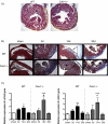
Figure 3. Visualization of the injured border zone and development of cardiac remodeling in cryoinjured heart samples
A) The injured border zone can be visualized by standard HE staining. The injured is outlined by yellow line on the representative sections. E: epicardium; LV: left ventricle; M: myocardium; RV: right ventricle. B) Representative photomicrographs of trichrome-stained heart sections from sham-operated and cryoinjured Cav1−/− and WT animals; The collagen network is stained in blue and the nuclei of the cells in purple. C) The procol1a2 and procol3a1 expression in cryoinjured mice increased after 3 days, with an even higher level at 14 days, and returned to that seen in sham-operated samples by 30 days (n = 7). *: p<0.05; **: p<0.01; and ***: p<0.001.
Figure 4
Figure 4. Increased collagen deposition in cryoinjured Cav1−/− hearts compared to WT animals
A) Representative photomicrographs of picrosirius red stained heart sections from cryoinjured WT and Cav1−/− mice. The collagen network is stained in red. The scar contained a large amount of collagen, which peaked at 14 days post injury in WT. The extent of collagen deposition decreased thereafter. By contrast, Cav1−/− mice had increased collagen deposition in the injured region that extended into the non-infarcted myocardium. This accumulation of collagen is still visible after 30 days of injury. B) Quantitative analysis demonstrated that Cav1−/− mice had significantly more collagen deposition than WT animals. *: p<0.05 (statistical analysis in the same strain) #: p<0.05 indicates a statistical difference between WT and Cav1−/− at the same time point.
Figure 5
Figure 5. Altered MMP activation in Cav1−/− mice after cryoinjury
A) MMP-2, −8, −9 and −13 mRNA abundance in WT and Cav1−/− heart after cryoablation. The level of MMP expression was determined in Cav1−/− and WT mice by qPCR with TaqMan probes specific to the murine cDNA −3d, −14d, or −30d post injury and compared to sham. The results were normalized to the GAPDH expression. The assays were performed in triplicate. B) Total MMP activity was measured. Data are expressed as mean ± SD. *: p<0.05; **: p<0.01; ***: p<0.001 (statistical analysis in the same strain) #: p<0.05; ##: p<0.01; ###: p<0.001 indicates a statistical difference between WT and Cav1−/− at the same time point.
Figure 6
Figure 6
Activation of TGF-β signaling following cryoinjury was assessed by measuring phosphorylation of Smad2 and ERK1 in mouse heart following cryoablation. Bar represents mean ± SD. **= p<0.01 between 2 time points for the same strain; # = p<0.05 and ## = p<0.01 between both strains for the same time point, n= 5-10.
Figure 7
Figure 7. Enhanced activity of TGF-β1 signaling in TGF–β1-stimulated lung fibroblasts lacking cav1 expression
The Smad response element construct was transfected into heart fibroblasts extracted from WT and Cav1−/−. These fibroblasts were transfected with 1μg of plasmid and 0.1 μg of pRL-SV40 plasmid for control purposes and cultured for 24 hr with 2 ng/ml of TGF-β1. Luciferase activity was normalized to the renilla luciferase activity. Each value represents the mean ± SD of at least three independent transfection experiments, each performed in triplicate. *: p<0.05; and **: p<0.01 (statistical analysis in the same strain) #: p<0.05 indicates a statistical difference between WT and Cav1−/− at the same time point.
Figure 8
Figure 8. Cav1 scaffolding domain (CSD) peptide treatment prevents increased collagen deposition in cryoinjured myocardium
Representative photomicrographs of picrosirius red stained heart sections from cryoinjured WT and Cav1−/− mice. The collagen network is stained in red. The scar contained a large amount of collagen at 14 days post injury when the mice were treated with the scrambled peptide. There is less collagen deposition in the hearts of mice treated with the CSD peptide for both strains. B) Qualitative analysis of picrosirius red stained heart sections demonstrated that Cav1−/− mice had significantly more collagen deposition than WT animals at 14 days when treated with the scrambled peptide. Administration of the CSD peptide prevented the accumulation of collagen. C) The procol1a2 expression in cryoinjured mice treated with CSD was reduced in both Cav1−/− and WT mice.
Figure 9
Figure 9. Representative Western Blot analysis of Smad2, pSmad2, ERK1/2, and pERK1/2 in WT and Cav1−/− heart samples treated with the Cav1 scaffolding domain (CSD) peptide
Bar represents the mean of the ratio of phosphorylated/total signaling proteins. Values are represented as mean ± SD. **: p<0.01; and ***: p<0.001 (statistical analysis in the same strain) #: p<0.05 indicates a statistical difference between WT and Cav1−/− at the same time point. Scr: scrambled peptide and CSD: caveolin 1 scaffolding domain

References

    1. Augustus AS, Buchanan J, Gutman E, Rengo G, Pestell RG, et al. Hearts lacking caveolin-1 develop hypertrophy with normal cardiac substrate metabolism. Cell Cycle. 2008;7:2509–2518. - PubMed
    1. Barth K, Gentsch M, Blasche R, Pfuller A, Parshyna I, et al. Distribution of caveolin-1 and connexin43 in normal and injured alveolar epithelial R3/1 cells. Histochem Cell Biol. 2005;123:239–247. - PubMed
    1. Bernatchez PN, Bauer PM, Yu J, Prendergast JS, He P, Sessa WC. Dissecting the molecular control of endothelial NO synthase by caveolin-1 using cell-permeable peptides. Proc Natl Acad Sci U S A. 2005;102:761–766. - PMC - PubMed
    1. Bucci M, Gratton JP, Rudic RD, Acevedo L, Roviezzo F, et al. In vivo delivery of the caveolin-1 scaffolding domain inhibits nitric oxide synthesis and reduces inflammation. Nat Med. 2000;6:1362–1367. - PubMed
    1. Bujak M, Frangogiannis NG. The role of TGF-beta signaling in myocardial infarction and cardiac remodeling. Cardiovasc Res. 2007;74:184–195. - PMC - PubMed

Publication types

MeSH terms