. 2011 Jul 29;286(30):26978-86.
doi: 10.1074/jbc.M111.249532. Epub 2011 Jun 3.

Oxidation of 3,4-dihydroxyphenylacetaldehyde, a toxic dopaminergic metabolite, to a semiquinone radical and an ortho-quinone

Affiliations

Oxidation of 3,4-dihydroxyphenylacetaldehyde, a toxic dopaminergic metabolite, to a semiquinone radical and an ortho-quinone

David G Anderson et al. J Biol Chem. .

Abstract

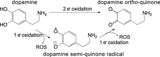
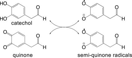
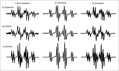
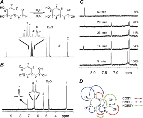
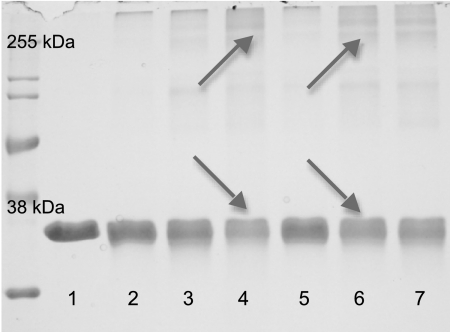
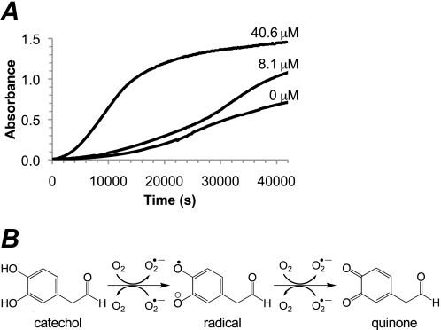
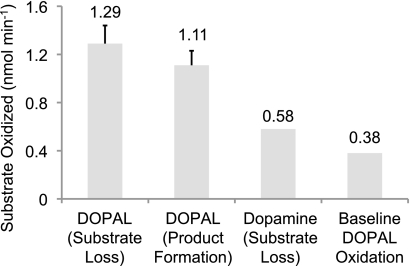
The oxidation and toxicity of dopamine is believed to contribute to the selective neurodegeneration associated with Parkinson disease. The formation of reactive radicals and quinones greatly contributes to dopaminergic toxicity through a variety of mechanisms. The physiological metabolism of dopamine to 3,4-dihydroxyphenylacetaldehyde (DOPAL) via monoamine oxidase significantly increases its toxicity. To more adequately explain this enhanced toxicity, we hypothesized that DOPAL is capable of forming radical and quinone species upon oxidation. Here, two unique oxidation products of DOPAL are identified. Several different oxidation methods gave rise to a transient DOPAL semiquinone radical, which was characterized by electron paramagnetic resonance spectroscopy. NMR identified the second oxidation product of DOPAL as the ortho-quinone. Also, carbonyl hydration of DOPAL in aqueous media was evident via NMR. Interestingly, the DOPAL quinone exists exclusively in the hydrated form. Furthermore, the enzymatic and chemical oxidation of DOPAL greatly enhance protein cross-linking, whereas auto-oxidation results in the production of superoxide. Also, DOPAL was shown to be susceptible to oxidation by cyclooxygenase-2 (COX-2). The involvement of this physiologically relevant enzyme in both oxidative stress and Parkinson disease underscores the potential importance of DOPAL in the pathogenesis of this condition.


Figures

SCHEME 1.
SCHEME 1.
Oxidation of dopamine.
FIGURE 1.
FIGURE 1.
UV-visible spectrophotometric characterization of DOPAL oxidation products. A, overlaid spectra of DOPAL oxidized by tyrosinase at pH 7.4 show the formation of two unique species. The early oxidation product is transient and centered at 520 nm, whereas the later species is relatively more stable and centered at 400 nm. Only the first several minutes are displayed for clarity. B, relative absorbance of the two oxidation products over time.
SCHEME 2.
SCHEME 2.
Comproportionation of quinone and catechol to radicals.
FIGURE 2.
FIGURE 2.
EPR spin stabilization spectra of the DOPAL semiquinone radical formed by a variety of methods. DOPAL oxidized by tyrosinase (A), NaIO4 (B), HRP-H2O2 (C), and K3Fe(CN)6 (D) in the presence of Mg2+ resulted in the formation of a semiquinone radical. The overall efficacy of oxidation methods for radical production under the experimental conditions utilized was tyrosinase > HRP-H2O2 > NaIO4 > K3Fe(CN)6. For both the enzymatic (A and C) and chemical (B and D) methods, two-electron oxidation and comproportionation (A and B) appeared to produce higher levels of the radical than direct one-electron oxidation (C and D).
FIGURE 3.
FIGURE 3.
EPR spectra of DA, DOPAL, and DOPAC semiquinone radicals. DA (A), DOPAL (B), and DOPAC (C) radical spectra were generated via high pH-induced auto-oxidation under non-spin-stabilized conditions (column i), Mg2+-stabilized tyrosinase oxidation at pH 7.4 (column ii), and WinSim computer simulation (column iii). The radicals of the dopaminergic compounds are spectrally similar under the same conditions. Also note the hyperfine splitting apparent under conditions of spontaneous oxidation (column i).
FIGURE 4.
FIGURE 4.
NMR analysis of the DOPAL quinone. A, 1H NMR of unoxidized DOPAL in aqueous media (D2O) shows an approximate 5:2 ratio of hydrate:aldehyde. Proton assignments are indicated. Aromatic signals are magnified for clarity. B, 1H NMR of DOPAL oxidized by 1.00 eq of NaIO4. As indicated, the resonances fit with the expected structure of the quinone. Aromatic signals are magnified for clarity. C, the stability of the quinone was investigated by measuring the intensity of selected peaks over time (25 °C, D2O). As the quinone disappeared, the hypothesized polymer (8.44 ppm) resonance increased. The percentage of quinone remaining is indicated for each of the spectra. D, NOESY- and two-dimensional NMR-based resonance assignments and NOE correlations verify the ortho-quinone hydrate structure. Correlations useful for structural determination are indicated by colored arrows (red, COSY; blue, HMBC; green, NOESY).
FIGURE 5.
FIGURE 5.
Oxidation of DOPAL-enhanced GAPDH cross-linking. SDS-PAGE and densitometry were used to compare the ability of DOPAL oxidized by various methods to induce cross-linking of the model protein, GAPDH. 0.3 mg/ml protein in 50 mm NaH2PO4, pH 7.4, was reacted with 50 μm DOPAL for 4 h at 37 °C. Cross-linking is apparent as a loss of control protein concurrent with the formation of higher molecular weight species (indicated by arrows). Conditions and results for each lane are as noted in Table 3.
FIGURE 6.
FIGURE 6.
Enhancement of the rate of DOPAL auto-oxidation by SOD indicates that superoxide is being formed. A, auto-oxidation of DOPAL at pH 7.8 proceeds slowly but is greatly accelerated in the presence of increasing concentrations of SOD. B, although superoxide is formed during spontaneous (i.e. air) oxidation of catechols and hydroquinones, the reverse reaction is much faster. Sufficient removal of superoxide by SOD allows the reaction to proceed much more quickly.
FIGURE 7.
FIGURE 7.
Summary of the initial linear slopes of DOPAL oxidized by COX-2. DOPAL (50 μm) was oxidized by 100 units/ml COX-2. The results, based on the concentration of substrate oxidized, were calculated using both product formation and substrate loss. DA (65 μm) was also shown for comparison using substrate disappearance to calculate the initial linear slope. The assay system without the added enzyme functioned as a control and allowed the results to be corrected for base-line DOPAL oxidation.

References

    1. Davie C. A. (2008) Br. Med. Bull. 86, 109–127 - PubMed
    1. Chinta S. J., Andersen J. K. (2008) Biochim. Biophys. Acta 1780, 1362–1367 - PMC - PubMed
    1. Fornstedt B., Brun A., Rosengren E., Carlsson A. (1989) J. Neural Transm. Park. Dis. Dement. Sect. 1, 279–295 - PubMed
    1. Collins M. A., Neafsey E. J. (2002) Neurotoxicol. Teratol. 24, 571–577 - PubMed
    1. Terland O., Almas B., Flatmark T., Andersson K. K., Sorlie M. (2006) Free Radic. Biol. Med. 41, 1266–1271 - PubMed

Publication types

MeSH terms