. 2011 Jun 21;108(25):10314-9.
doi: 10.1073/pnas.1017079108. Epub 2011 Jun 3.

Bidirectional plasticity in the primate inferior olive induced by chronic ethanol intoxication and sustained abstinence

Affiliations

Bidirectional plasticity in the primate inferior olive induced by chronic ethanol intoxication and sustained abstinence

John P Welsh et al. Proc Natl Acad Sci U S A. .

Abstract

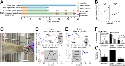
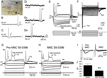
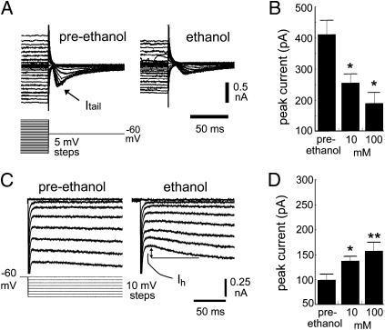
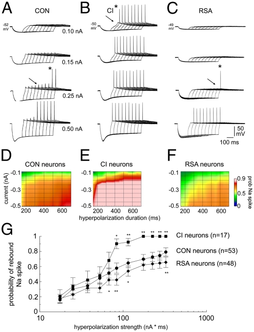
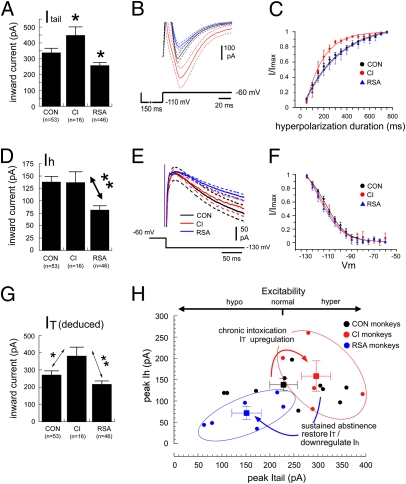
The brain adapts to chronic ethanol intoxication by altering synaptic and ion-channel function to increase excitability, a homeostatic counterbalance to inhibition by alcohol. Delirium tremens occurs when those adaptations are unmasked during withdrawal, but little is known about whether the primate brain returns to normal with repeated bouts of ethanol abuse and abstinence. Here, we show a form of bidirectional plasticity of pacemaking currents induced by chronic heavy drinking within the inferior olive of cynomolgus monkeys. Intracellular recordings of inferior olive neurons demonstrated that ethanol inhibited the tail current triggered by release from hyperpolarization (I(tail)). Both the slow deactivation of hyperpolarization-activated cyclic nucleotide-gated channels conducting the hyperpolarization-activated inward current and the activation of Ca(v)3.1 channels conducting the T-type calcium current (I(T)) contributed to I(tail), but ethanol inhibited only the I(T) component of I(tail). Recordings of inferior olive neurons obtained from chronically intoxicated monkeys revealed a significant up-regulation in I(tail) that was induced by 1 y of daily ethanol self-administration. The up-regulation was caused by a specific increase in I(T) which (i) greatly increased neurons' susceptibility for rebound excitation following hyperpolarization and (ii) may have accounted for intention tremors observed during ethanol withdrawal. In another set of monkeys, sustained abstinence produced the opposite effects: (i) a reduction in rebound excitability and (ii) a down-regulation of I(tail) caused by the down-regulation of both the hyperpolarization-activated inward current and I(T). Bidirectional plasticity of two hyperpolarization-sensitive currents following chronic ethanol abuse and abstinence may underlie persistent brain dysfunction in primates and be a target for therapy.


Conflict of interest statement

The authors declare no conflict of interest.

Figures

Fig. 1.
Fig. 1.
Alcoholic phenotype in cynomolgus monkeys. (A) Experimental timeline. (B) Mean ethanol intake for five CI monkeys in the month before necropsy and for six RSA monkeys during the 4 mo before their last withdrawal. (C) Reach-to-grasp task. The third metacarpophalangeal joint (red) was used to reference hand trajectory. (D) Plots of the reach (red), grasp (black), and return (blue) phases after a day of voluntary withdrawal (Fig. S1A) as spatial trajectories (Upper) and vertical position over time (Lower). (E) As in D but after a drinking binge. (F) Occurrence of three behaviors during intoxication, acute withdrawal, and sustained abstinence for RSA monkeys. *P < 0.05. (G) Normalized incidence of alcohol-withdrawal behaviors.
Fig. 2.
Fig. 2.
Electrophysiological properties of primate IO neurons. (A) Ventral surface of the monkey brainstem. Pyr, pyramidal tracts. (B and C) Voltage records of two IO neurons showing STOs, spontaneous action potentials, and a rebound response following hyperpolarization (arrow). (Diiii) Another IO neuron (Vrest −50 mV) showing voltage-insensitive STOs as tested by injecting depolarizing (Di) and hyperpolarizing (Diii) current. Arrows indicate spike doublets at the STO frequency. (E) Positive current triggers a sodium spike and an after-depolarizing potential (arrowhead); negative current triggers a depolarization sag; release from hyperpolarization triggers rebound spikes (arrow). (F) Currents underlying the sodium spike (INa), depolarization sag (Ih), and rebound response (Itail). (G and H) Voltage recordings of an IO neuron before (G) and after (H) addition of 30 μm NNC 55–0396. (I) Current recordings showing that NNC 55–0936 reduced Itail. (J) Mean reduction of Itail.by NNC 55–0396 (n = 4). **P < 0.01.
Fig. 3.
Fig. 3.
Acute effect of ethanol on pacemaking currents in primate IO neurons. (A) Inactivation protocol to study Itail. One hundred millimoles of ethanol reduced peak Itail in response to a range of voltage steps to −60 mV. (B) Effect of 10 and 100 mM ethanol on peak values of Itail (*P <0.05 compared with preethanol). (C) Protocol for testing effect of ethanol on Ih showing a robust augmentation of Ih by 100 mM ethanol. (D) Effect of 10 and 100 mM ethanol on peak values of Ih (*P < 0.05; **P < 0.01 compared to preethanol).
Fig. 4.
Fig. 4.
Changes in the voltage and time dependence of rebound firing with chronic ethanol intoxication and repeated abstinences in IO neurons. (AC) Voltage responses triggered by hyperpolarizing current in three neurons from CON (A), CI (B), and RSA (C) monkeys. Responses were triggered by injecting four hyperpolarizing currents (0.1–0.5 nA) for increasingly longer durations (150–600 ms). Arrows indicate the threshold for eliciting a low-threshold spike, and asterisks indicate the first occurrence of an action potential. (DF) Voltage dependence and time dependence of spiking for CON (n = 53) (D), CI (n = 17) (E), and RSA (n = 48) (F) neurons. (G) Semilog plot of the probability of a rebound spike vs. hyperpolarization strength. *P < 0.05; **P < 0.01 vs. CON.
Fig. 5.
Fig. 5.
Bidirectional plasticity of hyperpolarization-activated currents induced by chronic ethanol intoxication and subsequent abstinence. (A) Values of peak Itail for the three groups in response to a 50-mV hyperpolarizing step for 150 ms. *P < 0.05 vs. CON. (B) Mean (±1 SEM) traces of Itail for CON (black), CI (red), and RSA (blue) monkeys. (C) Mean Itail deinactivation curves for the three groups fit with exponential functions. τ: CON, 254 ms; CI, 146 ms; RSA, 296 ms. (D) Values of peak Ih for the three drinking groups in response to a 65-mV hyperpolarizing step. **P < 0.01. (E) Mean (±1 SEM) traces of Ih for CON, CI, and RSA neurons. (F) Mean Ih activation curves (V1/2 and k: CON, −117 mV and 2.2, respectively; CI, −118 mV and 2.3, respectively; RSA, −119 mV and 2.4, respectively). (G) Mean values of peak IT deduced for the three drinking groups. *P < 0.05; **P < 0.01. (H) Bidirectional plasticity of excitability induced by chronic ethanol intake. The excitability of CI monkey IO neurons (red) was enhanced by an up-regulation of IT without a change in Ih; the excitability of RSA monkey IO neurons (blue) was inhibited by the restoration of IT to normal and the suppression of Ih. Solid lines represent the mean trace and dotted lines represent 1 SEM from the means. The squares and error bars indicate the mean (±1 SEM) of the groups.

References

    1. Edwards G, Gross MM. Alcohol dependence: Provisional description of a clinical syndrome. BMJ. 1976;1:1058–1061. - PMC - PubMed
    1. Myrick H, Anton RF. Treatment of alcohol withdrawal. Alcohol Health Res World. 1998;22:38–43. - PMC - PubMed
    1. Harris RA. Ethanol actions on multiple ion channels: Which are important? Alcohol Clin Exp Res. 1999;23:1563–1570. - PubMed
    1. Yang L, Long C, Evans MS, Faingold C. Ethanol withdrawal results in aberrant membrane properties and synaptic responses in periaqueductal gray neurons associated with seizure susceptibility. Brain Res. 2002;957:99–108. - PubMed
    1. Grant KA, Bennett AJ. Advances in nonhuman primate alcohol abuse and alcoholism research. Pharmacol Ther. 2003;100:235–255. - PubMed

Publication types

MeSH terms