. 2011 Jul 1;187(1):424-33.
doi: 10.4049/jimmunol.1100750. Epub 2011 Jun 3.

Binding of flavivirus nonstructural protein NS1 to C4b binding protein modulates complement activation

Affiliations

Binding of flavivirus nonstructural protein NS1 to C4b binding protein modulates complement activation

Panisadee Avirutnan et al. J Immunol. .

Abstract

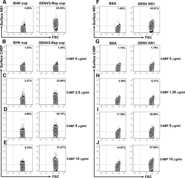
The complement system plays a pivotal protective role in the innate immune response to many pathogens including flaviviruses. Flavivirus nonstructural protein 1 (NS1) is a secreted nonstructural glycoprotein that accumulates in plasma to high levels and is displayed on the surface of infected cells but absent from viral particles. Previous work has defined an immune evasion role of flavivirus NS1 in limiting complement activation by forming a complex with C1s and C4 to promote cleavage of C4 to C4b. In this study, we demonstrate a second mechanism, also involving C4 and its active fragment C4b, by which NS1 antagonizes complement activation. Dengue, West Nile, or yellow fever virus NS1 directly associated with C4b binding protein (C4BP), a complement regulatory plasma protein that attenuates the classical and lectin pathways. Soluble NS1 recruited C4BP to inactivate C4b in solution and on the plasma membrane. Mapping studies revealed that the interaction sites of NS1 on C4BP partially overlap with the C4b binding sites. Together, these studies further define the immune evasion potential of NS1 in reducing the functional capacity of C4 in complement activation and control of flavivirus infection.


Figures

Figure 1
Figure 1. Flavivirus NS1 binds to C4BP
A-C. ELISA. Microtiter plates were coated with BSA or C4BP (5 μg/ml). After incubation with increasing concentrations of purified DENV NS1 (A), WNV NS1 (B), or YFV NS1 (C), bound NS1 was detected with specific mAbs. Error bars indicate standard error of the mean from three independent experiments and asterisks indicate statistical difference from the control BSA (*, P < 0.05; **, P < 0.01; ***, P < 0.0001). D-E. Binding of NS1 with C4BP is not affected by salt concentration. Microtiter plates were coated with BSA or C4BP (5 μg/ml). After incubation with 15 μg/ml purified DENV NS1 (D) or WNV NS1 (E), bound NS1 was detected with NS1-specific mAbs. Error bars indicate standard error of the mean from three independent experiments and asterisks indicate statistical difference from the control BSA (***, P < 0.0001). F-G. Co-immunoprecipitation studies. Serum-free supernatants from BHK-DENV2-Rep, BHK-WNV-Rep or control BHK cells were incubated with purified C4BP (15 μg/ml) and Western blots were performed after immunoprecipitation with anti-DENV NS1 2G6 mAb-Sepharose (F) or anti-WNV NS1 9NS1 mAb-Sepharose (G). Immunoprecipitates were probed with a rabbit polyclonal anti-human C4BP Ab. Western blot results are representative of two to three independent experiments.
Figure 2
Figure 2. Flavivirus NS1 binds to the α-chain of C4BP
A. Schematic diagrams of different forms of C4BP used in this study. Plasma-purified C4BP mostly consists of seven α-chains, one β-chain, and one molecule of protein S bound to the β-chain. Recombinant wild type C4BP consists of six α-chains, but lacks a β-chain and protein S (7, 17). The α-chain is composed of eight CCP domains with the carboxy terminal region that is responsible for multimerization of a single C4BP molecule. Deletion mutants lacking an individual repeating CCP domain are depicted as ΔCCP. B and C. Purity and electrophoretic mobility of recombinant wild type and mutant C4BP. Purified recombinant wild type (rWT) C4BP and deletion mutants lacking a single repeating CCP domain (ΔCCP1-ΔCCP8) (1 μg) were separated by non-reducing 4% SDS-PAGE (B) or reducing 12% SDS-PAGE (C) followed by silver staining. D. Microtiter plates were coated with BSA, recombinant wild type C4BP or deletion mutants lacking an individual CCP domain. Adsorbed proteins were detected using a rabbit polyclonal anti-C4BP Ab. E. Adsorbed rWT C4BP or deletion mutants lacking individual CCP domains were incubated with serum-free supernatants from BHK, BHK-DENV2-Rep or BHK-WNV-Rep cells, bound DENV and WNV NS1 were detected with specific mAbs. The data represent the mean ± standard error for three independent experiments and asterisks indicate statistical difference from the control BSA (***, P < 0.0001).
Figure 3
Figure 3. DENV and WNV NS1 recruit C4BP to degrade C4b in solution
A. Schematic depiction of the C4BP cofactor activity for factor I-mediated cleavage of C4b. Cleavage of C4b by factor I requires the presence of a specific cofactor, in this case C4BP. Factor I cleaves the α′-chain of C4b at two sites indicated by arrows. Cleavage at either one of the two sites generates fragments, α3-C4d (70 kDa) and α4 (14 kDa) or α3 (25 kDa) and C4d-α4 (59 kDa), which are seen after SDS-PAGE under reducing conditions. Further cleavage of the α′-chain of C4b yields the large soluble C4c (146 kDa) fragment and the small C4d (45 kDa) fragment which remains associated with targets. B-E. DENV and WNV recruit C4BP to degrade C4b. Serum-free supernatants from control BHK, BHK-DENV2-Rep (B) or BHK-WNV-Rep (D) cells were incubated with purified C4BP (15 μg/ml) and immunoprecipitated with anti-DENV NS1 2G6 mAb-Sepharose or anti-WNV NS1 9NS1 mAb-Sepharose, respectively. The NS1-charged beads were washed extensively prior to sequential addition of biotinylated C4b and factor I. Reactions were stopped after 15 min with SDS-reducing sample buffer. Cleavage of C4b was analyzed by 4-12% SDS-PAGE under reducing conditions and Western blot using HRP-conjugated Extravidin. A longer exposure of the C4d fragment is shown in the lower panel (D). Results are representative of three independent experiments. Intensity of the bands of the α′-chain of C4b in panel B for DENV NS1 (C) and panel D for WNV NS1 (E), from each individual experiment was measured using quantitative densitometry. Asterisks denote statistical differences from the control (***, P < 0.001). F and G. DENV NS1 recruits C4BP in human serum to degrade C4b. Serum-free supernatants from control BHK or BHK-DENV2-Rep were incubated with 7% normal human serum (NHS) or matched C4BP-depleted serum (C4BP-dp HS) followed by immunoprecipitation with anti-DENV NS1 2G6 mAb-Sepharose. After extensive washing, immunoprecipitates were divided: one half was mixed with unlabelled C4b and factor I and incubated for 15 min. C4b cleavage fragments were separated by 4-12% SDS-PAGE under reducing conditions followed by Western blotting using an anti-C4d mAb (F). A longer exposure of the C4d fragment is shown in the lower panel (F). The second half of the immunoprecipitate was subjected to 4-12% SDS-PAGE under non-reducing conditions followed by Western blotting using a rabbit anti-human C4BP antibody (G). H. DENV and WNV NS1 lack cofactor activity for factor I to degrade C4b. Purified DENV or WNV NS1 (20 μg/ml) (lanes 2 and 3), or C4BP (0.25 μg/ml, lane 4) was incubated with C4b (8.9 μg/ml) followed by addition of factor I (4.4 μg/ml). Reactions were stopped after 1 h with SDS-reducing sample buffer. Cleavage of C4b was analyzed as described in F. C4b fragments are labeled at the right of the gel. Cofactor activity was confirmed by the appearance of α3-C4d (70 kDa) or C4d (45 kDa). Results are representative of two to three independent experiments.
Figure 4
Figure 4. Soluble DENV NS1 recruits C4BP to the cell surface
Serum-free supernatants from BHK or BHK-DENV2-Rep cells (A-E) and BSA or purified DENV NS1 (30 μg/ml) (F-J) were incubated with the indicated concentrations of C4BP and 1 × 10⁶ BHK cells. Cell-surface NS1 and C4BP were assessed by staining with mAbs to DENV NS1 (A and F) or C4BP (B-E and G-J) and analyzed by flow cytometry. Examples of contour plots from three independent experiments are shown. The y axis indicates the levels of surface-associated NS1 or C4BP, and the x axis shows the forward scatter (FSC) of the cell population.
Figure 5
Figure 5. Soluble DENV NS1 recruits C4BP to degrade C4b on the cell surface
A. Serum-free supernatants from BHK (lanes 1, 3, and 5) and BHK-DENV2-Rep cells (lanes 2, 4, 6, and 7) were pre-incubated with 1.25 μg/ml (lanes 1 and 2) or 2.5 μg/ml (lanes 3, 4, and 6) C4BP. The mixture of DENV NS1 and C4BP was incubated with BHK cells. After three washes, the cells were incubated with C4b and factor I. After 15 min, cofactor activity was determined in the supernatants by the appearance of C4b cleavage products, a 70 kDa α3-C4d and a 45 kDa C4d after 4-12% SDS-PAGE under reducing condition followed by immunoblotting with anti-C4d mAb. A longer exposure of C4d fragments is shown in the lower panel (A). Results are representative of three independent experiments. Intensity of the bands of substrate α′-chain (B), the incomplete cleavage fragment α3-C4d (C) and the final cleavage product C4d (D) from each individual experiment was measured using quantitative densitometry. Asterisks denote statistical difference from the control (**, P < 0.005; ***, P < 0.0001). E and F. BHK cells were incubated with the mixture of DENV NS1 and C4BP (2.5 μg/ml) followed by three washes with medium. A small volume of washing buffer after each wash (wash 1, wash 2, and wash 3) was collected and subjected to a cofactor assay (F). Cells were subsequently incubated with an equal volume of buffer as in A, but without C4b and factor I. After 15 min, cofactor activity was determined in both supernatant and cell fractions as above (E). Results are representative of three independent experiments.
Figure 6
Figure 6. Model of complement antagonism of the classical pathway by Flavivirus NS1
At parenchymal sites of infection, virus-infected cells synthesize and secrete soluble NS1. High local levels of NS1 coupled with low levels of extravascular complement could result in attenuation of the classical pathway of complement activation by two mechanisms: (a) Flavivirus NS1 could bind C1s or proC1s produced by the infected cell or neighboring cells to inactivate C4 in the fluid phase. By forming a complex with C1s, oligomeric NS1 can promote continuous cleavage of C4 to C4b (48). In solution, nascently generated C4b is inactivated by hydrolysis in microseconds (74), and thereby limits the supply of native C4. (b) In some situations, C4b might still successfully deposit on targets. The interaction of soluble NS1 with C4BP could inactivate C4b on the cell surfaces and restrict the classical and lectin pathway C3 and C5 convertases by enhancing cofactor activity of C4BP. The result is protection of viruses and infected cells from complement attack.

References

    1. Avirutnan P, Mehlhop E, Diamond MS. Complement and its role in protection and pathogenesis of flavivirus infections. Vaccine. 2008;26(Suppl 8):I100–107. - PMC - PubMed
    1. Stoermer KA, Morrison TE. Complement and viral pathogenesis. Virology. 2011;411:362–373. - PMC - PubMed
    1. Lambris JD, Ricklin D, Geisbrecht BV. Complement evasion by human pathogens. Nat Rev Microbiol. 2008;6:132–142. - PMC - PubMed
    1. Dahlback B, Smith CA, Muller-Eberhard HJ. Visualization of human C4b-binding protein and its complexes with vitamin K-dependent protein S and complement protein C4b. Proc Natl Acad Sci U S A. 1983;80:3461–3465. - PMC - PubMed
    1. de Cordoba SR, Garcia OC, Sanchez-Corral P. C4b-binding protein. In: Morley BJ, Walport MJ, editors. The complement facts book. Academic Press; London: 2000. pp. 161–167.

Publication types

MeSH terms

Substances