Review
. 2011 Aug;17(8):478-99.
doi: 10.1093/molehr/gar044. Epub 2011 Jun 4.

Rediscovering sperm ion channels with the patch-clamp technique

Affiliations
Review

Rediscovering sperm ion channels with the patch-clamp technique

Yuriy Kirichok et al. Mol Hum Reprod. 2011 Aug.

Abstract

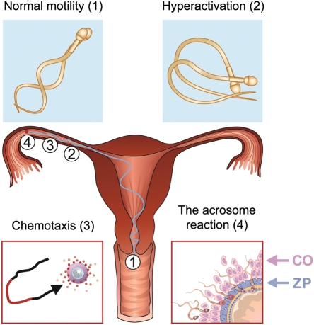
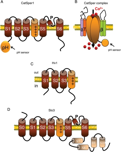
Upon ejaculation, mammalian spermatozoa have to undergo a sequence of physiological transformations within the female reproductive tract that will allow them to reach and fertilize the egg. These include initiation of motility, hyperactivation of motility and perhaps chemotaxis toward the egg, and culminate in the acrosome reaction that permits sperm to penetrate the protective vestments of the egg. These physiological responses are triggered through the activation of sperm ion channels that cause elevations of sperm intracellular pH and Ca(2+) in response to certain cues within the female reproductive tract. Despite their key role in sperm physiology and their absolute requirement for the process of fertilization, sperm ion channels remain poorly understood due to the extreme difficulty in application of the patch-clamp technique to spermatozoa. This review covers the topic of sperm ion channels in the following order: first, we discuss how the intracellular Ca(2+) and pH signaling mediated by sperm ion channels controls sperm behavior during the process of fertilization. Then, we briefly cover the history of the methodology to study sperm ion channels, which culminated in the recent development of a reproducible whole-cell patch-clamp technique for mouse and human cells. We further discuss the main approaches used to patch-clamp mature mouse and human spermatozoa. Finally, we focus on the newly discovered sperm ion channels CatSper, KSper (Slo3) and HSper (H(v)1), identified by the sperm patch-clamp technique. We conclude that the patch-clamp technique has markedly improved and shifted our understanding of the sperm ion channels, in addition to revealing significant species-specific differences in these channels. This method is critical for identification of the molecular mechanisms that control sperm behavior within the female reproductive tract and make fertilization possible.


Figures

Figure 1
Figure 1
The milestones of sperm journey toward the egg. The approximate locations where normal sperm motility (1), hyperactivated sperm motility (2), chemotaxis (3) and the acrosome reaction (4) occur within the female reproductive tract are shown by the corresponding numbers. Normal motility (1) is triggered upon ejaculation in the anterior vagina and is characterized by the low amplitude, symmetrical tail bending. Hyperactive motility (2) occurs after spermatozoa enter Fallopian tubes. It characterized by the high-amplitude asymmetrical tail bending and develops much higher thrust to penetrate through viscous environment of the female reproductive tract. Sperm chemotaxis (3) is believed to occur close to the site of fertilization in the ampulla of Fallopian tubes. Chemoattractant(s) (showed by the red dots) released by the cumulus cells surrounding the egg (or by the egg itself) help spermatozoa to find the egg among the epithelial folds of the Fallopian tube. The acrosome reaction (4) occurs either within the CO or upon the contact with zona pellucida. The hydrolytic enzymes released from the sperm head as the result of the acrosome reaction help spermatozoa to penetrate these egg's protective vestments.
Figure 2
Figure 2
The modes of the patch-clamp technique. To form the cell-attached mode of the patch-clamp technique, a portion of the plasma membrane must be gently driven into the tip of the patch pipette by light suction so that it forms an Ω-shaped invagination within the tip of the pipette and establishes a tight seal with the internal walls of the tip. Since the cell-attached configuration allows only for a limited control of the membrane potential and the recording solutions, it is rarely used for electrophysiological measurements. After formation of the cell-attached mode, the membrane patch under the patch pipette can be destroyed by high-amplitude voltage pulses (up to 1 V) and/or suction to form the whole-mitoplast mode of the patch-clamp technique that allows the recording from the whole-cell plasma membrane. Alternatively, the patch pipette can be withdrawn from the cell to form the inside-out mode of the patch-clamp technique to record single channels. Two electrodes, one in the pipette and one in the bath solution, are connected to the patch-clamp amplifier that controls potential across the cell membrane (V) and measures transmembrane currents (I). The amplifier is shown only for the whole-cell mode.
Figure 3
Figure 3
Molecular architecture of sperm ion channels characterized with the patch-clamp technique. (A) Predicted membrane topology of the pore-forming CatSper1 subunit of CatSper channel. Note the classic six transmembrane helix structure (S1–S6) with the positively charged voltage sensor helix S4 and the pore region (P) between transmembrane helices S5 and S6. The putative pH sensor is located in the histidine-rich N-terminal domain of CatSper1. Other pore-forming subunits of the CatSper channel (CatSpers2–4) have similar membrane topology, but contain less charge in the S4 transmembrane helix and lack the putative pH-sensor in the N-terminal domain. (B) The molecular composition of CatSper channel complex. The Ca2+-selective pore is formed by the four different CatSpers1–4 subunits. The auxiliary CatSper subunits have one (CatSperγ and CatSperδ) or two (CatSperβ) transmembrane helices and large extracellular domains that may be involved in regulation of the CatSper channel by cues of the female reproductive tract. (C) Predicted membrane topology of the Hv1 channel. Hv1 consists of four-transmembrane helices (S1–S4) homologous to the voltage-sensor domain S1–S4 of voltage-gated ion channels, but it lacks the pore forming S5–S6 segment of these channels. The remaining voltage-sensor domain only S1–S4 structure mediates transmembrane proton transport (Ramsey et al., 2010). (D) Predicted membrane topology of the Slo3 channel. It has seven predicted transmembrane helices S0–S6, with S1–S6 helices homologous to classic voltage-gated ion channels. The large intracellular C-terminal domain is likely to be involved in the pH-sensitivity of Slo3 (Xia et al., 2004).
Figure 4
Figure 4
The sperm cytoplasmic droplet: the only gateway for sperm patch-clamp. (A) Electron microphotograph of the cytoplasmic droplet of a ram spermatozoon isolated from cauda epididymis. Note the loose association of the plasma membrane with the underlying mitochondria and the axoneme. The most anterior part of the principal piece is indicated with ‘P’ Reproduced from (Bloom and Nicander 1961; Figure 3) with kind permission of Springer Science + Business Media. (B) Mouse spermatozoon isolated from corpus epididymis. Cytoplasmic droplet is indicated with the red arrow. Annulus (connection between the principal piece and the midpiece is indicated with the blue arrow. (C) Ejaculated human spermatozoa. Cytoplasmic droplet is indicated with the red arrow. Annulus (connection between the principal piece and the midpiece is indicated with the blue arrow. (D) Diagram demonstrating fractionation of the spermatozoon for patch-clamp recording.
Figure 5
Figure 5
Different effects of progesterone on mouse and human CatSper channels. (A) Representative monovalent (Cs+) whole-cell CatSper currents recorded from a human spermatozoon in the absence (blue) and presence (red) of 500 nM progesterone. Note that the human CatSper current is small (especially at the negative membrane potentials) but increases dramatically after addition of progesterone to the bath solution. Currents were recorded in the absence of divalent ions to allow Cs+ permeation through normally Ca2+-selective CatSper channel. Voltage protocol is shown above. Right: a human spermatozoon attached to the recording pipette. (B) Representative monovalent (Cs+) whole-cell CatSper currents recorded from a mouse spermatozoon in the absence (blue) and presence (red) of 500 nM P. Mouse CatSper current is overall larger than human and is not affected by progesterone. All conditions are the same as in (A). Right: mouse spermatozoon attached to the recording pipette. Reproduced with permission from (Lishko et al., 2011).
Figure 6
Figure 6
Sperm patch-clamp technique provides only limited control of the concentration of the permeable ion in the sperm flagellum. Representative whole-cell Ba2+ CatSper current recorded from a mouse spermatozoon isolated from corpus epididymis (red trace). The recording solutions contained no ions that can permeate through ion channels, except for 50 mM Ba2+ in the bath solution. In spite of the absence of Ba2+ in the pipette solution, an outward Ba2+ current was observed at positive transmembrane potentials (indicated with the blue arrow). This outward current is due to Ba2+ accumulation inside the flagellum during the negative part of the voltage-ramp protocol and subsequent efflux of the accumulated Ba2+ through the CatSper channel during the positive part of the voltage ramp. The voltage protocol is shown above. The baseline current was recorded in 50 mM of Mg2+ in the bath solution.

References

    1. Acott TS, Carr DW. Inhibition of bovine spermatozoa by caudal epididymal fluid: II. Interaction of pH and a quiescence factor . Biol Reprod. 1984;30:926–935. - PubMed
    1. Aitken RJ, Irvine S, Kelly RW. Significance of intracellular calcium and cyclic adenosine 3',5′-monophosphate in the mechanisms by which prostaglandins influence human sperm function. J Reprod Fertil. 1986;77:451–462. doi:10.1530/jrf.0.0770451. - DOI - PubMed
    1. Alabi AA, Bahamonde MI, Jung HJ, Kim JI, Swartz KJ. Portability of paddle motif function and pharmacology in voltage sensors. Nature. 2007;450:370–375. doi:10.1038/nature06266. - DOI - PMC - PubMed
    1. Arnoult C, Cardullo RA, Lemos JR, Florman HM. Activation of mouse sperm T-type Ca2+ channels by adhesion to the egg zona pellucida. Proc Natl Acad Sci USA. 1996;93:13004–13009. doi:10.1073/pnas.93.23.13004. - DOI - PMC - PubMed
    1. Austin CR. Observations on the penetration of the sperm in the mammalian egg. Aust J Sci Res B. 1951;4:581–596. - PubMed

Publication types

MeSH terms