. 2011 Jun 5;17(7):822-9.
doi: 10.1038/nm.2345.

Suppression of inflammatory and neuropathic pain by uncoupling CRMP-2 from the presynaptic Ca²⁺ channel complex

Affiliations

Suppression of inflammatory and neuropathic pain by uncoupling CRMP-2 from the presynaptic Ca²⁺ channel complex

Joel M Brittain et al. Nat Med. .

Abstract

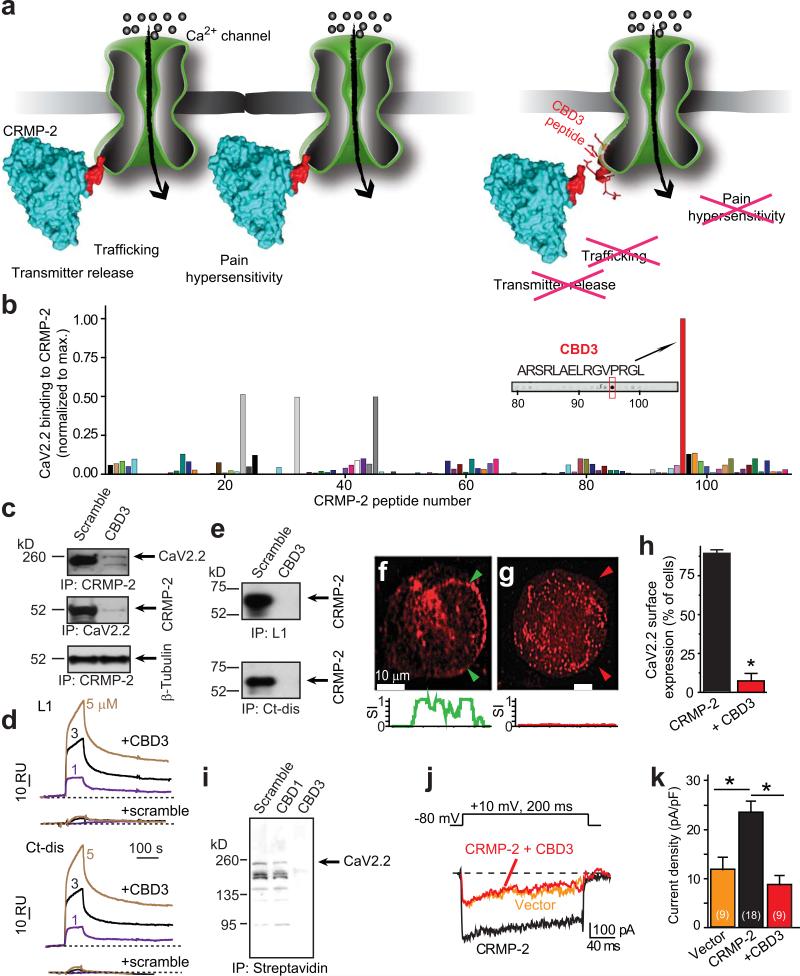
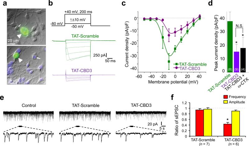
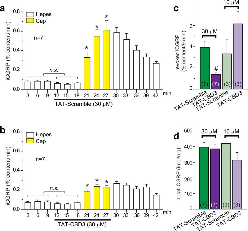
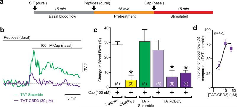
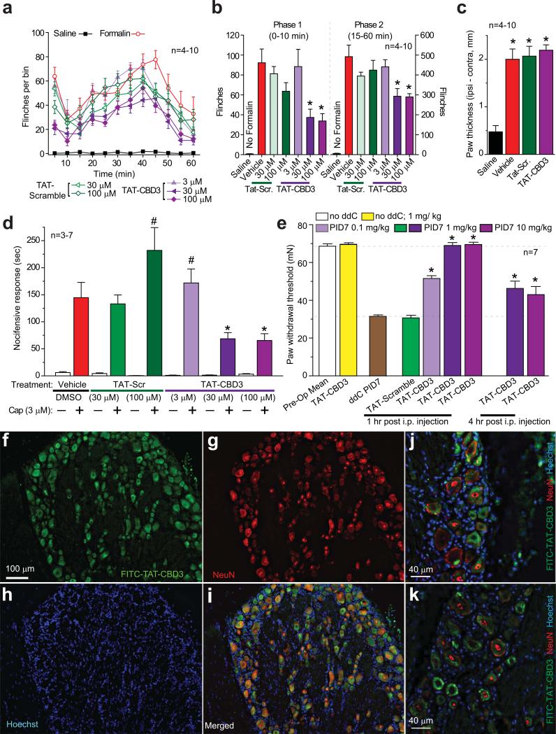
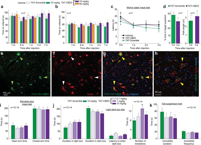
The use of N-type voltage-gated calcium channel (CaV2.2) blockers to treat pain is limited by many physiological side effects. Here we report that inflammatory and neuropathic hypersensitivity can be suppressed by inhibiting the binding of collapsin response mediator protein 2 (CRMP-2) to CaV2.2 and thereby reducing channel function. A peptide of CRMP-2 fused to the HIV transactivator of transcription (TAT) protein (TAT-CBD3) decreased neuropeptide release from sensory neurons and excitatory synaptic transmission in dorsal horn neurons, reduced meningeal blood flow, reduced nocifensive behavior induced by formalin injection or corneal capsaicin application and reversed neuropathic hypersensitivity produced by an antiretroviral drug. TAT-CBD3 was mildly anxiolytic without affecting memory retrieval, sensorimotor function or depression. At doses tenfold higher than that required to reduce hypersensitivity in vivo, TAT-CBD3 caused a transient episode of tail kinking and body contortion. By preventing CRMP-2-mediated enhancement of CaV2.2 function, TAT-CBD3 alleviated inflammatory and neuropathic hypersensitivity, an approach that may prove useful in managing chronic pain.


Figures

Figure 1
Figure 1
A CRMP-2 peptide suppresses the CaV2.2-CRMP-2 interaction in vitro. (a) Cartoon illustrating the main hypothesis. (b) Summary of normalized binding of CaV2.2 to 15 amino acid peptides (overlapping by 12 amino acids) encompassing full-length CRMP-2 overlaid with spinal cord lysates. Sequence of peptide #96, designated CBD3, is shown. (c) Immunoprecipitation (IP) with CRMP-2 antibody from spinal cord lysates in the presence of scramble or CBD3 peptides failed to pull-down CaV2.2 (top) and CRMP-2 (middle) but not β-tubulin (bottom). (d) Sensorgram of CBD3 (1/3/5 μM; solid traces) or scramble peptide (1/3/5 μM; dotted traces) binding to immobilized cytosolic loop 1 (L1) and distal C-terminus (Ct-dis) of CaV2.2. Dissociation was monitored for 4 min. RU, resonance units. (e) In vitro binding of L1-GST and Ct-dis-GST fusion proteins to CRMP-2 in the presence of scramble or CBD3 peptides (10 μM). CRMP-2 bound to L1 and Ct-dis was probed with a CRMP-2 antibody. CaV2.2 is detected on the surface of CAD cells (f) but not when CBD3 fused to GFP is over-expressed (g). Below, normalized surface intensity (SI) between arrows demarcating the surface of cells shown in f and g. (h) Summary of the percent of cells exhibiting surface CaV2.2 expression (n>100). (i) Immunoblots of biotinylated (surface) fractions of CAD cells expressing vector (scramble), an N-terminal region of CRMP-2 (CBD1), or CBD3 probed with a CaV2.2 antibody (n=3). (j) Top, voltage protocol. Bottom, exemplar traces from hippocampal neurons overexpressing vector (EGFP), CRMP-2 or CRMP-2 + CBD3. (k) Peak current density (pA/pF), at +10 mV, for CRMP-2- and CRMP-2 + CBD3-transfected neurons. *, p <0.05 versus CRMP-2, Student's t-test.
Figure 2
Figure 2
TAT-CBD3 reduces Ca2+ currents in DRGs and synaptic responses in lamina II neurons from spinal cord slices. (a) Representative differential interference contrast/fluorescence images showing robust penetration of FITC-TAT-CBD3 into DRGs (arrowheads) but not other cells (arrows). (b) Representative current traces from a DRG incubated for 15 min with TAT-Scramble (10 μM; green) or TAT-CBD3 (10 μM; purple) in response to voltage steps illustrated above the traces. (c) Current-voltage relationships for the currents shown in b fitted to a b-spline line. Peak currents were normalized to the cell capacitance. (d) Peak current density (pA/pF) measured at –10 mV for DRGs incubated with TAT-Scramble, TAT-CBD3 or TAT-CBD3 + 1 μM ω-CTX. The numbers in parentheses represent numbers of cell tested. *, p<0.05 versus TAT-Scramble. (e) Representative traces of spontaneous EPSCs (sEPSCs) in lamina II neurons in spinal cord slices before treatment (left traces), after application of 10 μM TAT-Scramble peptide (middle traces) or 10 μM TAT-CBD3 peptide (right traces). Lower panels are enlarged traces. Voltage-clamp recordings (holding voltage = –70 mV) were used to record synaptic responses. (f) Ratio of sEPSC frequency and amplitude. *, p < 0.05, compared with baseline. Note the significant decrease in the frequency but not amplitude of sEPSCs after the application of TAT-CBD3 peptide.
Figure 3
Figure 3
TAT-CBD3 peptide reduces capsaicin-stimulated release of iCGRP from spinal cord slices. iCGRP release from spinal cord slices stimulated by three 3-min exposures to Hepes buffer alone (white bars) or Hepes buffer containing 500 nM capsaicin (yellow bars) is expressed as mean percent total peptide content of iCGRP in the spinal cord slice±SEM (n=3–7 animals per condition). TAT-Scramble (a) or TAT-CBD3 (b), at 30 μM or 10 μM, was included in the six 3-min incubations indicated by lines, for a total exposure time of 18 min. *, p < 0.05 versus basal iCGRP release in the absence of capsaicin using an ANOVA with a Dunnett's post-hoc test. TAT-Scramble or TAT-CBD3 did not affect basal release of iCGRP (not significant; n.s.). For clarity, only the time course for the 30 μM experiments is shown. (c) Evoked release is compared between TAT treatments. The evoked release was obtained by subtracting iCGRP release during three basal fractions from that during the three capsaicin-stimulated fractions in each treatment group. In all cases, release stimulated by capsaicin was significantly higher than basal release. #, p < 0.05 versus TAT-Scramble using a Student's t-test. (d) Total content of iCGRP released during the perfusion and the amount remaining in the tissues measured at the end of the release experiments.
Figure 4
Figure 4
TAT-CBD3 reduces meningeal blood flow changes in response to capsaicin. (a) Experimental paradigm for the Laser Doppler flowmetry measurements. (b) Representative normalized traces of middle meningeal blood flow changes in response to nasally administered capsaicin (Cap, 100 nM) in the presence of TAT-Scramble (30 μM, green trace) or TAT-CBD3 pretreatment (30 μM, purple trace, applied durally 15 minute prior to Cap administration). Laser Doppler flowmetry measurements were collected at 1 Hz and binned by averaging every 10 samples for graphical representation. The data from each animal was normalized to the first 3 minutes of basal data and the horizontal dashed line indicates the calculated baseline. The ordinate represents red blood cell flux measurements in arbitrary units (AU). (c) Summary of blood flow changes following nasal administration of Cap in the absence or presence of previous administration of TAT-CBD3 or TAT-Scramble to the dura. The capsaicin-induced blood flow changes were CGRP-dependent as they could be blocked by prior dural administration of the CGRP antagonist, CGRP8-37. Values are mean ± S.E.M. *, p < 0.05 versus vehicle (unpaired Student's t-test). The number of animals tested for each condition is indicated in parentheses. (d) Dose response curve of percent inhibition (versus averaged TAT-scramble) of blood flow yields an IC50 of 3.1 ± 1.1 μM (n=4-5).
Figure 5
Figure 5
TAT-CBD3 reduces inflammatory and antiretroviral toxic neuropathic pain. (a) Time course of number of flinches following subcutaneous (dorsal surface of the paw) injection of formalin (2.5% in 50 μl saline) in animals pretreated with TAT-Scramble or TAT-CBD3 (3–100 μM; 20 μl dorsal surface of paw) 30 min before formalin (n=4–10). (b) The effect of TAT-Scramble or TAT-CBD3 on the total number of flinches in formalin-induced phase 1 (0-10 min; left) and phase 2 (15-60 min; right). *, p <0.05 versus formalin-injected animals. (c) Formalin (2.5%) induces paw edema. Paw thickness was measured 1h after injection of saline, formalin, and formalin + TAT peptides (100 μM). *, p <0.05 versus saline- injected animals. (d) Pretreatment with TAT-CBD3 peptide attenuates capsaicin-evoked nocifensive behavior. Vehicle (0.3% DMSO), TAT scramble (30–100 μM), or TAT-CBD3 (3–100 μM) in saline (40 μL) was applied in the right eye and nocifensive behavior was noted. Five minutes after treatment, capsaicin (Cap, 3 μM in 40 μL saline) was applied in the right eye and nocifensive behavior was noted by observers blinded to treatment condition. Data are shown as mean ± SEM (n = 3-7 per group; *, p<0.05 versus vehicle, ANOVA with Dunnett's post-hoc test). (e) Dose-dependent effect of TAT-CBD3 peptide on ddC-induced decreases in paw withdrawal threshold (PWT) in the rat at 1 h and 4 h post injection. Animals subjected to a single ddC injection exhibited a decrease in PWT that was abolished by i.p. administration of TAT-CBD3 peptide on post-injection day 7 (PID7). Data represent means ± S.E.M.; *, p < 0.05 versus ddC or TAT-Scramble (ANOVA with Dunnett's post-hoc test), n = 6 per condition. (f-k) DRGs were isolated 15 min post-injection with 20 mg/kg FITC-TAT-CBD3, cryosectioned, and subjected to immunohistochemistry using a monoclonal anti-NeuN antibody. TAT-CBD3 (f, green, FITC) accumulates in almost all neurons with the DRG. Neurons are indicated by NeuN (g, red). Nuclei of all cells are stained with Hoechst (h, blue). Merged images are shown in i-k. Scale bars, 100 μm (f-i); 40 μm (j-k).
Figure 6
Figure 6
TAT-CBD3 has no effects on sensorimotor and cognitive functions and despair but is mildly anxiolytic. (a, b) Time course of latency to fall off a rod rotating at a slow (a) or fast (b) acceleration at various time points after i.p. administration of 10 mg/ kg or 50 mg/kg peptides as indicated or vehicle (n=7 each). There were no significant differences in both low and fast accelerating rotarod performances between groups (ANOVA with Dunnett's post-hoc test). (c) Time course of latency (in sec) for mice to successfully find a hidden platform in the Morris water maze at several time points follow i.p. administration of 10 mg/kg peptides or vehicle (n=7). (d) Percent time in target quadrant and path length performance in the Morris water maze test. The platform was removed after the last day of hidden platform testing and mice were placed in the pool for a single 60-sec trial. There were no significant differences in percent time spent in target quadrant or path length between groups (Student's t-test). (e-h) Immunohistochemistry in dorsal horn of the spinal cord. Spinal cord was isolated 15 min post-injection with 20 mg/kg FITC-TAT-CBD3, cryosectioned, and subjected to immunohistochemistry using a monoclonal anti-NeuN antibody. TAT-CBD3 (e, green, FITC) accumulates in motor neurons (arrowheads) which co-label with NeuN (f, red). Nuclei of all cells are stained with Hoechst (blue). Merged images demonstrate co-labeling of FITC-TAT-CBD3-containing neurons (green) with NeuN (red) and Hoescht (blue) at low (g) and high magnification (h). Scale bars, 100 μm (e-g); 40 μm (h). (i) Elevated plus maze test to evaluate anxiety-associated behaviors. Neither the time spent in the open (F(3,48)=0.6, p=0.625) or closed (F(3,48)=0.04, p=0.987) arm, nor the frequency of entries into the (F(3,48)=1.3, p=0.282) or closed (F(3,48)=1.5, p=0.219) arms were altered by any of the 3 doses of TAT-CBD3, compared to the TAT-Scramble. (j) Light dark box test for anxiety-associated behaviors. TAT-CBD3 did not alter time spent in the light box compared to the dark box or aversion to first entering the dark box. The number of transitions between the light and dark box was increased with 1 mg/kg dose of TAT-CBD3 (F(3,47) =3.6, p=.020, *). (k) Tail suspension test of depression or despair-associated behaviors. Duration and frequency of immobility was not altered by i.p. injection of TAT-CBD3. Data reflect mean ± SEM.

Comment in

References

    1. Costigan M, Scholz J, Woolf CJ. Neuropathic pain: a maladaptive response of the nervous system to damage. Annu. Rev. Neurosci. 2009;32:1–32. 1-32. - PMC - PubMed
    1. Crofford LJ. Adverse effects of chronic opioid therapy for chronic musculoskeletal pain. Nat. Rev. Rheumatol. 2010;6:191–197. - PubMed
    1. Staats PS, et al. Intrathecal ziconotide in the treatment of refractory pain in patients with cancer or AIDS: a randomized controlled trial. JAMA. 2004;291:63–70. - PubMed
    1. Rauck RL, et al. A randomized, double-blind, placebo-controlled study of intrathecal ziconotide in adults with severe chronic pain. J. Pain Symptom. Manage. 2006;31:393–406. - PubMed
    1. Wallace MS, et al. Intrathecal ziconotide for severe chronic pain: safety and tolerability results of an open-label, long-term trial. Anesth. Analg. 2008;106:628–37. table. - PubMed

Publication types

MeSH terms

Substances