. 2011 Dec 8;30(49):4855-63.
doi: 10.1038/onc.2011.200. Epub 2011 Jun 6.

Phosphorylation and interaction of myopodin by integrin-link kinase lead to suppression of cell growth and motility in prostate cancer cells

Affiliations

Phosphorylation and interaction of myopodin by integrin-link kinase lead to suppression of cell growth and motility in prostate cancer cells

Y-P Yu et al. Oncogene. .

Abstract

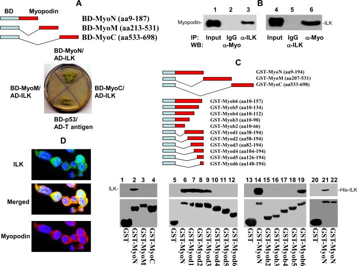
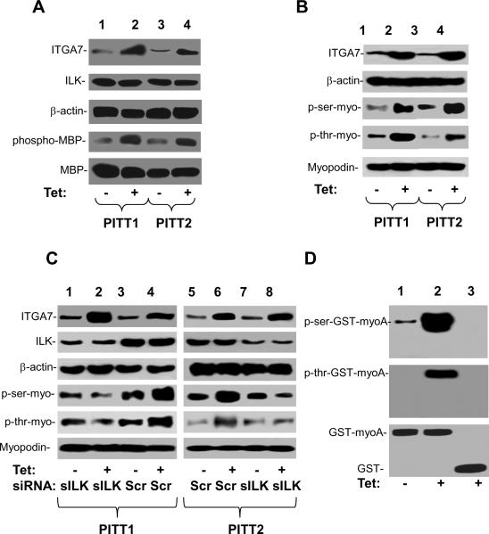
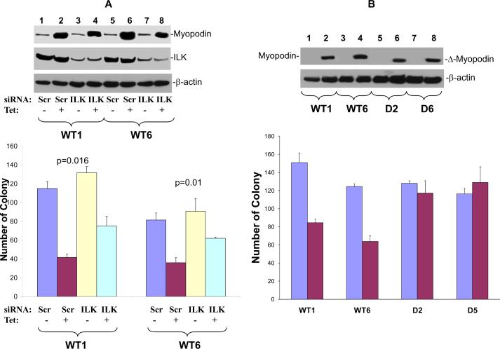
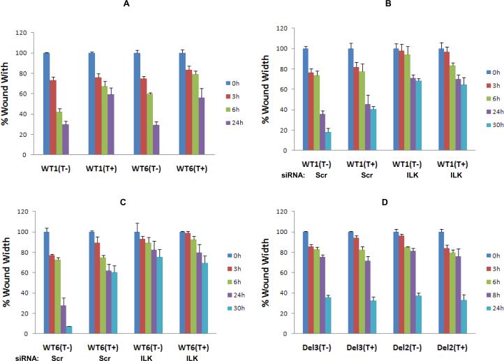
Myopodin is a tumor-suppressor gene that suppresses growth of prostate and urothelial carcinomas. However, the mechanism of myopodin tumor-suppressor activity or signaling that leads to activation of myopodin remains unclear. In this report, we showed that the N-terminus of myopodin binds integrin-linked kinase (ILK) both in vivo and in vitro. An ILK interaction motif of 78 amino acids (amino acids 82-157) was identified in the N-terminus region of myopodin. Induction of ILK-dependent kinase activity by integrin α7 led to phosphorylation of myopodin both in vivo and in vitro. Knocking down ILK dramatically reduced the inhibition of cell growth and motility mediated by myopodin. A mutant of myopodin lacking the ILK interaction motif is inactive in suppressing the growth and motility of PC3 cells. As a result, this study showed a novel and critical signaling pathway that leads to activation of myopodin.


Figures

Figure 1
Figure 1. Interaction of N-terminus of myopodin with ILK
(A) Constructs of N- terminus (BD-myoN), M-segment (BD-myoM) and C-terminus (BD-myoC) of myopodin with bait domain in yeast two hybrid analysis. Co-transformant of pBD-myoN, pBD-myoM, and pBD-myoC with pACT2-ILK on SD agar palte with high strigent nutrient selection (SD-leu-Trp-His-Ade). (B) Co-immunoprecipitation of ILK or myopodin using antibodies specific for myopodin (α-Myo) or ILK (α-ILK). The immunoprecipitates were blotted with the indicated antibodies. (C) Mapping binding motif of ILK on myopodin. Top: Constructs of series of myopodin deletion mutant with GST expression vectors. Bottom: Binding assays on GST or GST-myopodin deletion mutants with ILK from PC3 cells (lanes 1–19) or HisTag-ILK (lanes 20–21) or pET28a lysate (lane 22). The bound ILK was blotted with ILK antibodies. (D) Immunofluorescence staining of I4 cells with antibody against myopodin bound by rhodamin conjugated secondary antibody for rabbit, and antibody specifically against ILK recognized by FITC conjugated secondary antibody for goat.
Figure 2
Figure 2. Phosphorylation of myopodin by ILK
(A) ILK kinase activity induced by integrin α7. ILk was immunopurified from PITT1 and PITT2 cells induced with or without tetracycline. ILK was then subjected to the MBP phosphorylation analysis. Top 3 panels: immunoblots of lysates of PITT1 and PITT2 cells. Bottom 2 panels: immunoblots of kinase assay using myelin basic protein as substrate on ILK immunoprecipitates. (B) Immunopurified myopodin from PITT1 and PITT2 cells induced with or without tetracycline were blotted with antibody against phosphorylated serine or phosphorylated threonine. Top 2 panels: immunoblots of lysates of PITT1 and PITT2 cells. Bottom three panels: immunoblots of myopodin immunoprecipitates. (C) Immunopurified myopodin from PITT1 and PITT2 cells, induced with or without tetracycline and transfected with scramble siRNA or siRNA against ILK, were blotted with antibody against phosphorylated serine or phosphorylated threonine. Top 2 panels: immunoblots of lysates of PITT1 and PITT2 cells. Bottom three panels: immunoblots of myopodin immunoprecipitates. (D) ILK dependent phosphorylation assays on GST-myoN (N-terminus of myopodin) purified through Glutathione sepherose 4B. ILK immunopurified from PITT 1 cells induced with (Tet +) or without (Tet −) tetracycline were incubated with purified GST-MyoN in a kinase reaction. The results were analyzed with antibody specific for phospho-serine or phosphothreonine. GST was used as negative control (lane 3).
Figure 3
Figure 3. Down regulation of ILK and deletion of ILK binding motif abrogates myopodin cell growth suppression activity
(A) Colony formation assays for PC3 cell clones expressing wild type (WT1, WT6) treated with tetracycline and transfected with shRNA against ILK (ILK) or scramble control (Scr). Each condition was in triplicates. Top panel: Immunoblots of myopodin, ILK and β-actin of representative samples. Bottom panel: Colony formation assays. (B) Colony formation assays for PC3 cell clones expressing wild type (WT1, WT6) or ILK motif deletion mutants (D2 and D5) of myopodin treated with tetracycline. One thousand cells of wt or mutant myopodin were plated to each well of 6 well plates and colony formation was evaluated. Cells were treated with or without tetracycline. Each condition was in triplicates. Top panel: Immunoblots of myopodin and β-actin of representative samples. Bottom panel: Colony formation assays.
Figure 4
Figure 4. Down-regulation of ILK and deletion of ILK binding motif abrogates myopodin cell motility suppression activity
(A) Wound healing assays for PC3 cell clones expressing wild type (WT1, WT6) treated with or without tetracycline for 3, 6 and 24 hours (h). (B) Wound healing assays for WT1 cells treated with or without tetracycline and transfected with shRNA specific for ILK for 3, 6, 24 and 30 hours (h). (C) Wound healing assays for WT6 cells treated with or without tetracycline and transfected with shRNA specific for ILK for 3, 6, 24 and 30 hours (h). (D) Wound healing assays of PC3 cells transfected with mutant (D2 and D5) myopodin. The wound was measured at 0h, 6h, 8h and 24h. Triplicates were prepared for each cell condition.

References

    1. Acconcia F, Barnes CJ, Singh RR, Talukder AH, Kumar R. Phosphorylation-dependent regulation of nuclear localization and functions of integrin-linked kinase. Proc Natl Acad Sci U S A. 2007;104:6782–7. - PMC - PubMed
    1. Apte U, Gkretsi V, Bowen WC, Mars WM, Luo JH, Donthamsetty S, et al. Enhanced liver regeneration following changes induced by hepatocyte-specific genetic ablation of integrin-linked kinase. Hepatology. 2009;50:844–51. - PMC - PubMed
    1. Cebrian V, Alvarez M, Aleman A, Palou J, Bellmunt J, Gonzalez-Peramato P, et al. Discovery of myopodin methylation in bladder cancer. J Pathol. 2008;216:111–9. - PubMed
    1. Deng JT, Van Lierop JE, Sutherland C, Walsh MP. Ca2+-independent smooth muscle contraction. a novel function for integrin-linked kinase. J Biol Chem. 2001;276:16365–73. - PubMed
    1. Donthamsetty S, Bowen W, Mars W, Bhave V, Luo JH, Wu C, et al. Liver Specific Ablation of Integrin Linked Kinase (ILK) in Mice Results in Enhanced and Prolonged Cell Proliferation and Hepatomegaly after Phenobarbital Administration. Toxicol Sci. 2009 - PMC - PubMed

Publication types

Substances