. 2011 Nov 17;30(46):4678-86.
doi: 10.1038/onc.2011.185. Epub 2011 Jun 6.

Acquisition of p53 mutations in response to the non-genotoxic p53 activator Nutlin-3

Affiliations

Acquisition of p53 mutations in response to the non-genotoxic p53 activator Nutlin-3

M H Aziz et al. Oncogene. .

Abstract

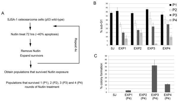
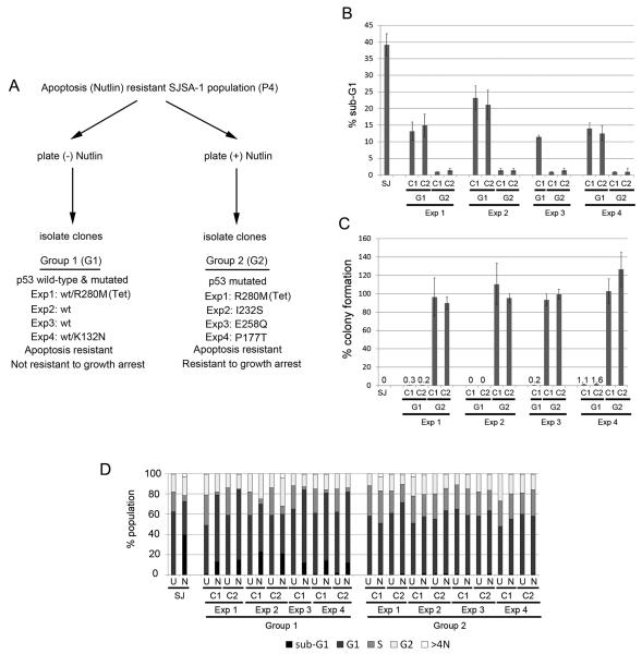
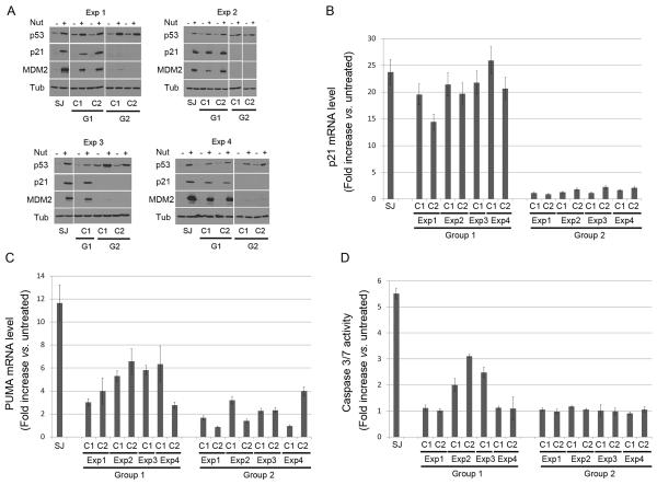
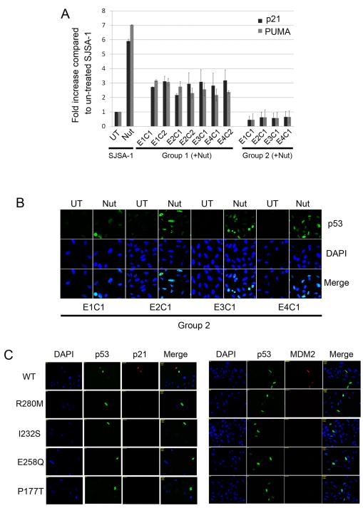
Wild-type p53 is a stress-responsive tumor suppressor and potent growth inhibitor. Genotoxic stresses (for example, ionizing and ultraviolet radiation or chemotherapeutic drug treatment) can activate p53, but also induce mutations in the P53 gene, and thus select for p53-mutated cells. Nutlin-3a (Nutlin) is pre-clinical drug that activates p53 in a non-genotoxic manner. Nutlin occupies the p53-binding pocket of murine double minute 2 (MDM2), activating p53 by blocking the p53-MDM2 interaction. Because Nutlin neither binds p53 directly nor introduces DNA damage, we hypothesized Nutlin would not induce P53 mutations, and, therefore, not select for p53-mutated cells. To test this, populations of SJSA-1 (p53 wild-type) cancer cells were expanded that survived repeated Nutlin exposures, and individual clones were isolated. Group 1 clones were resistant to Nutlin-induced apoptosis, but still underwent growth arrest. Surprisingly, while some Group 1 clones retained wild-type p53, others acquired a heterozygous p53 mutation. Apoptosis resistance in Group 1 clones was associated with decreased PUMA induction and decreased caspase 3/7 activation. Group 2 clones were resistant to both apoptosis and growth arrest induced by Nutlin. Group 2 clones had acquired mutations in the p53-DNA-binding domain and expressed only mutant p53s that were induced by Nutlin treatment, but were unable to bind the P21 and PUMA gene promoters, and unable to activate transcription. These results demonstrate that non-genotoxic p53 activation (for example, by Nutlin treatment) can lead to the acquisition of somatic mutations in p53 and select for p53-mutated cells. These findings have implications for the potential clinical use of Nutlin and other small molecule MDM2 antagonists.


Figures

Figure 1
Figure 1. Selection of Nutlin-resistant SJSA-1 populations
A. The procedure used to select SJSA-1 populations resistant to Nutlin-induced apoptosis. B. SJSA-1 (SJ) was obtained from American Type Culture Collection and grown in RPMI1640 medium (100 units/mL penicillin, 100 μg/mL streptomycin, 10% FBS). SJSA-1 and Nutlin-selected populations (P1, P2, P3 and P4) were treated with 10 μM Nutlin-3 (Nutlin, Sigma, USA) for 72 hrs. Cells were harvested, fixed in 25% ethanol, stained with propidium iodide and subjected to FACS analysis as previously described (Shen et al., 2008). Percentage of cells with sub-G1 DNA content was determined from the DNA profile histogram using FlowJo (Treestar Inc., USA). The mean of three independent experiments is shown ± S.E. (error bars). C. SJSA-1 (SJ) and Nutlin-resistant populations (P4) were plated at low density (1×10² - 1×10⁵ cells / 10cm dish). Cells were either untreated or treated with 10 μM Nutlin and allowed to grow for a 2-3 week period. Colonies were stained with 0.5% Crystal Violet and counted. The plating efficiency for untreated sample was set at 100%. The mean of three independent experiments is shown ± S.E. (error bars).
Figure 2
Figure 2. Cell cycle and colony formation analysis in selected Nutlin-Resistant clones
A. Selection of Nutlin-Resistant SJSA-1 cell clones. P4 populations from Experiments 1-4 were plated in the absence or presence of Nutlin for 14 days. Individual colonies were then isolated and expanded. Group 1 clones (isolated in the absence of Nutlin) undergo growth arrest in the presence of Nutlin but are resistant to Nutlin-induced apoptosis. Group 2 clones (isolated in the presence of Nutlin) are resistant to both apoptosis and growth arrest induced by Nutlin. The p53 status of the Group 1 and Group 2 clones from each experiment that were used in subsequent experiments was determined by cDNA sequencing and is indicated. The p53 sequence of all clones isolated in each experiment is listed in Supplemental Table 1. B, D. SJSA-1 (SJ) and Nutlin-resistant Group 1 (G1) and Group 2 (G2) clones were untreated or treated with 10 μM Nutlin for 72 hrs and subjected to FACS analysis. The percentage of cells with sub-G1 DNA content (B) and cell cycle distribution (D) were determined from DNA profile histograms using FlowJo. U: untreated; N: Nutlin treated. C. SJSA-1 (SJ) and Nutlin-resistant Group 1 and Group 2 clones were plated at low density (1×10² - 1×10⁵ cells / 10cm dish) and either untreated or treated with 10 μM Nutlin for 14-21 days. Colony forming ability was determined as described in Figure 1.
Figure 3
Figure 3. P53 transcriptional activity and caspase activation in Nutlin-resistant clones
SJSA-1 (SJ) and Nutlin-resistant Group 1 and Group 2 clones were untreated or treated with 10 μM Nutlin for 24 hrs. A. Cell lysates were analyzed by immunoblotting with antibodies against p53, MDM2, and p21, as previously described (Shen et al 2008). Tubulin levels were used as a loading control. Representative immunoblots are shown. B, C. Quantitative real-time PCR was performed to measure mRNA levels of p21 (B) and PUMA (C) in SJSA-1 and Nutlin-resistant clones. The complementary DNA (cDNA) was synthesized after treatment. The quantitative real-time PCR reaction was run in a 7300 Real Time PCR System (Applied Biosystems, Foster, CA) using SybrGreen PCR master mix, (Applied Biosystems, Foster City, CA) following manufacturer’s instructions. Thermocycling was done in a final volume of 20 μL containing 2 μL of cDNA and 400 nmol/L of primers (Primers are listed in Supplemental Table 2). All samples were amplified in triplicate using the following cycle scheme: 95°C for 2 minutes, 40 cycles of 95°C for 15 seconds and 55°C for 60 seconds. Fluorescence was measured in every cycle and mRNA levels were normalized using the GAPDH values in all samples. A single peak was obtained for targets, supporting the specificity of the reaction. The fold increase (compared to untreated levels) in p21 and PUMA mRNA was determined after 24hr Nutlin treatment. Data is presented as the mean fold increase ± S.E. (n=3). D. Caspase-Glo-3/7 Assay was performed with untreated and Nutlin-treated (10 μM Nutlin for 24 hrs) SJSA-1 and Nutlin-resistant clones with the Caspase-Glo-3/7 Assay (Promega Biotech, Madison, WI) per manufacture’s instruction. Results are presented as the mean (signal-to-noise ratios) ± SEM of the triplicate assays.
Figure 4
Figure 4. Mutant p53s in Nutlin-resistant clones lack DNA binding ability and transcription activity
A. SJSA-1, Group 1 and Group 2 clones were untreated or Nutlin-treated (10 μM) for 24 hrs. Chromatin immunoprecipitation (ChIP) was performed using Human p53 ChampionChIP Antibody kit (SABiosciences/Qiagen, Frederick, MD). Briefly, formaldehyde cross-linking was performed for 10 min, and samples were sonicated to obtain DNA fragments with average size of 400–500 bp. Protein–DNA complexes were immunoprecipitated using p53 antibody (SABiosciences/Qiagen, Frederick, MD). DNAs were purified and subjected to quantitative real-time PCR amplification. The primers used are listed in Supplemental Table 2. The fold-increase ± S.E. in p53 binding to the P21 and PUMA gene promoters after 24 hrs Nutlin treatment is plotted (n=3). The level of p53 binding to each promoter in untreated SJSA-1 parental cells was given a value of 1.0, and everything else scored relative to that. B. Group 2 clones were untreated or Nutlin-treated (10 μM) for 24 hrs. Cells were fixed with 4% formaldehyde and subjected to immunofluorescence with anti-p53 antibody as described (Shen et al., 2008). Cell nuclei were counterstained with DAPI (blue). Representative images were captured at ×40 magnification and are shown. UT: untreated; Nut: Nutlin treated. C. Site directed mutations in wild-type (WT) p53 plasmid were constructed using QuikChange mutagenesis kit (Stratagene La Jolla, CA) for the mutations R280M, I232S, E258Q and P177T (Primers used for mutagenesis listed in Supplemental Table 2). Plasmids encoding wild-type p53, p53R280M, p53I232S, p53E258Q and p53P177T were transfected into Saos-2 (p53-null) cells using FuGENE-6 transfection reagent. Saos-2 cells were fixed 48 hrs after transfection and subjected to immunofluorescence with indicated antibodies.

References

    1. Ahmed AA, Etemadmoghadam D, Temple J, Lynch AG, Riad M, Sharma R, Stewart C, Fereday S, Caldas C, Defazio A, Bowtell D, Brenton JD. Driver mutations in TP53 are ubiquitous in high grade serous carcinoma of the ovary. J Pathol. 2010;221:49–56. - PMC - PubMed
    1. Ashkenazi A. Targeting the extrinsic apoptosis pathway in cancer. Cytokine Growth Factor Rev. 2008;19:325–31. - PubMed
    1. Blagosklonny MV. Oncogenic resistance to growth-limiting conditions. Nat Rev Cancer. 2002;2:221–5. - PubMed
    1. Blagosklonny MV. Carcinogenesis, cancer therapy and chemoprevention. Cell Death Differ. 2005;12:592–602. - PubMed
    1. Brown L, Boswell S, Raj L, Lee SW. Transcriptional targets of p53 that regulate cellular proliferation. Crit Rev Eukaryot Gene Expr. 2007;17:73–85. - PubMed

Publication types

MeSH terms