. 2011 Jun;11(6):1236-47.
doi: 10.1111/j.1600-6143.2011.03566.x.

Mechanisms of donor-specific tolerance in recipients of haploidentical combined bone marrow/kidney transplantation

Affiliations

Mechanisms of donor-specific tolerance in recipients of haploidentical combined bone marrow/kidney transplantation

G Andreola et al. Am J Transplant. 2011 Jun.

Abstract

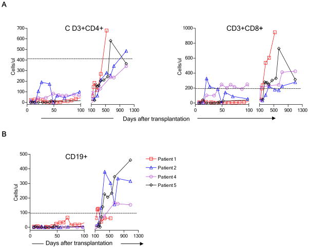
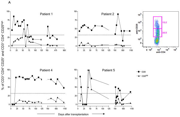
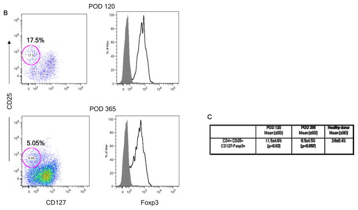
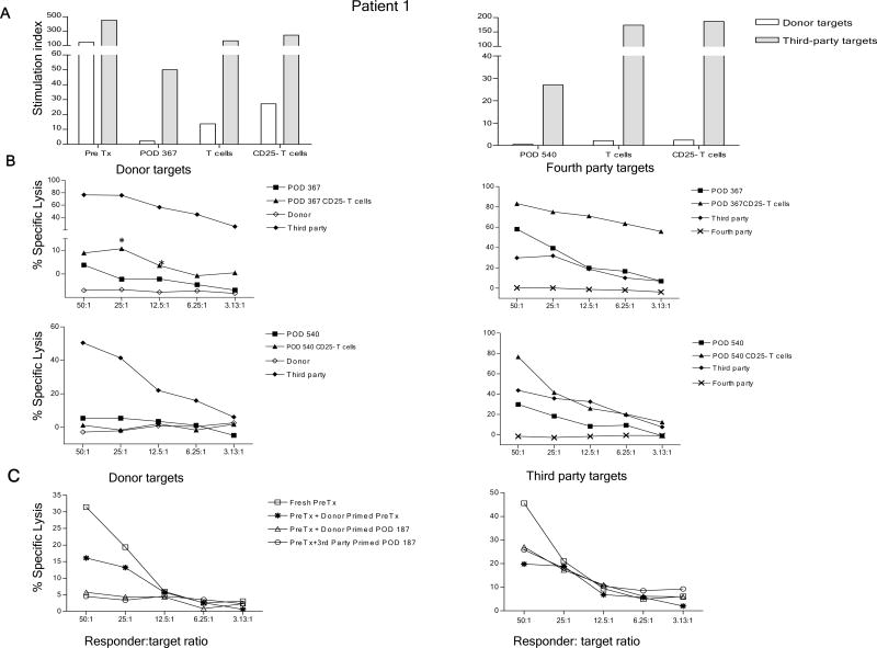
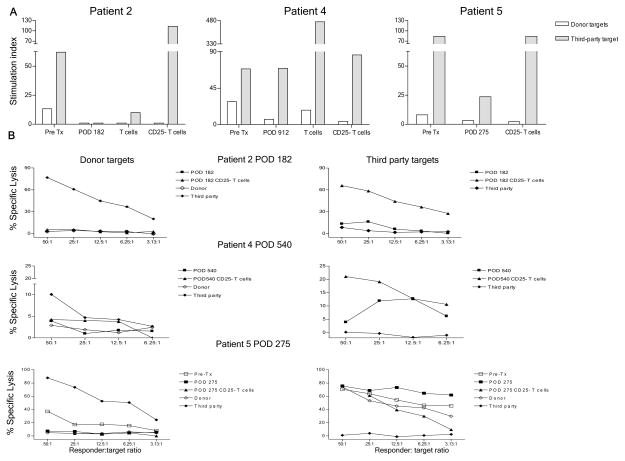
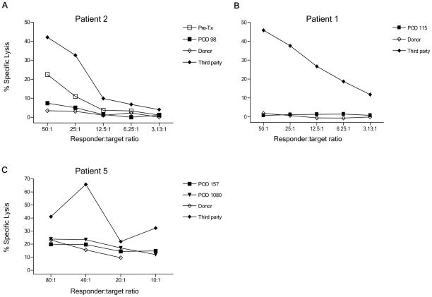
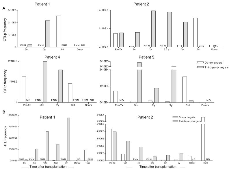
We recently reported long-term organ allograft survival without ongoing immunosuppression in four of five patients receiving combined kidney and bone marrow transplantation from haploidentical donors following nonmyeloablative conditioning. In vitro assays up to 18 months revealed donor-specific unresponsiveness. We now demonstrate that T cell recovery is gradual and is characterized by memory-type cell predominance and an increased proportion of CD4⁺ CD25⁺ CD127⁻ FOXP3⁺ Treg during the lymphopenic period. Complete donor-specific unresponsiveness in proliferative and cytotoxic assays, and in limiting dilution analyses of IL-2-producing and cytotoxic cells, developed and persisted for the 3-year follow-up in all patients, and extended to donor renal tubular epithelial cells. Assays in two of four patients were consistent with a role for a suppressive tolerance mechanism at 6 months to 1 year, but later (≥ 18 months) studies on all four patients provided no evidence for a suppressive mechanism. Our studies demonstrate, for the first time, long-term, systemic donor-specific unresponsiveness in patients with HLA-mismatched allograft tolerance. While regulatory cells may play an early role, long-term tolerance appears to be maintained by a deletion or anergy mechanism.


Conflict of interest statement

Disclosure

The authors of this manuscript have no conflicts of interest as described by the American Journal of Transplantation

Figures

Figure 1
Figure 1
Time course of T and B cell recovery in four patients following non-myeloablative conditioning. (A) CD3+CD4+, CD3+CD8+ and (B) CD19+ recovery. Peripheral blood concentrations of each cell type in fresh blood samples are shown at the indicated time point. The horizontal dotted lines denote the 10th percentile for normal adults.
Figure 2
Figure 2
Enrichment of CD4+CD25+ cells after transplant. (A) Recovery of CD25+ and CD25high cells in fresh blood samples in each of the four patients among total gated CD3+CD4+ T cells. Dotted and solid lines, respectively, denote the median value of CD4+CD25+ and of CD4+CD25high cells found in normal controls. The inset panel shows CD25low and CD25high populations in a representative sample from Patient 5. (B) Expression of FoxP3 (right panel), open histograms. Shaded histograms represent unstained controls on gated cryopreserved/thawed CD4+CD25+CD127− cells (left panel), at POD 120 and 365. Plots from Patient 5 are representative of results from all 4 patients. (C) Percentages of CD25+CD127−FOXP3+ cells among gated cryopreserved/thawed CD4+ cells in patients’ peripheral blood at POD 120 and 1 year compared to healthy donors (HD) (n=4 patients at POD 120 and n=3 at 1 year and 5 normal controls).
Figure 2
Figure 2
Enrichment of CD4+CD25+ cells after transplant. (A) Recovery of CD25+ and CD25high cells in fresh blood samples in each of the four patients among total gated CD3+CD4+ T cells. Dotted and solid lines, respectively, denote the median value of CD4+CD25+ and of CD4+CD25high cells found in normal controls. The inset panel shows CD25low and CD25high populations in a representative sample from Patient 5. (B) Expression of FoxP3 (right panel), open histograms. Shaded histograms represent unstained controls on gated cryopreserved/thawed CD4+CD25+CD127− cells (left panel), at POD 120 and 365. Plots from Patient 5 are representative of results from all 4 patients. (C) Percentages of CD25+CD127−FOXP3+ cells among gated cryopreserved/thawed CD4+ cells in patients’ peripheral blood at POD 120 and 1 year compared to healthy donors (HD) (n=4 patients at POD 120 and n=3 at 1 year and 5 normal controls).
Figure 3
Figure 3
Role of CD25+ T cells in donor-specific unresponsiveness in Patient 1. (A) Pre-transplant and post-transplant (POD 367 and POD 540) whole PBMC, T cell and CD25− fractions were tested against irradiated donor and third party targets in MLR; (B) Whole PBMC and CD25− fraction from POD 367 and POD 540 were tested at various ratios against irradiated donor and fourth party stimulators and targets (unrelated to third party or patient) in CML assay. CD25+ cell depletion reveals an anti-donor CML response (*p<.05) in Patient 1 at POD 367 but not at POD 540 (Panel B, left). Normal third-party control killing of donor targets is seen in the same assay; (C) Suppression of anti-donor CTL responses by Patient 1 cells at POD 187 in co-culture assay. Pre-transplant or POD 187 PBMC were stimulated for 5 days with donor or third party PBMC stimulators, then tested for the ability to suppress anti-donor or 3rd party CML responses.
Figure 4
Figure 4
Donor-specific MLR and CML unresponsiveness in Patients 2, 4, and 5 is not dependent on CD25+ T cells. (A) Pre-transplant and POD 182 whole PBMC, T cells and CD25− fractions in Patient 2, pre-transplant and POD 912 whole PBMC, T cells and CD25− fractions in Patient 4, pre-transplant and POD 275 whole PBMC and CD25− fractions in Patient 5 were tested against irradiated donor and third party stimulators in MLR; (B) POD 182 whole PBMC and CD25− fraction in Patient 2, POD 540 whole PBMC and CD25− fraction in Patient 4, pre-transplant and POD 275 whole PBMC and CD25− fraction in Patient 5 were stimulated for 5 days with irradiated PBMC, then tested at different ratios for cytotoxicity against donor and third party targets in CML assays.
Figure 5
Figure 5
Lack of killing of donor RTECs by patient NK cell-depleted PBMC in a Cr51 realease cytotoxicity assay. (A) Patient 2, third party and donor responses to donor RTEC targets; (B) Patient 1, third party and donor responses to donor RTECs. (C) Patient 5, third party and donor responses to donor RTECs.
Figure 6
Figure 6
CTL precursor and IL-2-producing cell frequencies as detected by limiting dilution assays against donor and third party PBMC stimulators and targets at various time points after transplant. (A) Summary of CTL precursor frequencies as calculated with the Poisson distribution. Responders and time points are indicated on the x axes. Stimulators and targets were donor and third party PBMC; (B) Frequencies of anti-donor IL-2-producing cells (helper T lymphocyte, HTL) in Patients 1 and 2 at the indicated time points, as calculated by Poisson distribution from IL-2 production measured in supernatants from limiting dilution cultures of patient and control PBMC stimulated with donor or 3rd party PBMC. FNM (Frequency not measurable), ND (Not Done)
Figure 7
Figure 7
Donor-specific unresponsiveness in Patient 5 at 6 and 9 months is not dependent on CD25+ T cells. (A) Frequencies of anti-donor and third party CTLp of indicated cell populations as calculated from Poisson distribution analysis of the limiting dilution cultures; (B) Frequencies of anti-donor and third party IL-2-producing cell (helper T lymphocyte, HTL) of indicated cell populations as calculated from Poisson distribution analysis of the IL-2 content of supernatants from limiting dilution cultures. FMN (Frequency not measurable), ND (Not Done)

References

    1. Sharabi Y, Sachs DH. Mixed chimerism and permanent specific transplantation tolerance induced by a non-lethal preparative regimen. J Exp Med. 1989;169:493–502. - PMC - PubMed
    1. Kawai T, Cosimi AB, COLVIN RB, Powelson J, Eason J, Kozlowski T, et al. Mixed allogeneic chimerism and renal allograft tolerance in cynomologous monkeys. Transplantation. 1995;59:256–62. - PubMed
    1. Sharabi Y, Sachs DH, Sykes M. T cell subsets resisting induction of mixed chimerism across various histocompatibility barriers. In: Gergely J, Benczur M, Falus A, Fust Gy, Medgyesi G, Petranyi Gy, et al., editors. Progress in Immunology VIII; Proceedings of the Eighth International Congress of Immunology; Budapest. 1992; Heidelberg: Springer-Verlag; 1992. pp. 801–5.
    1. Ohdan H, Yang Y-G, Shimizu A, Swenson KG, Sykes M. Mixed bone marrow chimerism induced without lethal conditioning prevents T cell and anti-Gala1,3Gal-mediated graft rejection. J Clin Invest. 1999;104:281–90. - PMC - PubMed
    1. Fudaba Y, Spitzer TR, Shaffer J, Kawai T, Fehr T, Delmonico F, et al. Myeloma Responses and Tolerance Following Combined Kidney and Nonmyeloablative Marrow Transplantation: In Vivo and In Vitro Analyses. Am J Transplant. 2006 Jun 22;6(9):2121–33. - PubMed

Publication types