. 2011 Jun 7;18(1):37.
doi: 10.1186/1423-0127-18-37.

Sphingosine-1-phosphate promotes the differentiation of human umbilical cord mesenchymal stem cells into cardiomyocytes under the designated culturing conditions

Affiliations

Sphingosine-1-phosphate promotes the differentiation of human umbilical cord mesenchymal stem cells into cardiomyocytes under the designated culturing conditions

Zhenqiang Zhao et al. J Biomed Sci. .

Abstract

Background: It is of growing interest to develop novel approaches to initiate differentiation of mesenchymal stem cells (MSCs) into cardiomyocytes. The purpose of this investigation was to determine if Sphingosine-1-phosphate (S1P), a native circulating bioactive lipid metabolite, plays a role in differentiation of human umbilical cord mesenchymal stem cells (HUMSCs) into cardiomyocytes. We also developed an engineered cell sheet from these HUMSCs derived cardiomyocytes by using a temperature-responsive polymer, poly(N-isopropylacrylamide) (PIPAAm) cell sheet technology.

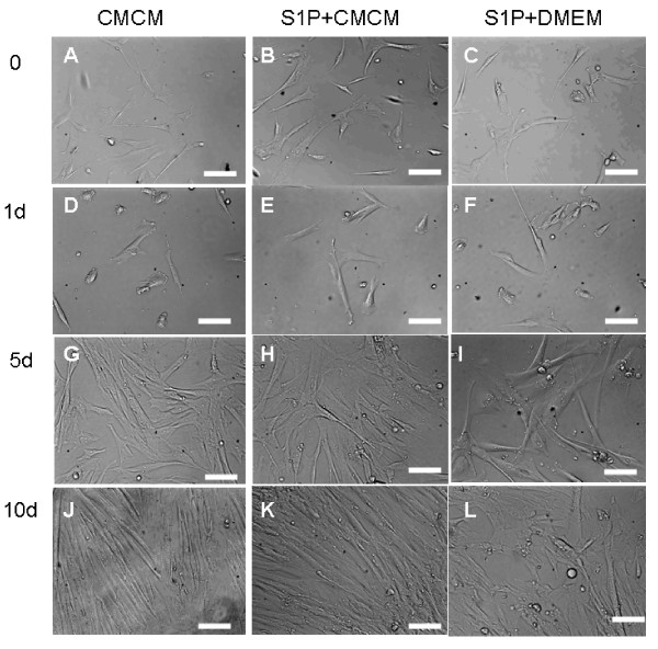
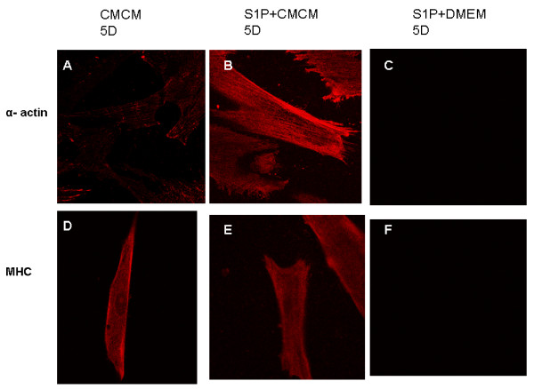
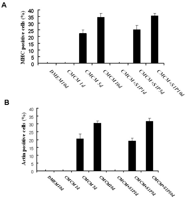
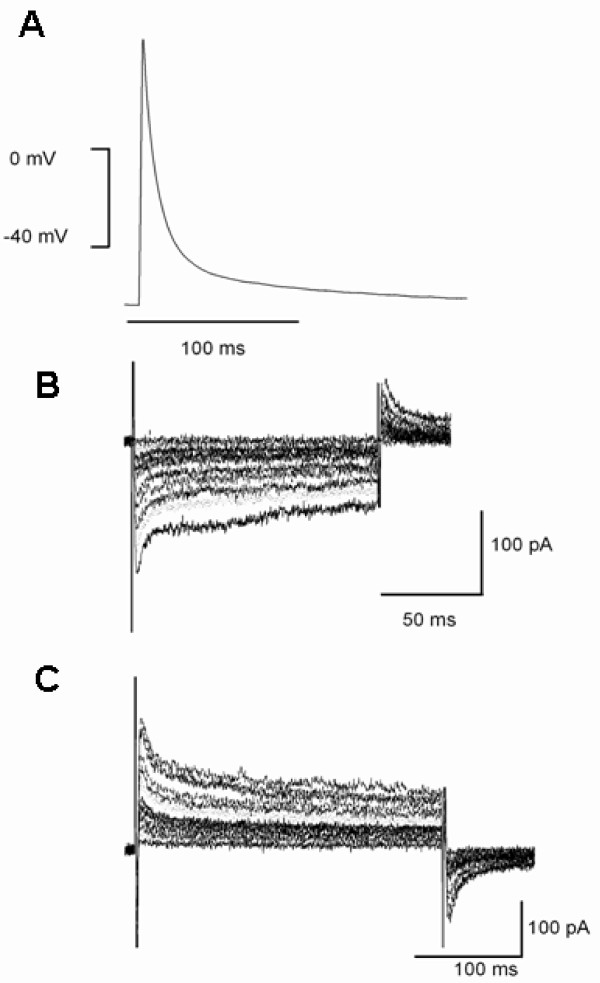
Methods: Cardiomyogenic differentiation of HUMSCs was performed by culturing these cells with either designated cardiomyocytes conditioned medium (CMCM) alone, or with 1 μM S1P; or DMEM with 10% FBS + 1 μM S1P. Cardiomyogenic differentiation was determined by immunocytochemical analysis of expression of cardiomyocyte markers and patch clamping recording of the action potential.

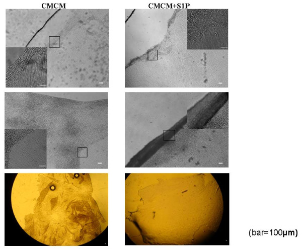
Results: A cardiomyocyte-like morphology and the expression of α-actinin and myosin heavy chain (MHC) proteins can be observed in both CMCM culturing or CMCM+S1P culturing groups after 5 days' culturing, however, only the cells in CMCM+S1P culture condition present cardiomyocyte-like action potential and voltage gated currents. A new approach was used to form PIPAAm based temperature-responsive culture surfaces and this successfully produced cell sheets from HUMSCs derived cardiomyocytes.

Conclusions: This study for the first time demonstrates that S1P potentiates differentiation of HUMSCs towards functional cardiomyocytes under the designated culture conditions. Our engineered cell sheets may provide a potential for clinically applicable myocardial tissues should promote cardiac tissue engineering research.


Figures

Figure 1
Figure 1
UC-MSC cells showed a fibroblast-like morphology before conditioned culturing (AC); the induced cells change their morphology with time. In cells treated with CMCM or CMCM+S1P, HUMSCs displayed a cardiomyocyte-like morphology such as myotube-like shape between 5-7 days (D, E, G, H); At around 10 days, the cells became elongated and lined up in CMCM and CMCM+S1P groups (J, K), and the alignment of the cells appeared in an ordered perpendicular terrace-pattern, like intercalated disc in CMCM+S1P groups. (K). But the cells had no similar change in S1P+DMEM groups (F, I), and the alignment looked random. (L)
Figure 2
Figure 2
Immunostaining of anti-α-actinin and anti-α MHC in cells at different time points of culturing. A strong expression of both α-actinin and MHC proteins (A, B, D, E, G, H, J, K) was observed in CMCM and CMCM+S1P groups, but not in cells from the DMEM+S1P group(C, F, I, L).
Figure 3
Figure 3
Histograms showing the percentage of human umbilical mesenchymal cells expressing α-actin (A) and sarcomeric α/β myosin cardiac heavy chain (B) after CMCM or CMCM+S1P treatment. The results are expressed as mean ± SE of ten randomly selected microscopic fields each from two different experiments. At least 200 cells were counted in each experiment. A statistical difference at *P < 0.05 compared with DMEM-only group and 1 day; *P < 0.05 compared with 5 days. B statistical difference at *P < 0.05 compared with DMEM-only group and 1 day; *P < 0.05 compared with 5 days.
Figure 4
Figure 4
Representative recordings of action potential (A) and whole cell voltage gated inward (B) and outward currents (C) by whole cell patch clamping in myocytes of CMCM+S1P group. The currents were recorded during 200 ms step depolarization pulses from a holding potential of -50 mV to a range of potential between -40 mV and +50 mV.
Figure 5
Figure 5
Human cardiac myocyte cell film (left) or differentiated HUMSCs (right) in CMCM+S1P group detachment under cooling at the ambient temperature of 20°C. Top panel shows the local detachment, middle panel shows a large cell sheet peered off and the bottom panel shows a large pile of cell sheets.

References

    1. Can A, Karahuseyinoglu S. Concise Review: Human Umbilical Cord Stroma with Regard to the Source of Fetus-Derived Stem Cells. Stem cells. 2007;25:2886–2895. doi: 10.1634/stemcells.2007-0417. - DOI - PubMed
    1. Wu KH, Mo XM, Zhou B, Lu SH, Yang SG, Liu YL, Han ZC. Cardiac potential of stem cells from whole human umbilical cord tissue. J Cellular Biochem. 2009;107:926–932. doi: 10.1002/jcb.22193. - DOI - PubMed
    1. Yang C-C, Shih Y-H, Ko M-H, Hsu S-Y, Cheng H, Fu Y-S. Transplantation of Human Umbilical Mesenchymal Stem Cells from Wharton's Jelly after Complete Transection of the Rat Spinal Cord. PLoS ONE. 2008;3:e3336. doi: 10.1371/journal.pone.0003336. - DOI - PMC - PubMed
    1. Avery K, Avery S, Shepherd J, Heath PR, Moore H. Sphingosine-1-Phosphate Mediates Transcriptional Regulation of Key Targets Associated with Survival, Proliferation, and Pluripotency in Human Embryonic Stem Cells. Stem Cells Dev. 2008;17:1195–1206. doi: 10.1089/scd.2008.0063. - DOI - PubMed
    1. Rodgers A, Mormeneo D, Long JS, Delgado A, Pyne NJ, Pyne S. Sphingosine 1-Phosphate Regulation of Extracellular Signal-Regulated Kinase-1/2 in Embryonic Stem Cells. Stem Cells Dev. 2009;18:1319–1330. doi: 10.1089/scd.2009.0023. - DOI - PubMed

Publication types

MeSH terms