. 2011 Jun 8;19(6):810-20.
doi: 10.1016/j.str.2011.02.017.

Domain orientation in the N-Terminal PDZ tandem from PSD-95 is maintained in the full-length protein

Affiliations

Domain orientation in the N-Terminal PDZ tandem from PSD-95 is maintained in the full-length protein

James J McCann et al. Structure. .

Abstract

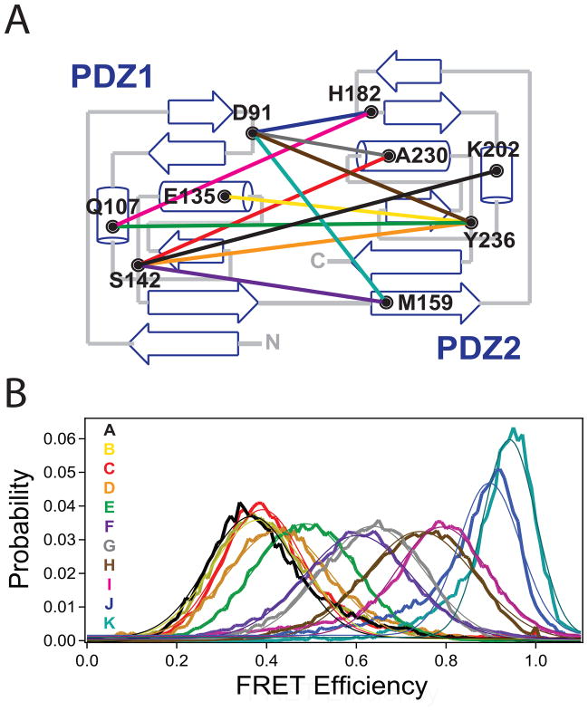
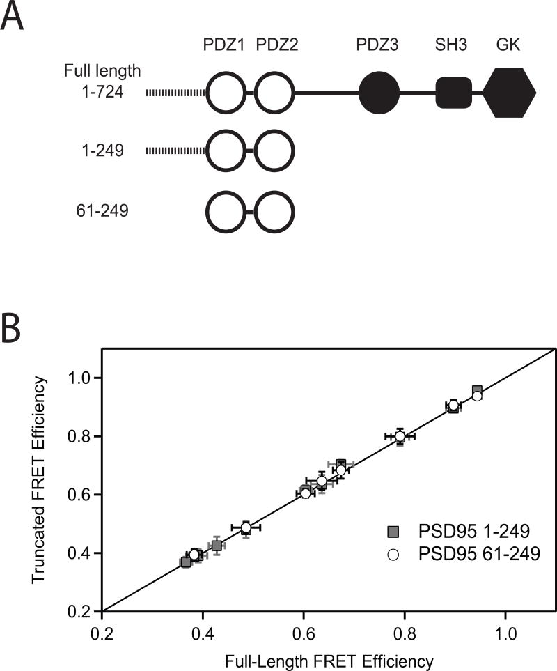
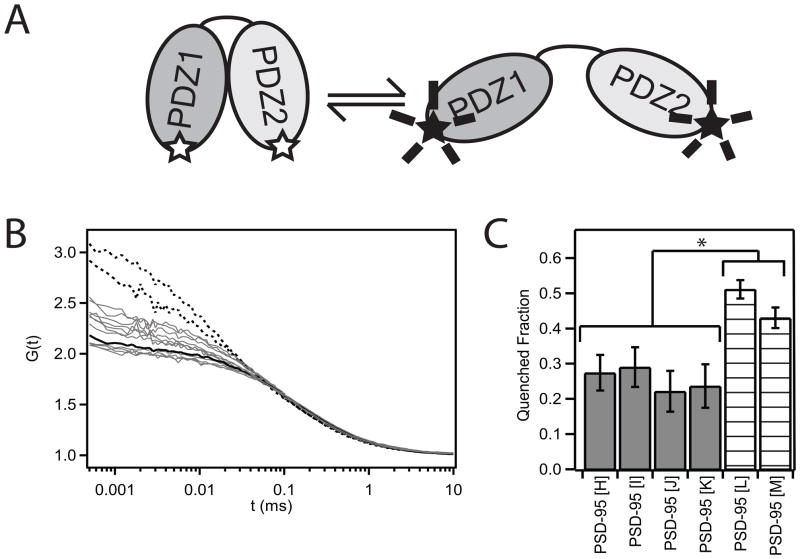
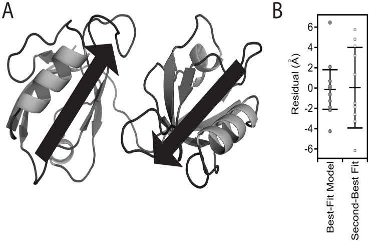
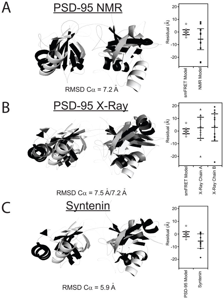
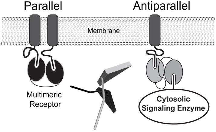
Tandem PDZ domains have been suggested to form structurally independent supramodules. However, dissimilarity between crystallography and NMR models emphasize their malleable conformation. Studies in full-length scaffold proteins are needed to examine the effect of tertiary interactions within their native context. Using single-molecule fluorescence to characterize the N-terminal PDZ tandem in PSD-95, we provide the first direct evidence that PDZ tandems can be structurally independent within a full-length scaffold protein. Molecular refinement using our data converged on a single structure with an antiparallel alignment of the ligand-binding sites. Devoid of interaction partners, single-molecule conditions captured PSD-95 in its unbound, ground state. Interactions between PDZ domains could not be detected while fluctuation correlation spectroscopy showed that other conformations are dynamically sampled. We conclude that ultra-weak interactions stabilize the conformation providing a "low-relief" energy landscape that allows the domain orientation to be flipped by environmental interactions.


Figures

Figure 1
Figure 1. Single Molecule FRET Measurements between PDZ1 and PDZ2 in Full Length PSD-95
(A) Topology diagram showing the position of the cysteine labeling sites in each domain and the eleven combinations of labeling sites used for fluorescence studies. A representation of the labeling sites on the ribbon diagram of the two domains is shown in Figure S1. (B) smFRET histograms for all eleven FRET pairs made in full-length PSD-95. Letter codes (indicated within the panel) were assigned to each mutant in order of increasing FRET efficiency. The coloring of the FRET distributions corresponds to the colored lines denoting labeling combinations in panel A. Thin lines indicate the fit to a single Gaussian function. See also Figure S1.
Figure 2
Figure 2. Truncation of the PDZ Tandem Does Not Alter Domain Orientation
(A) Cartoon representation of the constructs used in this experiment. Construct 1-724 is full length PSD-95. Construct 1-249 is truncated after PDZ2 but retains the 60 residue disordered N-terminal domain. Construct 61-249 encompasses only the PDZ tandem. (B) The mean smFRET efficiency measured in the two truncated fragments (y-axis) is plotted against the mean FRET efficiency between the same labeling sites in the full-length protein (x-axis). Mean smFRET for the 1-249 truncations are shown in gray while those for the 61-249 truncation are colored white. The solid line represents equality between mean smFRET measured in the full length protein and measured in the truncations. Error bars indicate the standard deviation in mean FRET from three or more replicate histograms.
Figure 3
Figure 3. Fluorescence Correlation Spectroscopy Measurement of Structural Dynamics
(A) Cartoon illustrating the tetramethylrhodamine (TMR) self-quenching assay. The fluorescence intensity for a protein doubly-labeled with TMR dyes will decrease as the dyes are brought into close proximity. (B) Autocorrelation curves for TMR-labeled PSD-95. Singly-labeled PSD-95 is shown as a black solid line. PDZ1-PDZ2 mutants used in the FRET studies are all colored gray. Mutants labeled in PDZ1 and the disordered N-terminal region are shown as dashed lines. All experiments were performed using the 1-249 construct. (C) Amplitude of the relaxation term required to fit the FCS autocorrelation. Samples were only included if the F-ratio test showed a statistical improvement in the fit by incorporating the additional terms. The amplitude is reported in terms of the fraction in the dark (quenched) state. (* P < 0.01 based on standard deviation from replicate measurements) See also Figure S2.
Figure 4
Figure 4. FRET Model for the Tandem PDZ Domains in Full-length PSD-95
(A) Cartoon representation of the best fit model to the eleven FRET distance restraints. The model shows a relatively compact orientation without interdomain contacts. The position and direction of canonical PDZ ligand binding sites are represented as arrows. (B) Goodness of fit for the first and second best-fit models based on the smFRET distance restraints. Each model was assessed by plotting the residual (xFRET − xModel) for each of the 11 labeling-site pairs. The structure of the second best model is shown in Figure S3. See also Figure S3.
Figure 5
Figure 5. Comparison the smFRET Model to Other Tandem PDZ Structures
Only residues 61-249 are shown. The best-fit smFRET model is shown in white. (A) Alignment with the representative NMR model of the PDZ tandem from PSD-95 (black). This representative model was kindly provided by Dr. Mingjie Zhang but other models for domain orientation were also compatible with their data (Long et al., 2003). (B) Alignment with the two conformers observed in the crystal structure for the PDZ tandem from PSD-95 (PDB ID: 3GSL) Chains A and B are colored gray and black, respectively. (C) Alignment with the crystal structure for the PDZ tandem of syntenin (black) (PDB ID: 1N99). Dye positions were simulated for the other models and the goodness of fit with the FRET efficiency data is plotted next to the alignment. RMSD values are for the best overall alignment. See also Figure S4.
Figure 6
Figure 6. Effect of Linker Sequence on Interdomain Positioning
(A) Sequence alignment of the PDZ1-2 linker in the four synaptic MAGUK homologues in Rattus novegicus. The last structured residue of each PDZ domain is indicated by the arrows. The well-conserved positively charged tripeptide at the beginning of the linker was mutated to alanines (RRK150-152AAA). The diproline sequence in the center of the PSD-95 linker was mutated to glycines (PP153,154GG). (B) smFRET measurements between identical sites on PSD-95 (1-249) containing mutations within interdomain linker were compared to wildtype (solid black). RRK150-152AAA (dashed black) and PP153,154GG (gray) mutants. See also Figure S5.
Figure 7
Figure 7. Domain Orientation of Tandem PDZ Domains Can Influence Function
PDZ domains are depicted as ovals attached to C-terminal ligand peptides at the cell surface. The relative alignment of tandem PDZ binding sites determines the geometry of higher order complexes at the scaffold. Left: Parallel orientation (black) selects for proteins originating from the same side of the scaffold, such as multimeric transmembrane receptors. Right: The antiparallel configuration (grey) would allow coordinate binding of membrane receptors and cytosolic signaling enzymes. Domain positioning could be a key element in understanding the functions of scaffold proteins such as PSD-95. Center: Relative orientation of the canonical peptide ligand in the antiparallel (grey) and co-aligned (black) PDZ configurations in the smFRET and representative NMR models. The ligand site in PDZ1 was aligned as shown in Figure S2A. The peptide was modeled in PDZ2 based on the peptide-bound NMR structure (PDB ID: 2KA9).

References

    1. Benítez JJ, Keller AM, Chen P, Nils GW. Methods in Enzymology. Academic Press; 2010. Nanovesicle Trapping for Studying Weak Protein Interactions by Single-Molecule FRET; pp. 41–60. - PMC - PubMed
    1. Bernacchi S, Mély Y. Exciton interaction in molecular beacons: a sensitive sensor for short range modifications of the nucleic acid structure. Nucleic Acids Research. 2001;29:e62. - PMC - PubMed
    1. Bhatnagar J, Freed JH, Crane BR. Rigid body refinement of protein complexes with long-range distance restraints from pulsed dipolar ESR. Methods Enzymol. 2007;423:117–133. - PubMed
    1. Bhattacharyya RP, Remenyi A, Yeh BJ, Lim WA. Domains, motifs, and scaffolds: the role of modular interactions in the evolution and wiring of cell signaling circuits. Annu Rev Biochem. 2006;75:655–680. - PubMed
    1. Bonnet G, Krichevsky O, Libchaber A. Kinetics of conformational fluctuations in DNA hairpin-loops. Proc Natl Acad Sci U S A. 1998;95:8602–8606. - PMC - PubMed

Publication types

MeSH terms

LinkOut - more resources