. 2011 Sep 1;39(16):7049-57.
doi: 10.1093/nar/gkr420. Epub 2011 Jun 6.

UV stalled replication forks restart by re-priming in human fibroblasts

Affiliations

UV stalled replication forks restart by re-priming in human fibroblasts

Ingegerd Elvers et al. Nucleic Acids Res. .

Abstract

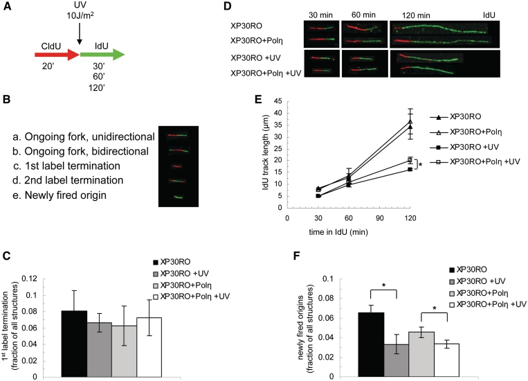
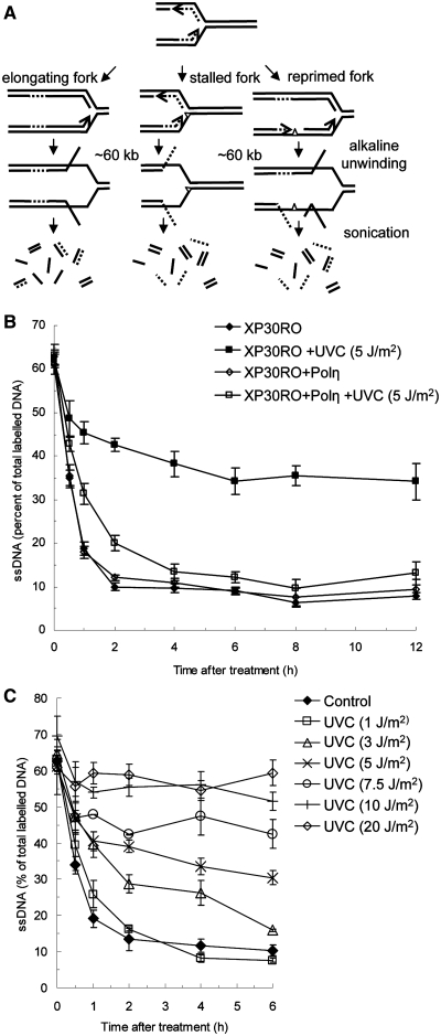
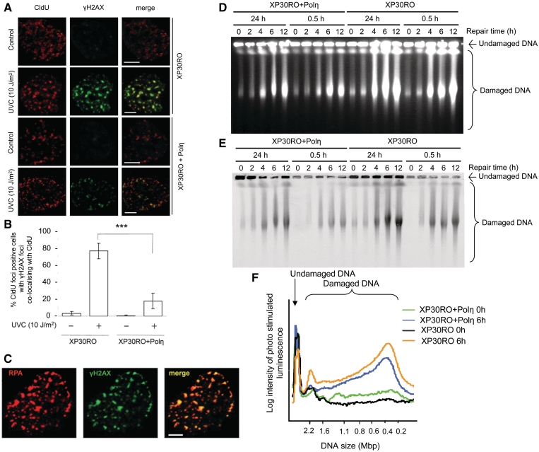
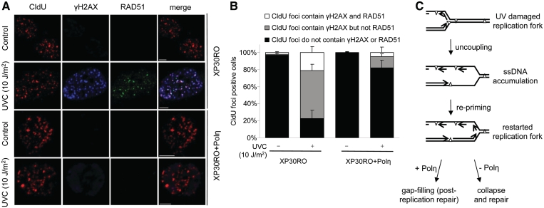
Restarting stalled replication forks is vital to avoid fatal replication errors. Previously, it was demonstrated that hydroxyurea-stalled replication forks rescue replication either by an active restart mechanism or by new origin firing. To our surprise, using the DNA fibre assay, we only detect a slightly reduced fork speed on a UV-damaged template during the first hour after UV exposure, and no evidence for persistent replication fork arrest. Interestingly, no evidence for persistent UV-induced fork stalling was observed even in translesion synthesis defective, Polη(mut) cells. In contrast, using an assay to measure DNA molecule elongation at the fork, we observe that continuous DNA elongation is severely blocked by UV irradiation, particularly in UV-damaged Polη(mut) cells. In conclusion, our data suggest that UV-blocked replication forks restart effectively through re-priming past the lesion, leaving only a small gap opposite the lesion. This allows continuation of replication on damaged DNA. If left unfilled, the gaps may collapse into DNA double-strand breaks that are repaired by a recombination pathway, similar to the fate of replication forks collapsed after hydroxyurea treatment.


Figures

Figure 1.
Figure 1.
Replicating DNA fibres continues to grow in similar manners in UV irradiated Polηmut and restored cells. (A) Experimental setup. (B) Different structures observed. (C) Fraction of first label terminations of the replication structures, after 30 min IdU labelling. Mean and s.e.m. of three independent experiments. (D) Representative fibres after UV exposure and different times with IdU label. (E) IdU incorporation in UV exposed and unirradiated Polηmut and restored cells. Mean and s.e.m. of three independent experiments. (F) Fraction of newly fired origins among the replication structures, after 30 min IdU labelling. Mean and s.e.m. of three independent experiments. Statistical significance determined in t-test is indicated with one star (P < 0.05).
Figure 2.
Figure 2.
Continuous replication fork elongation is disrupted in Polηmut cells, but not in restored cells, after UV exposure. (A) Schematic illustration of the alkaline DNA unwinding assay used to monitor continuous replication fork progression. Directly before UV exposure, ongoing replication forks are pulse labelled (0.5 h) with 3H-TdR. As replication forks progress the labelled DNA is located further away from the DNA ends at the fork and is not released into the ssDNA fraction by the alkaline unwinding that is initiated from ssDNA ends. However, by this technique, ssDNA will also be released if gaps are formed during replication. (B) Replication progression of replication forks in UV irradiated (5 J/m2) Polηmut XP30RO and restored cells. Replication progression is monitored as loss of 3H activity in the ssDNA fraction (C) Continuous replication fork progression in Polηmut XP30RO cells is slowed down in a dose dependent manner following UV irradiation.
Figure 3.
Figure 3.
Replication forks stalled by UV-induced damage collapse into replication-associated DSBs. (A) Representative images of UV-induced γH2AX foci at replication forks labelled with CldU in Polηmut XP30RO and restored cells, 6 h following UV treatments. One nucleus is shown, bar is 5 μm. (B) Percentage of CldU foci positive cells where γH2AX foci co-localize with CldU. Mean and s.e.m. of four independent experiments is depicted. Statistical significance determined in t-test is indicated with three stars (P < 0.001). (C) UV-induced γH2AX foci in a Polηmut XP30RO cell, co-localizing with RPA 6 h after irradiation (10 J/m2). Bar is 5 μm. (D) UV-induced DSBs produced in Polηmut XP30RO and restored cells after exposure to 10 J/m2, measured by PFGE, and visualized by ethidium bromide or (E) autoradiography. (F) Quantification of the intensity of radioactively labelled DNA fragments released from the 0.5-h pulse labelled regions of XP30RO and XP30RO + Polη cells, 0 h (black and green line, respectively) and 6 h (blue and yellow line, respectively) after UV exposure, depicting remaining damage at previously replicated forks.
Figure 4.
Figure 4.
RAD51 co-localizes with replication sites of after UV exposure of Polηmut cells. (A) Representative images of UV-induced γH2AX and RAD51 foci at replication forks in Polηmut XP30RO and restored cells pulse labelled (10 min) with CldU directly before UV exposure (10 J/m2), and allowed to repair for 6 h before fixation. γH2AX was used to visualize DSBs and RAD51 as an indicator of homologous recombination. Bar is 5 μm. (B) Quantification of co-localization of γH2AX and RAD51 with CldU foci, in cells fixed 6 h after UV exposure (10 J/m2). Only cells with distinct CldU foci were checked for co-localization, to only study cells replicating when irradiated. Mean and s.e.m. of four independent experiments are shown. (C) Model for the continuation of DNA replication on damaged DNA. When the replication fork runs into a UV-induced DNA lesion, the PCNA molecule is left and can be ubiquitinated to allow bypass with Polη. Replication is resumed on the 5′ side of the lesion, leaving a gap. In the absence of Polη, the gaps are not bypassed, but collapsed into DSBs that are repaired with a pathway involving RAD51.

References

    1. Guo Z, Kumagai A, Wang SX, Dunphy WG. Requirement for Atr in phosphorylation of Chk1 and cell cycle regulation in response to DNA replication blocks and UV-damaged DNA in Xenopus egg extracts. Genes Dev. 2000;14:2745–2756. - PMC - PubMed
    1. Hekmat-Nejad M, You Z, Yee MC, Newport JW, Cimprich KA. Xenopus ATR is a replication-dependent chromatin-binding protein required for the DNA replication checkpoint. Curr. Biol. 2000;10:1565–1573. - PubMed
    1. Zou L, Elledge SJ. Sensing DNA damage through ATRIP recognition of RPA-ssDNA complexes. Science. 2003;300:1542–1548. - PubMed
    1. Pacek M, Walter JC. A requirement for MCM7 and Cdc45 in chromosome unwinding during eukaryotic DNA replication. EMBO J. 2004;23:3667–3676. - PMC - PubMed
    1. Byun TS, Pacek M, Yee MC, Walter JC, Cimprich KA. Functional uncoupling of MCM helicase and DNA polymerase activities activates the ATR-dependent checkpoint. Genes Dev. 2005;19:1040–1052. - PMC - PubMed

Publication types