. 2011 Aug 1;589(Pt 15):3775-87.
doi: 10.1113/jphysiol.2011.210807. Epub 2011 Jun 6.

Distinct cellular properties of identified dopaminergic and GABAergic neurons in the mouse ventral tegmental area

Affiliations

Distinct cellular properties of identified dopaminergic and GABAergic neurons in the mouse ventral tegmental area

Billy Chieng et al. J Physiol. .

Abstract

The midbrain ventral tegmental area (VTA) contains neurons largely with either a dopaminergic (DAergic) or GABAergic phenotype. Physiological and pharmacological properties of DAergic neurons have been determined using tyrosine hydroxylase (TH) immunohistochemistry but many properties overlap with non-DAergic neurons presumed to be GABAergic. This study examined properties of GABAergic neurons, non-GABAergic neurons and TH-immunopositive neurons in VTA of GAD67-GFP knock-in mice. Ninety-eight per cent of VTA neurons were either GAD-GFP or TH positive,with the latter being five times more abundant. During cell-attached patch-clamp recordings, GAD-GFP neurons fired brief action potentials that could be completely distinguished from those of non-GFP neurons. Pharmacologically, the μ-opioid agonist DAMGO inhibited firing of action potentials in 92% of GAD-GFP neurons but had no effect in non-GFP neurons. By contrast, dopamine invariably inhibited action potentials in non-GFP neurons but only did so in 8% of GAD-GFP neurons. During whole-cell recordings, the narrower width of action potential in GAD-GFP neurons was also evident but there was considerable overlap with non-GFP neurons. GAD-GFP neurons invariably failed to exhibit the potassium-mediated slow depolarizing potential during injection of positive current that was present in all non-GFP neurons. Under voltage-clamp the cationic current, I(h), was found in both types of neurons with considerable overlap in both amplitude and kinetics. These distinct cellular properties may thus be used to confidently discriminate GABAergic and DAergic neurons in VTA during in vitro electrophysiological recordings.


Figures

Figure 1
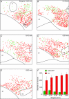
Figure 1. Distribution of TH positive and GAD-GFP positive neurons throughout the VTA
AE, rostro-caudal series of confocal images of coronal sections containing VTA (distance from bregma estimated from Franklin & Paxinos, 1997) in a GAD67-GFP mouse. VTA is marked by intense TH labelling (red). GAD-GFP neurons are green and TH neurons are red. MT is medial terminal nucleus of accessory optic tract, SNc is substantial nigra pars compacta, IP is interpeduncular nucleus, ml is medial lemniscus. F, a representative confocal image of VTA taken from a separate section in the same mouse (60× oil objective). Scale bar A–E, 300 μm and F, 30 μm.
Figure 2
Figure 2. Scored distribution of TH positive and GAD-GFP positive neurons in VTA
AE, distribution of GAD-GFP (green dots) and TH (red dots) neurons along the rostro-caudal axis of VTA that correspond to sections shown in Figure 1. F, summary of numbers of GAD-GFP and TH positive neurons identified per section along the rostrocaudal axis of VTA. Neurons were counted from confocal images 300 × 400 μm from single 30 μm sections sampled from each level in 4 mice.
Figure 3
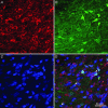
Figure 3. Most neurons in VTA are either TH positive or GAD-GFP positive
Confocal images (45 adjacent 0.5 μm optical sections merged) of VTA neurons in a single brain section triple labelled with TH (A), GAD-GFP (B) and NeuN (C). D, a merged image of A–C. Arrows indicate neurons positive only for NeuN (non-TH and non-GFP positive). Scale bar is 50 μm.
Figure 4
Figure 4. Post hoc identification of TH positive and GAD-GFP positive VTA neurons
A, confocal images of two biocytin labelled neurons combined with post hoc immunohistochemistry. B, a TH positive neuron (arrow) was also labelled with biocytin in A. C, a GFP positive neuron (arrow) was also labelled with biocytin in A. D, diameters (longest axis) of biocytin-labelled GAD-GFP (n = 18) and TH positive (n = 25) neurons. E and F, anatomical locations of recorded VTA neurons plotted in horizontal brain slice orientation at two bregma levels. Green and red dots represent individual GFP and non-GFP neurons, respectively. MT is medial terminal nucleus, SN is substantial nigra, IPF is interpeduncular fossa, ml is medial lemniscus, fr is fasciculus retroflexus, MG is midline group and 3n is oculomotor nerve.
Figure 5
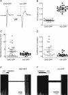
Figure 5. Physiological and pharmacological properties of VTA neurons recorded in cell-attached patch-clamp mode
A, representative examples of action potentials of a GAD-GFP neuron (left) and non-GFP neuron (right). Duration of action potential was measured within the dotted line in horizontal axis. B, summary of action potential durations of GAD-GFP and non-GFP neurons. Circles represent neurons sampled with Axopatch 200A amplifier and triangles represent neurons recorded using a Multiclamp 700B. C, spontaneous action potential frequency for GAD-GFP and non-GFP neurons sampled for 60 s. D, coefficient of variation for spontaneous action potential frequency calculated by dividing SD of interspike interval by the mean of interspike interval. E, representative profile of most spontaneously active GAD-GFP neurons showing no response to superfusion of dopamine (DA, 100 μm) but complete, reversible inhibition of action potential activity by DAMGO (1 μm). F, representative profile of all spontaneously active non-GFP neurons showing no response to superfusion of DAMGO (1 μm) but complete, reversible inhibition of action potential activity by dopamine (100 μm).
Figure 6
Figure 6. Physiological properties of VTA neurons recorded in whole-cell patch-clamp mode
A, action potentials of a GAD-GFP neuron (left) and a non-GFP neuron (right). Duration of action potential was measured within the dotted line in horizontal axis. B, summary of action potential durations of GAD-GFP and non-GFP neurons. Each circle represents a neuron sampled with Axopatch 200A amplifier and triangles represent neurons recorded using a Multiclamp 700B. C, action potential rise time (10–90%) for GAD-GFP and non-GFP neurons. D, correlation plot of action potential durations for GAD-GFP (open circles) and non-GFP (filled circles) neurons under cell-attached and whole-cell modes. The linear regression line is fitted. It has a slope of 0.96 and a correlation coefficient of 0.88. E, under whole-cell current clamp, action potentials from GAD-GFP (left) and non-GFP (middle) neurons were evoked by injection of a positive current. Each cell was held at −90 mV. Non-GFP VTA neurons typically exhibited a slow initial rising phase before triggering an action potential. Panel on the right shows that slow initial rising phase in the same non-GFP neuron was blocked by superfusion of 4-aminopyridine (4AP; 4 mm).
Figure 7
Figure 7. Ih currents in VTA neurons
Aa–d, under whole-cell voltage clamp, neurons were held at −70 mV stepped to more negative potentials in 20 mV increments. Examples of GAD-GFP (a and b) and non-GFP neurons (c and d), respectively, that showed either very little or substantial Ih regardless of neurotransmitter phenotype. B, amplitude of time-dependent rectification was calculated for each neuron during voltage steps to −130 mV (ba, as shown in Ad). C, time constants (tau) of tail currents from a single exponential fit of peak tail current during relaxations from −130 mV to −70 mV.

References

    1. Cameron DL, Wessendorf MW, Williams JT. A subset of ventral tegmental area neurons is inhibited by dopamine, 5-hydroxytryptamine and opioids. Neuroscience. 1997;77:155–166. - PubMed
    1. Ford CP, Mark GP, Williams JT. Properties and opioid inhibition of mesolimbic dopamine neurons vary according to target location. J Neurosci. 2006;26:2788–2797. - PMC - PubMed
    1. Franklin KB, Paxinos G. The Mouse Brain in Stereotaxic Coordinates. Academic Press; 1997.
    1. Garzon M, Pickel VM. Plasmalemmal mu-opioid receptor distribution mainly in nondopaminergic neurons in the rat ventral tegmental area. Synapse. 2001;41:311–328. - PubMed
    1. Grace AA, Bunney BS. The control of firing pattern in nigral dopamine neurons: single spike firing. J Neurosci. 1984;4:2866–2876. - PMC - PubMed

Publication types

MeSH terms

LinkOut - more resources