Review
. 2011 Sep 1;589(17):4125-36.
doi: 10.1113/jphysiol.2011.210294. Epub 2011 Jun 6.

Targeting the Nrf2-Keap1 antioxidant defence pathway for neurovascular protection in stroke

Affiliations
Review

Targeting the Nrf2-Keap1 antioxidant defence pathway for neurovascular protection in stroke

Alessio Alfieri et al. J Physiol. .

Abstract

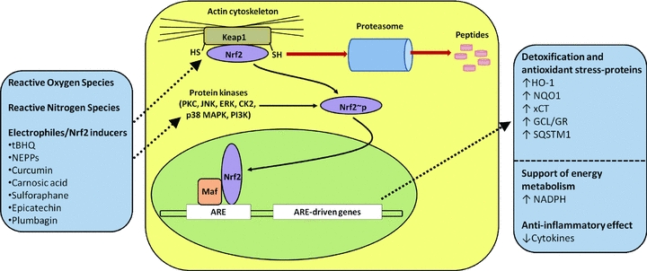
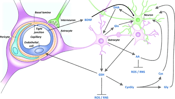
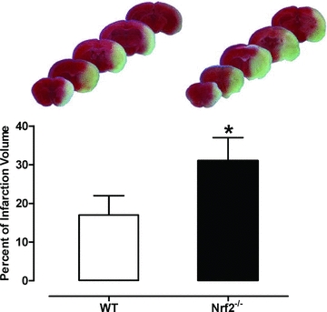
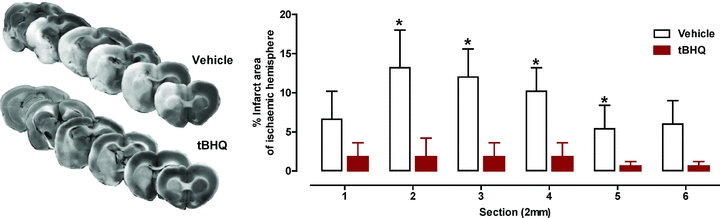
Endogenous defence mechanisms by which the brain protects itself against noxious stimuli and recovers from ischaemic damage are a key target of stroke research. The loss of viable brain tissue in the ischaemic core region after stroke is associated with damage to the surrounding area known as the penumbra. Activation of the redox-sensitive transcription factor nuclear factor erythroid 2-related factor 2 (Nrf2) plays a pivotal role in the cellular defence against oxidative stress via transcriptional upregulation of phase II defence enzymes and antioxidant stress proteins. Although recent evidence implicates Nrf2 in neuroprotection, it is not known whether activation of this pathway within the neurovascular unit protects the brain against blood-brain barrier breakdown and cerebrovascular inflammation. Targeting the neurovascular unit should provide novel insights for effective treatment strategies and facilitate translation of experimental findings into clinical therapy. This review focuses on the cytoprotective role of Nrf2 in stroke and examines the evidence that the Nrf2-Keap1 defence pathway may serve as a therapeutic target for neurovascular protection.


Figures

Figure 1
Figure 1. Activation of the Nrf2–Keap1 defence pathway in oxidative stress
Nrf2 is normally retained in the cytosol bound to the actin-binding protein Keap1 and targeted for proteasomal degradation. Reactive oxygen species, reactive nitrogen species and endogenous and exogenous electrophiles/activators (see text for more details) can alter the Nrf2–Keap1 complex by modifying cysteine (-SH) residues on Keap1. Subsequent phosphorylation of Nrf2 by cytoplasmic kinases may increase its nuclear translocation, where it binds with small Maf proteins to the antioxidant response element (ARE). Induction of ARE-driven genes results in upregulation of haem oxygenase-1 (HO-1), NAD(P)H-quinone oxidoreductase-1 (NQO1), glutamate-cysteine ligase (GCL), glutathione reductase, sequestosome-1 (SQSTM1) and the cystine/glutamate anionic amino acid transporter (xCT). Nrf2/ARE-linked detoxification and antioxidant stress proteins restore the basal redox status in cells exposed to oxidative stress and inflammatory mediators. tBHQ, tert-butylhydroquinone; NEPPs, neurite outgrowth-promoting prostaglandins. Adapted from Itoh et al. (1999); Ishii et al. (2000); Kensler et al. (2007); Innamorato et al. (2008); Siow & Mann (2010).
Figure 2
Figure 2. Communication between astrocytes, neurons and brain endothelial cells in the defence against oxidative stress
Glutathione (GSH) and its precursors, ascorbic acid (AA) and dehydroascorbate (DHA), and brain-derived neurotrophic factor (BDNF) provide protection of the neurovascular unit against free radical-mediated injury (see text for more details). Cys, cysteine; Gln, glutamine; Gly, glycine; RNS, reactive nitrogen species. Modified from Fig. 2 in Abbott et al. (2006) with permission from the Nature Publishing Group.
Figure 3
Figure 3. Cerebral infarct volumes in wild-type (WT) and Nrf2-deficient mice subjected to 90 min MCAO and 24 h reperfusion
Upper panel: representative images of serial brain sections stained with TTC (2,3,5-triphenyltetrazolium chloride). The light areas denote the infarct region. Lower panel: quantification of infarct areas in WT and Nrf2−/− (mean ± SEM, P < 0.01; n = 8 per group). Adapted from Fig. 1 in Shah et al. (2007) with permission from Elsevier.
Figure 4
Figure 4. Reduction in cerebral infarct area in stroke following pre-treatment with the Nrf2-inducer tert-butylhydroquinone
Left panel: representative images of serial brain sections stained with TTC. Right panel: effect of intracerebral ventricular delivery of tert-butylhydroquinone (tBHQ, 1 mm for 72 h from pump implant) before 90 min MCAO and 24 h of reperfusion. Infarct areas are shown between section 1 (anterior) and section 2 (posterior). Data are expressed as mean ± SEM, *P < 0.05; n = 7–9. Adapted from Fig. 2 in Shih et al. (2005) with permission from The Journal of Neuroscience.
Figure 5
Figure 5. Keap1 and Nrf2-positive cells in the peri-infarct and infarct region of mouse brains after MCAO
A, brain sections were immunostained at different times of reperfusion after 60 min of MCAO (mean ± SEM. *P < 0.05 and **P < 0.01 vs. sham; #P < 0.05 and ##P < 0.01 vs. infarct region. n = 5 per group). Double immunofluorescent staining for the neural marker NeuN and Keap1 (B) and NeuN and Nrf2 (C) in brain sections from control (sham) mice and in the peri-infarct region of mice subjected to 60 min MCAO and 8 h of reperfusion. DAPI (4',6-diamidino-2-phenylindole) was used for nuclear staining. Adapted from Figs 1 and 2 in Tanaka et al. (2011) with permission from Elsevier.

References

    1. Abbott NJ, Patabendige AA, Dolman DE, Yusof SR, Begley DJ. Structure and function of the blood-brain barrier. Neurobiol Dis. 2010;37:13–25. - PubMed
    1. Abbott NJ, Ronnback L, Hansson E. Astrocyte-endothelial interactions at the blood-brain barrier. Nat Rev Neurosci. 2006;7:41–53. - PubMed
    1. Adibhatla RM, Hatcher JF. Lipid oxidation and peroxidation in CNS health and disease: from molecular mechanisms to therapeutic opportunities. Antioxid Redox Signal. 2010;12:125–169. - PubMed
    1. Allen CL, Bayraktutan U. Oxidative stress and its role in the pathogenesis of ischaemic stroke. Int J Stroke. 2009;4:461–470. - PubMed
    1. Arai K, Lo EH. An oligovascular niche: cerebral endothelial cells promote the survival and proliferation of oligodendrocyte precursor cells. J Neurosci. 2009;29:4351–4355. - PMC - PubMed