. 2011 Jun 21;108(25):10190-5.
doi: 10.1073/pnas.1107402108. Epub 2011 Jun 6.

Tubular network formation protects mitochondria from autophagosomal degradation during nutrient starvation

Affiliations

Tubular network formation protects mitochondria from autophagosomal degradation during nutrient starvation

Angelika S Rambold et al. Proc Natl Acad Sci U S A. .

Abstract

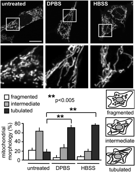
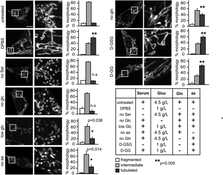
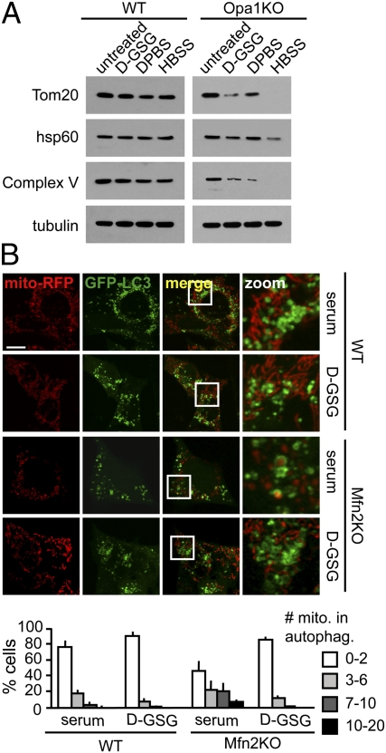
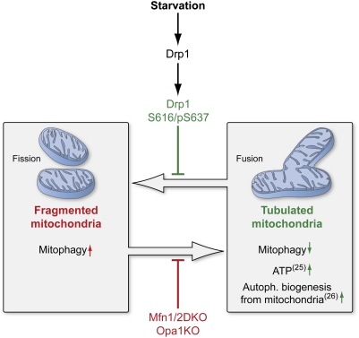
Mitochondria are highly dynamic organelles that mediate essential cell functions such as apoptosis and cell-cycle control in addition to their role as efficient ATP generators. Mitochondrial morphology changes are tightly regulated, and their shape can shift between small, fragmented units and larger networks of elongated mitochondria. We demonstrate that mitochondrial elements become significantly elongated and interconnected shortly after nutrient depletion. This mitochondrial morphological shift depends on the type of starvation, with an additive effect observed when multiple nutrients are depleted simultaneously. We further show that starvation-induced mitochondrial elongation is mediated by down-regulation of dynamin-related protein 1 (Drp1) through modulation of two Drp1 phosphorylation sites, leading to unopposed mitochondrial fusion. Finally, we establish that mitochondrial tubulation upon nutrient deprivation protects mitochondria from autophagosomal degradation, which could permit mitochondria to maximize energy production and supply autophagosomal membranes during starvation.


Conflict of interest statement

The authors declare no conflict of interest.

Figures

Fig. 1.
Fig. 1.
Starvation induces mitochondrial tubulation. MEFs were transfected with mito-YFP. (Upper) Cells were starved 16 h after transfection for 2 h, and live images were acquired. (Images in the second row are magnified views of the boxed areas in first row.) (Lower) Mitochondrial morphology was scored as follows: fragmented, mainly small and round; intermediate, mixture of round and shorter tubulated; and tubulated, long and higher interconnectivity. The percentage of cells with indicated mitochondrial morphologies was determined as a percentage of the total number of transfected cells counted (≥100 cells per experiment). n = 6 independent experiments. (Scale bar: 15 μm.)
Fig. 2.
Fig. 2.
Mitochondrial tubulation is more efficiently induced by additive nutrient deprivation. MEFs were transfected with mito-YFP and starved for 2 h (DPBS, D-GSG, and no Ser) or 6 h (no glc, low glc, no aa, no gln, and D-GG). Media compositions are given in the table. Mitochondrial morphology was scored as described in Fig. 1 (n ≥ 3). Representative images of cells are shown in the first column. Images in the second column are magnified views of the boxed areas in the first column. glc, glucose; gln, glutamine; aa, amino acids. (Scale bar: 15 μm.)
Fig. 3.
Fig. 3.
Unopposed fusion mediates mitochondrial tubulation during starvation. (A, C, and D) MEFs were treated with the media indicated for 2.5 h (A) or 5 h (C and D) and lysed in ice-cold lysis buffer A, and samples were subjected to immunoblotting against Mfn1, Mfn2, Opa1, Drp1, and pS616-Drp1 as indicated (whole-cell lysates were probed in A and C; isolated mitochondria were probed in D). (B) At 16 h after transfection with the indicated YFP-tagged Drp1 constructs, cells were treated with full medium or starved in D-GSG for 2 h. Mitochondrial morphology was scored as described in Fig. 1 (n = 3).
Fig. 4.
Fig. 4.
Mitochondrial tubulation ameliorates autophagosomal degradation of mitochondria during starvation. (A) WT and Opa1KO MEFs were treated with the indicated media for 5 h, and equal numbers of cells were lysed. Whole-cell lysates were subjected to immunoblotting against the proteins indicated. (B) Mfn2KO cells show increased mitophagy during serum starvation, which can be rescued through mitochondrial tubulation. WT and Mfn2KO MEF cells were transfected with GFP-LC3 and mito-RFP and starved for serum or D-GSG for 4 h in the presence of bafilomycin A1, and live images were acquired. Cells were grouped into categories according to the number of mitochondria in autophagosomes and are presented as percentage of the total number of transfected cells counted (n = 3). (Scale bar: 15 μm.)
Fig. 5.
Fig. 5.
Elongation of mitochondria is mediated by posttranslational modification of Drp1 and prevents mitophagy during nutrient starvation. Model shows the protective effect of tubulation on mitochondria upon nutrient deprivation. Tubulation of mitochondria is mediated through modifications of Drp1. Lack of Mfn1/2 or Opa1 inhibits the mitochondria's fusion capacity during starvation. Reference numbers are indicated in parentheses. (This figure was prepared by Medical Illustration, National Institutes of Health Medical Arts Design Section.)

References

    1. Bereiter-Hahn J. Behavior of mitochondria in the living cell. Int Rev Cytol. 1990;122:1–63. - PubMed
    1. Nunnari J, et al. Mitochondrial transmission during mating in Saccharomyces cerevisiae is determined by mitochondrial fusion and fission and the intramitochondrial segregation of mitochondrial DNA. Mol Biol Cell. 1997;8:1233–1242. - PMC - PubMed
    1. Santel A, et al. Mitofusin-1 protein is a generally expressed mediator of mitochondrial fusion in mammalian cells. J Cell Sci. 2003;116:2763–2774. - PubMed
    1. Santel A, Fuller MT. Control of mitochondrial morphology by a human mitofusin. J Cell Sci. 2001;114:867–874. - PubMed
    1. Chen H, et al. Mitofusins Mfn1 and Mfn2 coordinately regulate mitochondrial fusion and are essential for embryonic development. J Cell Biol. 2003;160:189–200. - PMC - PubMed

Publication types