. 2011 Jun 21;108(25):10296-301.
doi: 10.1073/pnas.1101118108. Epub 2011 Jun 6.

Mimivirus shows dramatic genome reduction after intraamoebal culture

Affiliations

Mimivirus shows dramatic genome reduction after intraamoebal culture

Mickaël Boyer et al. Proc Natl Acad Sci U S A. .

Erratum in

  • Proc Natl Acad Sci U S A. 2011 Oct 11;108(41):17234

Abstract

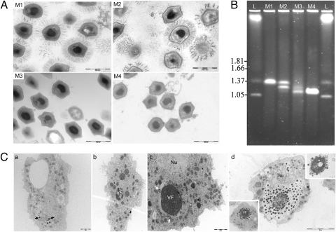
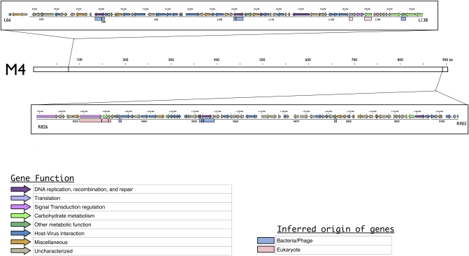
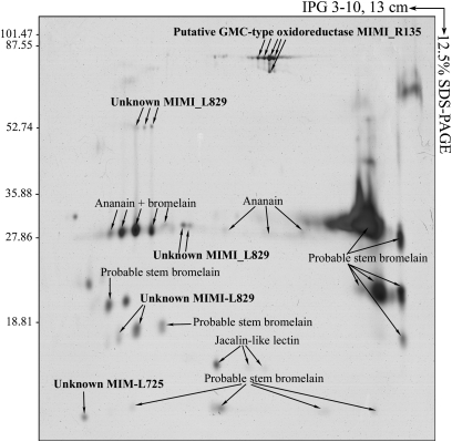
Most phagocytic protist viruses have large particles and genomes as well as many laterally acquired genes that may be associated with a sympatric intracellular life (a community-associated lifestyle with viruses, bacteria, and eukaryotes) and the presence of virophages. By subculturing Mimivirus 150 times in a germ-free amoebal host, we observed the emergence of a bald form of the virus that lacked surface fibers and replicated in a morphologically different type of viral factory. When studying a 0.40-μm filtered cloned particle, we found that its genome size shifted from 1.2 (M1) to 0.993 Mb (M4), mainly due to large deletions occurring at both ends of the genome. Some of the lost genes are encoding enzymes required for posttranslational modification of the structural viral proteins, such as glycosyltransferases and ankyrin repeat proteins. Proteomic analysis allowed identification of three proteins, probably required for the assembly of virus fibers. The genes for two of these were found to be deleted from the M4 virus genome. The proteins associated with fibers are highly antigenic and can be recognized by mouse and human antimimivirus antibodies. In addition, the bald strain (M4) was not able to propagate the sputnik virophage. Overall, the Mimivirus transition from a sympatric to an allopatric lifestyle was associated with a stepwise genome reduction and the production of a predominantly bald virophage resistant strain. The new axenic ecosystem allowed the allopatric Mimivirus to lose unnecessary genes that might be involved in the control of competitors.


Conflict of interest statement

The authors declare no conflict of interest.

Figures

Fig. 1.
Fig. 1.
(A) Ultrastructure of the Mimivirus progeny virions stained by ruthenium red at the start of the experiment (M1), at the 100th passage (M2), at the 150th passage (M3), and after 0.40-μm filtering on the 150th amoebal subcultured supernatant (M4). (Scale bar, 500 nm.) (B) Comparative PFGE of intact genomic DNA from M1, M2, M3, and M4. Electrophoresis was performed in 1% agarose in 0.5× TBE buffer and the pulse time was ramped between 50 and 250 s for 22 h at a voltage of 5 V/cm at 14 °C. Hansenula wingei chromosomes (lane L) were used as size markers. Sizes are indicated on the Left in megabase pairs. (C) Ultrastructure of the Mimivirus M4 replication cycle in A. polyphaga at several time points postinfection. At 0.5 h p.i. (a), some viral particles (black arrows) were seen inside phagocytic vacuoles. At 4 h p.i. (b), the early virion factory (VF) appears as a dense structure with multilobed aspects. At 6 h p.i. (c), first steps of particle assembly (white arrows) were observed around the VF. Nu, cell nucleus. At 11 h p.i. (d), virions accumulated around the VF, sometimes imitating a wire (Lower Left Inset). According to our observations, this is the last step before particle release. After this point, free VF were often seen outside the cell (Upper Right Inset).
Fig. 2.
Fig. 2.
Electron micrographs of frozen-hydrated Mimivirus M4 particles. (A) Individual particles, some of which show starfish-like structures (arrow) at a special vertex. The particles are lacking the fibrous layer of the M1 Mimivirus. (Scale bar, 500 nm.) (B) 3D cryo-EM reconstruction of the M4 passaged Mimivirus. (Scale bar, 50 nm.)
Fig. 3.
Fig. 3.
Genetic map of the two regions deleted at both ends of the M4 genome. The M4 genome is represented by a white box and its coordinates are indicated in kilobases. The two frames indicate the two regions deleted from the M1 virus. These are represented by genetic maps, including colored arrows representing CDS according to their putative function and colored boxes representing the phylogenetic origin of each gene inferred from previous phylogenetic analyses (9).
Fig. 4.
Fig. 4.
Representative 2D differential gel electrophoresis (DIGE) analysis of Mimivirus proteins. Each individual sample from the M1 and M4 strains and a pooled reference sample were labeled with Cy5, Cy3, and Cy2, respectively, and were then separated on the same gel using the 2D DIGE system. Three images were obtained from each gel and an overlay of dye scan images was also obtained. Selected protein spots (green) exhibiting an ANOVA score lower or close to 0.05 and a change of at least 1.9-fold intensity are indicated by circles and spot numbers as indicated in Table S3.
Fig. 5.
Fig. 5.
2D electrophoresis of the fiber fraction from the M1 Mimivirus strain. Purified fibers were resolved in pI values ranging from 3 to 10 in the first dimension followed by 12.5% SDS-PAGE in the second dimension, after which the proteins were detected by silver staining. Each polypeptide spot detected was identified by MALDI MS as previously described (18). Proteins identified as originating from M1 are indicated in bold.

References

    1. La Scola B, et al. A giant virus in amoebae. Science. 2003;299:2033. - PubMed
    1. Raoult D, et al. The 1.2-megabase genome sequence of Mimivirus. Science. 2004;306:1344–1350. - PubMed
    1. Filée J, Pouget N, Chandler M. Phylogenetic evidence for extensive lateral acquisition of cellular genes by nucleocytoplasmic large DNA viruses. BMC Evol Biol. 2008;8:320. - PMC - PubMed
    1. Colson P, Raoult D. Gene repertoire of amoeba-associated giant viruses. Intervirology. 2010;53:330–343. - PubMed
    1. Suhre K. Gene and genome duplication in Acanthamoeba polyphaga Mimivirus. J Virol. 2005;79:14095–14101. - PMC - PubMed

Publication types

Associated data