. 2012 Apr;90(4):396-403.
doi: 10.1038/icb.2011.50. Epub 2011 Jun 7.

CD4 T cells play important roles in maintaining IL-17-producing γδ T-cell subsets in naive animals

Affiliations

CD4 T cells play important roles in maintaining IL-17-producing γδ T-cell subsets in naive animals

Jeong-Su Do et al. Immunol Cell Biol. 2012 Apr.

Abstract

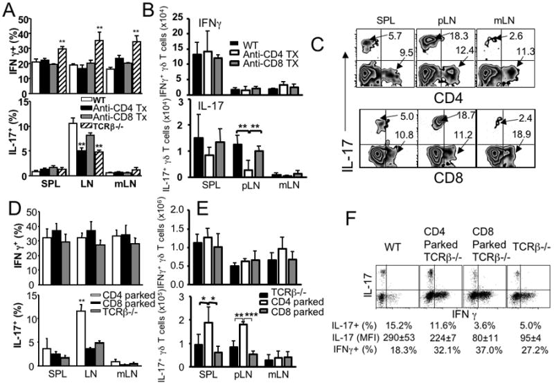
A proportional balance between αβ and γδ T-cell subsets in the periphery is exceedingly well maintained by a homeostatic mechanism. However, a cellular mechanism underlying the regulation remains undefined. We recently reported that a subset of developing γδ T cells spontaneously acquires interleukin (IL)-17-producing capacity even within naive animals through a transforming growth factor (TGF)β1-dependent mechanism, thus considered 'innate' IL-17-producing cells. Here, we report that γδ T cells generated within αβ T cell (or CD4 T cell)-deficient environments displayed altered cytokine profiles; particularly, 'innate' IL-17 expression was significantly impaired compared with those in wild-type mice. Impaired IL-17 production in γδ T cells was directly related to CD4 T-cell deficiency, because depletion of CD4 T cells in wild-type mice diminished and adoptive CD4 T-cell transfer into T-cell receptor β-/- mice restored IL-17 expression in γδ T cells. CD4 T cell-mediated IL-17 expression required TGFβ1. Moreover, Th17 but not Th1 or Th2 effector CD4 T cells were highly efficient in enhancing γδ T-cell IL-17 expression. Taken together, our results highlight a novel CD4 T cell-dependent mechanism that shapes the generation of IL-17+ γδ T cells in naive settings.


Figures

Figure 1
Figure 1. γδ T cells in various T cell-deficient mice
(A) Groups of wild type (filled symbol) and TCRβ−/− (open symbol) mice were sacrificed and the total numbers of γδ T cells in the indicated lymphoid tissues (SPL, spleen; pLN, peripheral LN; mLN, mesenteric LN, and thymus) were enumerated by FACS analysis. Each symbol represents individual mouse. (B) Total numbers of γδ T cells in WT, MHC II−/−, and β2m−/− mice were calculated by FACS analysis. Each symbol represents individually tested mouse. (C) Peripheral LN cells were stained for γδ TCR plus indicated Vγ chains. The mean ± SD was calculated from four individually tested mice.
Figure 2
Figure 2. Cytokine and proliferation profiles of γδ T cells
(A) IFNγ and IL-17 production from the indicated lymphoid tissues of wild type and TCRβ−/− mice was measured after in vitro stimulation with PMA plus ionomycin as described in the Materials and Methods. Representative FACS dot plots of intracellular IL-17- and IFNγ-expression of γδ T cell gated population the indicated tissues are shown. The mean ± SD was calculated from three to ten independent experiments. **, p<0.01. (B) Absolute numbers of cytokine producing γδ T cells of the indicated tissues were calculated. (C) mRNA expression of IL-17 and IFNγ in pLN γδ T cells FACS sorted from wild type or TCRβ−/− mice was examined by real time PCR. (D) CFSE labeled γδ T cells (from wild type or TCRβ−/− mice) were transferred into Rag1−/− recipients. CFSE dilution was determined 7 days post transfer. CFSE profiles shown are representative from two independent experiments.
Figure 3
Figure 3. Profiles of cytokine producing γδ T cells
(A) Cytokine expression of γδ T cells from mice of different age. Cells from the indicated lymphoid tissues (thymus or pLN) of wild type and TCRβ−/− mice of the indicated age were in vitro stimulated with PMA plus ionomycin. The proportion of as well as the absolute numbers of IFNγ and IL-17 production was determined by intracellular cytokine staining. The mean ± SD of cytokine expressing γδ T cells from 4–5 individually tested mice are shown. (B) Surface phenotypes of IFNγ- and IL-17- producing γδ T cells. Cells from the indicated lymphoid tissues of wild type, TCRβ−/−, and TCRα−/− mice were in vitro stimulated with PMA plus ionomycin and stained for CD27 and cytokines. Data shown are representative from 2–3 individually tested mice. Similar results were obtained from another independent experiment. N.D., not done.
Figure 4
Figure 4. γδ T cell cytokine expression from MHCI and MHCII deficient mice
(A) Cells from the indicated tissues of MHC II−/− and β2m−/− mice were stimulated with PMA plus ionomycin and cytokine expression was determined by intracellular cytokine staining as described above. The mean ± SD was calculated from 7–10 individually tested mice. *, p<0.05; **, p<0.01. (B) Groups of TCRβ−/− mice (n=3) were injected with anti-NK1.1 or control Ab every 3 days. IL-17 production by γδ T cells were determined as described above.
Figure 5
Figure 5. γδ T cell IL-17 expression altered by CD4 T cells
(A) Groups of B6 mice received anti-CD4 or anti-CD8 Ab (250μg per injection) every 3 days. At day 21, mice were sacrificed and γδ T cell cytokine expression was determined as described above. Untreated wild type and TCRβ−/− mice were included as control groups. **, p<0.01. (B) The total numbers of IL-17+ and IFNγ+ γδ T cells were calculated in WT mice that were injected with anti-CD4 or anti-CD8 Abs as described above. **, p<0.01. (C) Representative FACS plots of IL-17, CD4, and CD8 expression of γδ T cells are shown. Experiments were repeated three times and similar results were observed. (D) Groups of TCRβ−/− mice were transferred with FACS sorted CD44low naïve CD4 or CD8 T cells (3 × 10⁶ cells per recipient). At day 21, mice were sacrificed and γδ T cell cytokine expression was determined. The mean ± SD was calculated from 4–6 individually tested mice. **, p<0.01. (E) The total numbers of IL-17+ and IFNγ+ γδ T cells were calculated in TCRβ−/− mice that received CD4 or CD8 T cells as described above. *, p<0.05; **, p<0.01; ***, p<0.001. (F) γδ T cell IL-17 production increases by CD4 T cells on a per cell basis. FACS sorted CD44low naïve CD4 and CD8 T cells (3 × 10⁶ cells per recipient) were transferred into groups of TCRβ−/− mice. At day 21, mice were sacrificed and IL-17 expression by γδ T cells was determined. Dot plots of IL-17/IFNγ expression of γδ T cells in different settings are shown. The mean fluorescent intensity (mean ± SD) of IL-17 determined by FACS analysis is shown. The percentile shown represents the proportion of IL-17+ and IFNγ+ γδ T cells. Similar data was observed from 5–9 individually tested mice in 2–3 independent experiments.
Figure 6
Figure 6. CD4-mediated increase in γδ T cell IL-17 expression is TGFβ1-dependent
Groups of TCRβ−/− mice were transferred with FACS sorted naïve CD4 T cells (3 × 10⁶ cells per recipient). The mice were i.p. injected with 500μg anti-TGFβ1 or rat IgG every 3 days. The mice were sacrificed 3 weeks post T cell transfer, and indicated lymphoid tissue cells were stimulated as described above. The proportion (A) and the total numbers (B) of IL-17+ and IFNγ+ γδ T cells were calculated as described above. The mean ± SD was calculated from 3 individually tested mice. *, p<0.05; **, p<0.01; ***, p<0.001.
Figure 7
Figure 7. Th17 CD4 T cell mediated increase in γδ T cell IL-17 expression
CD4 T cells were in vitro stimulated with soluble anti-CD3 plus anti-CD28 Ab as described in Materials and Methods. Th1, Th2, and Th17 phenotype effector CD4 T cells were generated. (A) Prior to transfer, differentiation status of the resulting effector cells was examined by measuring IFNγ, IL-4, and IL-17 expression. (B) 3 × 10⁶ CD4 T cells were adoptively transferred into TCRβ−/− recipients, and γδ T cell cytokine expression was determined 2 weeks post transfer. FACS dot plots shown are representative of cytokine expression of γδ T cells from three individually tested recipients.

Comment in

  • γδ T cells are not alone.
    Prinz I, Föhse L. Prinz I, et al. Immunol Cell Biol. 2012 Apr;90(4):370-1. doi: 10.1038/icb.2011.61. Epub 2011 Jul 12. Immunol Cell Biol. 2012. PMID: 21747406 No abstract available.

References

    1. Shibata K, Yamada H, Nakamura R, Sun X, Itsumi M, Yoshikai Y. Identification of CD25+ gamma delta T cells as fetal thymus-derived naturally occurring IL-17 producers. J Immunol. 2008;181(9):5940–7. - PubMed
    1. Haas JD, Gonzalez FH, Schmitz S, Chennupati V, Fohse L, Kremmer E, et al. CCR6 and NK1.1 distinguish between IL-17A and IFN-gamma-producing gammadelta effector T cells. Eur J Immunol. 2009;39(12):3488–97. - PubMed
    1. Do JS, Fink PJ, Li L, Spolski R, Robinson J, Leonard WJ, et al. Cutting Edge: Spontaneous Development of IL-17-Producing {gamma}{delta} T Cells in the Thymus Occurs via a TGF-{beta}1-Dependent Mechanism. J Immunol. 184(4):1675–9. - PMC - PubMed
    1. Kisielow J, Kopf M, Karjalainen K. SCART scavenger receptors identify a novel subset of adult gammadelta T cells. J Immunol. 2008;181(3):1710–6. - PubMed
    1. Zhu J, Yamane H, Paul WE. Differentiation of effector CD4 T cell populations (*) Annu Rev Immunol. 28:445–89. - PMC - PubMed

Publication types

Substances

LinkOut - more resources