. 2011;6(5):e20341.
doi: 10.1371/journal.pone.0020341. Epub 2011 May 27.

Identification of miRs-143 and -145 that is associated with bone metastasis of prostate cancer and involved in the regulation of EMT

Affiliations

Identification of miRs-143 and -145 that is associated with bone metastasis of prostate cancer and involved in the regulation of EMT

Xinsheng Peng et al. PLoS One. 2011.

Abstract

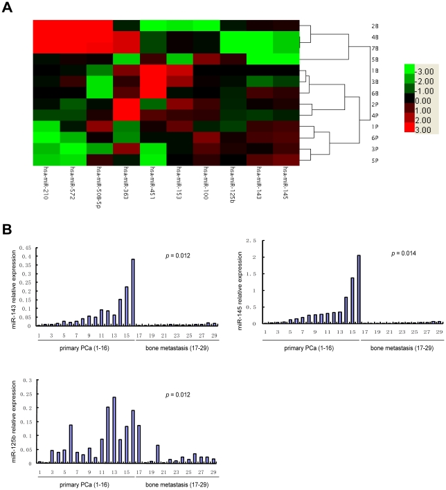
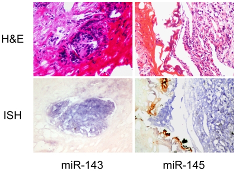
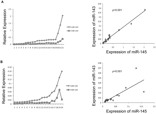
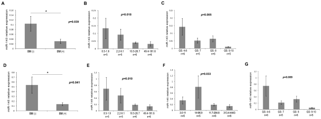
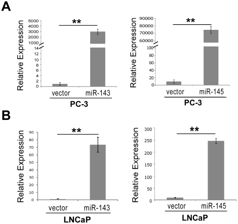
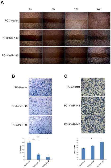
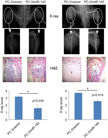
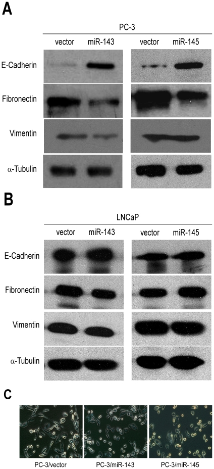
The principal problem arising from prostate cancer (PCa) is its propensity to metastasize to bone. MicroRNAs (miRNAs) play a crucial role in many tumor metastases. The importance of miRNAs in bone metastasis of PCa has not been elucidated to date. We investigated whether the expression of certain miRNAs was associated with bone metastasis of PCa. We examined the miRNA expression profiles of 6 primary and 7 bone metastatic PCa samples by miRNA microarray analysis. The expression of 5 miRNAs significantly decreased in bone metastasis compared with primary PCa, including miRs-508-5p, -145, -143, -33a and -100. We further examined other samples of 16 primary PCa and 13 bone metastases using real-time PCR analysis. The expressions of miRs-143 and -145 were verified to down-regulate significantly in metastasis samples. By investigating relationship of the levels of miRs-143 and -145 with clinicopathological features of PCa patients, we found down-regulations of miRs-143 and -145 were negatively correlated to bone metastasis, the Gleason score and level of free PSA in primary PCa. Over-expression miR-143 and -145 by retrovirus transfection reduced the ability of migration and invasion in vitro, and tumor development and bone invasion in vivo of PC-3 cells, a human PCa cell line originated from a bone metastatic PCa specimen. Their upregulation also increased E-cadherin expression and reduced fibronectin expression of PC-3 cells which revealed a less invasive morphologic phenotype. These findings indicate that miRs-143 and -145 are associated with bone metastasis of PCa and suggest that they may play important roles in the bone metastasis and be involved in the regulation of EMT Both of them may also be clinically used as novel biomarkers in discriminating different stages of human PCa and predicting bone metastasis.


Conflict of interest statement

Competing Interests: The author Tiejian Liu is employed by a commercial company, Laura Biotech Co., Ltd., and joined the authors' research group for private reasons. Because of his work in the research group, he will gain significant experience which will be helpful for his application for his PhD. This paper has no commercial relevance in terms of Laura Biotech, and the data and results detailed herein bear no relation to that company. The authors confirm that this does not alter their adherence to all the PLoS ONE policies on sharing data and materials.

Figures

Figure 1
Figure 1. Validation of select miRNAs predicted to be downregulated in prostate cancer.
A, The certified result of microarray analysis. B, Real-time RT–PCR assays on miR-143 (left panel), miR-145 (right panel), and miR-125b (bottom panel) in 16 primary prostate cancer and 13 bone metastasis tissues. The order of the tissue samples is the same for all three plots. p-values by t-test.
Figure 2
Figure 2. Detection of miRs-143 and -145 in primary PCa tissues by ISH.
Sections at upper panel showed the identification of tumor cells and stromal cells by H&E-stainning. Sections at lower panel showed the location of miR-143 (left) and miR-145 (right) in PCa cells by LNA-ISH. Signals of miRs-143 and -145 were in purple blue. Pictures were taken under microscope 200×.
Figure 3
Figure 3. miRs-143 and -145 showed similar expression tendencies in clinical specimens.
A–B, Expressions of miRs-143 and -145 and their relationships in primary prostate cancer samples (A) and bone metastasis samples (B).
Figure 4
Figure 4. Down-regulation of miRs-143 and -145 is associated with clinicopathological features of primary PCa.
A and D, The expression of miRs-143 or -145 in the patients with bone metastases was significantly lower than that without metastases (t-test, p = 0.039, p = 0.041, respectively). B and E, Tumor samples were divided into four groups with approximately equal sample sizes based on level of free PSA. The level of free PSA in patients with the primary tumor is presented on the x axis. The y axis is the mean of miRs-143 or -145 within each group. The bars represent the standard errors. There was a statistically significant Spearman correlation that characterized an inverse relationship between miRs-143 or -145 expression and free PSA (Spearman correlation = −0.501, p = 0.018; Spearman correlation = −0.536, p = 0.010). F, The level of total PSA is also correlated with miR-145 (Spearman correlation = −0.456, p = 0.033). C and G, The Gleason scores of primary tumor group are presented on the x axis. The y axis is the mean of miRs-143 or -145 within each group. The bars represent the standard errors. There was a statistically significant Spearman correlation that characterized an inverse relationship between miRs-143 or -145 expression and the Gleason scores (Spearman correlation = −0.574; p = 0.005; Spearman correlation = −0.546, p = 0.009).
Figure 5
Figure 5. Construction of stable cell lines.
A–B, Ectopic expression of miRs-143 or -145 in PC-3 (A) and LNCaP (B) were verified by qRT-PCR (t-test, p<0.01).
Figure 6
Figure 6. Ectopic expression of miRs-143 or -145 repressed metastasis and increased adhesion of PCa cells.
A, The migration actions of over-expressing miR-143 were analyzed by wound healing assay. PC-3 cell lines transfected with special plasmids showed significantly different healing conditions. B, The invasive properties of indicated cells were tested in invasion assay in a Transwell insert coated with Matrigel. Penetrated cells were counted and analyzed in histogram. C, The ability of adhesion of indicated cells were tested in fibronectin-coated plate assay. Adhered cells were counted and analyzed in histogram.
Figure 7
Figure 7. Both of miRs-143 and -145 repressed the development and invasion of PC-3 cells in bone.
Male SCID mice were inoculated with PC-3 cells through the intra-tibial route. Skeletal lesions in radiographs are demonstrated by arrows (upper panel), and histologic analysis was carried by H&E-staining in which tumors were lined out by dashed line and marked as “T” (middle panel, taken under microscope 40×). Lesion scores of control and experimental specimens were shown in lower panels, where results were showed by means ± SEM of each group, p = 0.035 and p = 0.014 respectively.
Figure 8
Figure 8. miRs-143 and -145 mediates bone metastasis of prostate cancer via regulating the EMT.
A–B, The levels of E-Cadherin, Fibronectin and Vimentin were shown in PC-3 (A) and LNCaP (B) cells. α-Tubulin was shown as loading control. C, The phenotype of PC-3 cell lines were photographed under microscope (200×).

References

    1. Nelson WG, De Marzo AM, Isaacs WB. Prostate cancer. N Engl J Med. 2003;349:366–381. - PubMed
    1. Roodman GD. Mechanisms of bone metastasis. N Engl J Med. 2004;350:1655–1664. - PubMed
    1. Mundy GR. Metastasis to bone: causes, consequences and therapeutic opportunities. Nat Rev Cancer. 2002;2:584–593. - PubMed
    1. Ye L, Kynaston HG, Jiang WG. Bone metastasis in prostate cancer: molecular and cellular mechanisms (Review). Int J Mol Med. 2007;20:103–111. - PubMed
    1. Lai EC. Micro RNAs are complementary to 3′ UTR sequence motifs that mediate negative post-transcriptional regulation. Nat Genet. 2002;30:363–364. - PubMed

Publication types