. 2011 Oct;63(10):2884-93.
doi: 10.1002/art.30493.

Characterization of interleukin-7 and interleukin-7 receptor in the pathogenesis of rheumatoid arthritis

Affiliations

Characterization of interleukin-7 and interleukin-7 receptor in the pathogenesis of rheumatoid arthritis

Sarah R Pickens et al. Arthritis Rheum. 2011 Oct.

Abstract

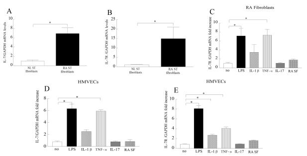
Objective: To characterize the expression of interleukin-7 (IL-7) and IL-7 receptor (IL-7R) in rheumatoid arthritis (RA) synovial tissue and to examine their regulation and pathogenic role in macrophages, endothelial cells, and synovial tissue fibroblasts in RA.

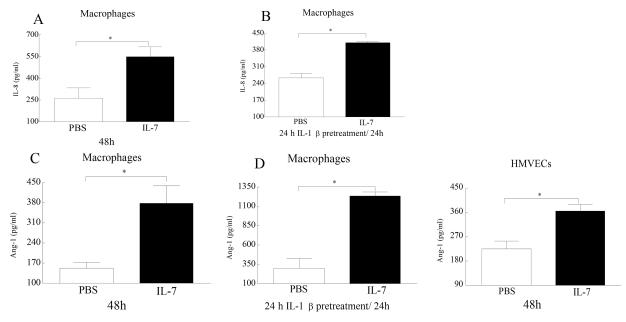
Methods: Expression of IL-7 and IL-7R in RA and normal synovial tissue was demonstrated by immunohistochemistry. Expression and regulation of IL-7 and IL-7R in RA peripheral blood in vitro-differentiated macrophages, RA synovial tissue fibroblasts, and human microvascular endothelial cells (HMVECs) were determined by real-time reverse transcription-polymerase chain reaction and/or flow cytometry. Enzyme-linked immunosorbent assay was used to examine production of proangiogenic factors by IL-7-activated macrophages, RA fibroblasts, and endothelial cells.

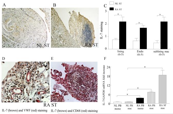
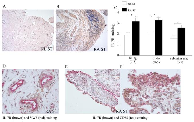
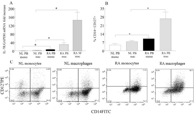
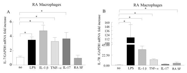
Results: IL-7 and IL-7R were coexpressed on RA synovial tissue lining and sublining macrophages and endothelial cells. Expression of IL-7 and its receptor was significantly elevated in RA synovial fluid and peripheral blood macrophages as well as RA fibroblasts, compared to normal cells. Toll-like receptor 4 ligation (with lipopolysaccharide) and tumor necrosis factor α (TNFα) stimulation modulated expression of IL-7 and IL-7R on RA macrophages and HMVECs. However, in RA fibroblasts, lipopolysaccharide and TNFα activation increased expression of IL-7R only. IL-7 also mediated RA pathogenesis by inducing production of potent proangiogenic factors from macrophages and endothelial cells.

Conclusion: We have identified, for the first time, regulators of IL-7 and IL-7R expression in RA fibroblasts, RA peripheral blood in vitro-differentiated macrophages, and endothelial cells. Our results document a novel role of IL-7 in RA angiogenesis.


Figures

Figure 1
Figure 1. IL-7 is increased in RA synovial tissue (ST) compared to normal (NL) ST and macrophages from RA peripheral blood and synovial fluid are an important source for IL-7 expression
NL (A) and RA ST (B) were stained with anti-human IL-7 (original magnification × 200) and positive immunostaining was scored on a 0-5 scale (C). ST lining and sublining macrophages and endothelial immunostaining are shown as mean ±SEM, (n=8-15). RA synovial tissues were stained for IL-7 (brown staining) and Von wille brand factor (fast red staining) (original magnification × 400) (D) or for IL-7 (brown staining) and CD68 (fast red staining) (original magnification × 400) (E) in order to distinguish endothelial cells or macrophages that express IL-7. IL-7 (F) mRNA levels were determined in NL and RA peripheral blood (PB) monocytes and macrophages as well as in RA synovial fluid (SF) macrophages by employing real-time RT-PCR (n=8-22). The data are shown as fold increase above NL PB monocytes and are normalized to GAPDH. * represents p <0.05.
Figure 2
Figure 2. IL-7R is elevated in RA ST compared to NL ST
NL (A) and RA ST (B) were stained with anti-human IL-7R (original magnification × 200) and positive immunostaining was scored on a 0-5 scale (C). ST lining and sublining macrophages and endothelial immunostaining are shown as mean ±SEM, (n=8-15). RA ST were stained for IL-7R (brown staining) and Von wille brand factor (fast red staining) (original magnification × 400) (D) or for IL-7R (brown staining) and CD68 (fast red staining) (original magnification × 200) (E) and (original magnification × 400) (F) in order to distinguish endothelial cells or macrophages that express IL-7R.
Figure 3
Figure 3. IL-7R is upregulated in RA SF and PB macrophages compared to NL PB macrophages
A. IL-7R mRNA levels were determined in NL and RA PB monocytes and macrophages as well as in RA SF macrophages by employing real-time RT-PCR (n=8-22). The data are shown as fold increase above NL PB monocytes and are normalized to GAPDH. Values demonstrate mean ± SEM. B. Normal and RA PB monocyte and macrophages were immunostained with CD14 and CD127 in order to determine % IL-7R positive cells. The values are presented as mean ± SEM of % CD14+IL-7R+ in each cell population (n=4-5). C. Representative flow cytometry histogram showing CD14+CD127+ NL and RA monocytes and macrophages. * represents p<0.05.
Figure 4
Figure 4. Proinflammatory factors induce the expression of IL-7 and IL-7R in RA PB in vitro differentiated macrophages
RA PB in vitro differentiated macrophages were untreated (PBS) or treated with LPS (10 ng/ml), IL-1β (10 ng/ml), TNF-α (10 ng/ml), IL-17 (50 ng/ml), or RA SF (1:4 dilution) and expression of IL-7 (A) and IL-7R (B) was measured by real-time RT-PCR (n=4-9). The data are shown as fold increase above untreated RA PB macrophages and are normalized to GAPDH. Values demonstrate mean ± SEM, * represents p<0.05.
Figure 5
Figure 5. RA ST fibroblasts have elevated levels of IL-7 and IL-7R and expression levels of IL-7 and IL-7R are modulated by TLR4 ligation and TNF-α activation in HMVECs
IL-7 (A) and IL-7R (B) mRNA levels were determined in NL and RA ST fibroblasts employing real-time RT-PCR (n=7-15). The data are shown as fold increase above NL ST fibroblasts and are normalized to GAPDH. RA ST fibroblasts were untreated (PBS) or treated with LPS (10 ng/ml), IL-1β (10 ng/ml), TNF-α (10 ng/ml), IL-17 (50 ng/ml) or RA SF (1:4 dilution) and expression of IL-7R (C) was measured by real-time RT-PCR (n=4-9). Since IL-7 expression levels were unaffected in stimulated RA fibroblasts, the data is not shown. The data are shown as fold increase above untreated RA ST fibroblasts and are normalized to GAPDH. HMVECs were similarly treated as RA fibroblasts mentioned above in C and expression of IL-7 (D) and IL-7R (E) was measured by real-time RT-PCR (n=4-6). The data are shown as fold increase above untreated HMVECs and are normalized to GAPDH. Values demonstrate mean ± SEM, * represents p<0.05.
Figure 6
Figure 6. IL-7 activates production of proangiogenic factors from macrophages and HMVECs
Macrophages (A-C) or HMVECs (E) were treated with PBS or IL-7 (10 ng/ml) for 24-48h and levels of IL-8 (A), and Ang-1 (C and E) were detected by ELISA utilizing the conditioned media. To determine whether elevated levels of IL-7R on macrophages would al1ow earlier detection or increase in proangiogenic factors cells were pretreated with IL-1β (10ng/ml; for 24h) prior to IL-7 (10 ng/ml) treatment. Production levels of IL-8 (B) and Ang-1 (D) was determined in condition media collected following 24h IL-7 treatment employing ELISA. Proangiogenic factors were not detected in IL-7 activated RA fibroblasts (data not shown). Values are the mean ± SEM, (n=5). * represents p <0.05.

Comment in

References

    1. Golden-Mason L, Kelly AM, Traynor O, McEntee G, Kelly J, Hegarty JE, et al. Expression of interleukin 7 (IL-7) mRNA and protein in the normal adult human liver: implications for extrathymic T cell development. Cytokine. 2001;14(3):143–51. - PubMed
    1. Kroncke R, Loppnow H, Flad HD, Gerdes J. Human follicular dendritic cells and vascular cells produce interleukin-7: a potential role for interleukin-7 in the germinal center reaction. Eur J Immunol. 1996;26(10):2541–4. - PubMed
    1. Sorg RV, McLellan AD, Hock BD, Fearnley DB, Hart DN. Human dendritic cells express functional interleukin-7. Immunobiology. 1998;198(5):514–26. - PubMed
    1. de Saint-Vis B, Fugier-Vivier I, Massacrier C, Gaillard C, Vanbervliet B, Ait-Yahia S, et al. The cytokine profile expressed by human dendritic cells is dependent on cell subtype and mode of activation. J Immunol. 1998;160(4):1666–76. - PubMed
    1. van Roon JA, Verweij MC, Wijk MW, Jacobs KM, Bijlsma JW, Lafeber FP. Increased intraarticular interleukin-7 in rheumatoid arthritis patients stimulates cell contact-dependent activation of CD4(+) T cells and macrophages. Arthritis Rheum. 2005;52(6):1700–10. - PubMed

Publication types

MeSH terms