. 2011 Jul;240(7):1826-40.
doi: 10.1002/dvdy.22669. Epub 2011 Jun 3.

Gene expression profile of the regeneration epithelium during axolotl limb regeneration

Affiliations

Gene expression profile of the regeneration epithelium during axolotl limb regeneration

Leah J Campbell et al. Dev Dyn. 2011 Jul.

Abstract

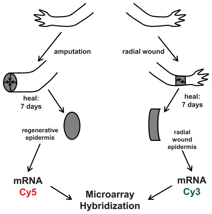
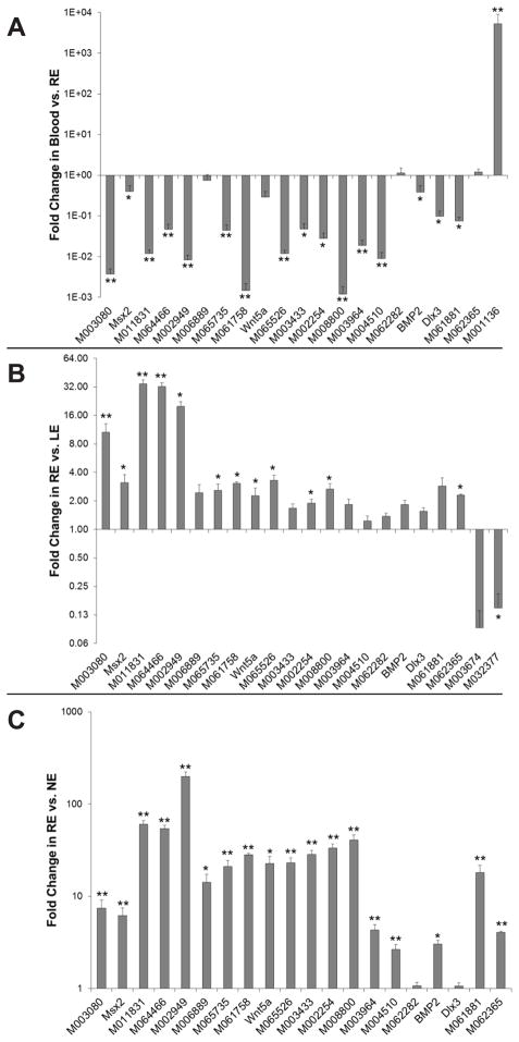
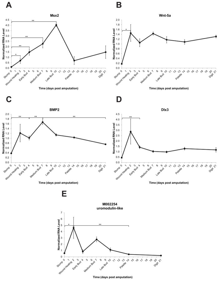
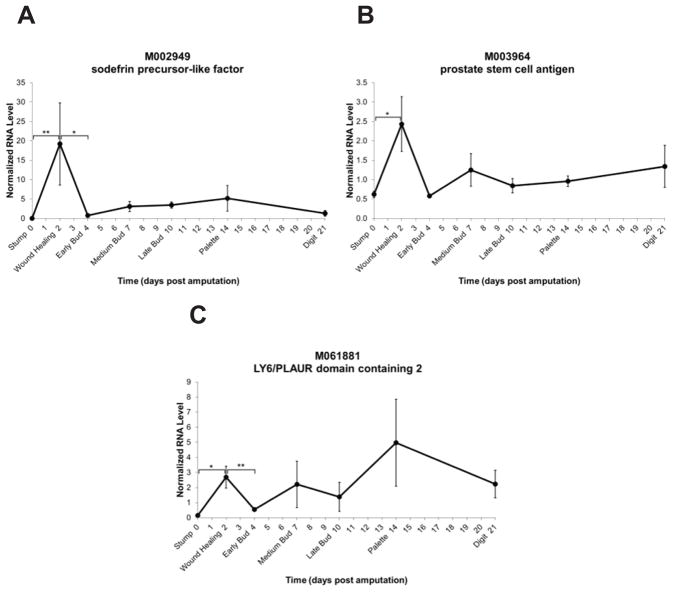
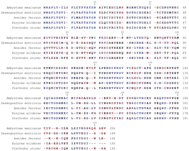
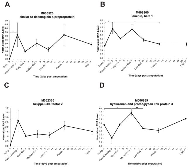
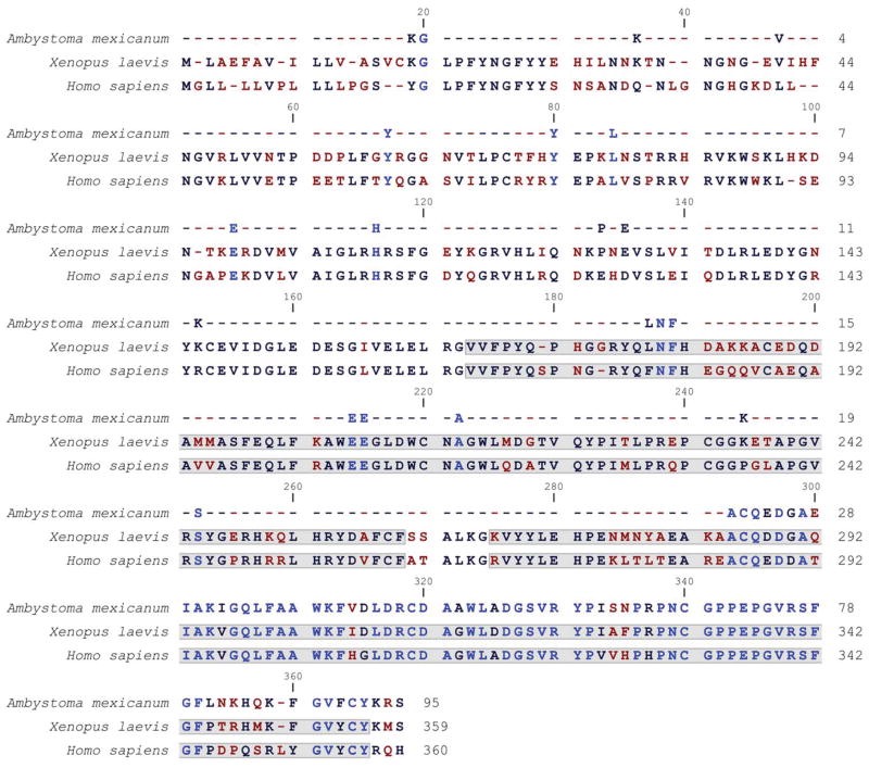
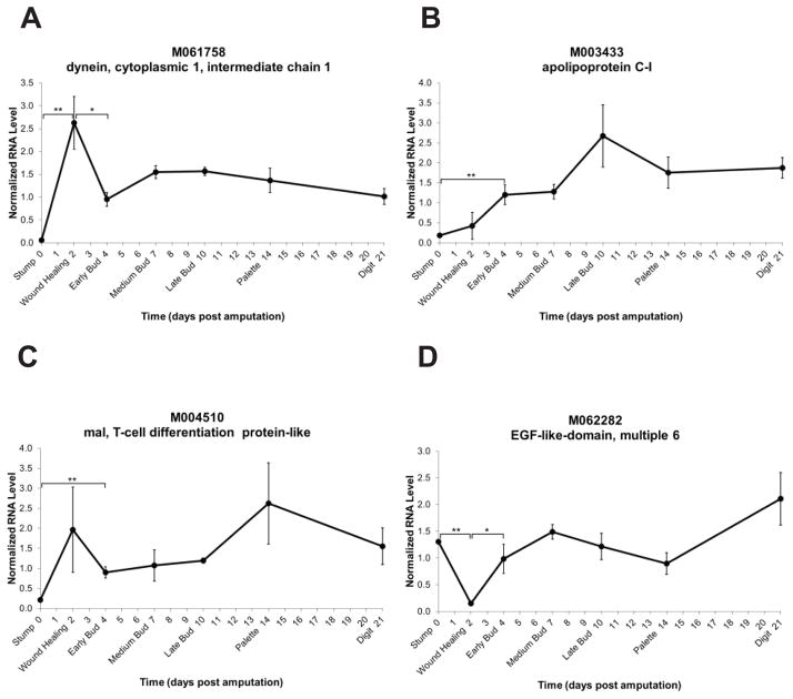
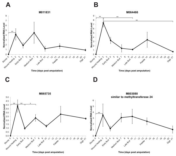
Urodele amphibians are unique among adult vertebrates in their ability to regenerate missing limbs. The process of limb regeneration requires several key tissues including a regeneration-competent wound epidermis called the regeneration epithelium (RE). We used microarray analysis to profile gene expression of the RE in the axolotl, a Mexican salamander. A list of 125 genes and expressed sequence tags (ESTs) showed a ≥1.5-fold expression in the RE than in a wound epidermis covering a lateral cuff wound. A subset of the RE ESTs and genes were further characterized for expression level changes over the time-course of regeneration. This study provides the first large scale identification of specific gene expression in the RE.


Figures

Fig. 1
Fig. 1
Schematic depicting tissue dissection and RNA isolation for microarray hybridization. Axolotl limbs were amputated or wounded with a lateral cuff wound. After 7 days of healing, the RE and the LE were dissected and RNA was isolated. mRNA was labeled with Cy3 and Cy5 dyes and hybridized to custom microarray slides designed from EST assemblies for A. mexicanum, A. tigrinum, and other salamander species.
Fig. 2
Fig. 2
qPCR validation of fold change gene expression of RE upregulated hits. A: Fold change expressions in blood over RE. B: Fold change expressions in RE over LE. C: Fold change expressions in RE over NE. Genes are represented by Sal ID or gene name as in Table 1. Data represents the mean of three biological replicates and standard error of the mean. *p≤0.05; **p≤0.01.
Fig. 3
Fig. 3
Whole mount in situ hybridization analysis of M002949 (sodefrin precursor factor) expression in 7 day post amputation regenerating axolotl limbs. All panels are forelimbs with distal at the top. A: M002949 expression covering distal tip of regenerating limb indicated in blue as determined with anti-sense probe. B: Negative control using M002949 sense probe. C, D: Sister sections of tissue from panel A. Dashed line represents level of amputation. Tissue sectioned at 14 μm. C: Expression in the regeneration epithelium indicated in blue. D: Hematoxylin and eosin. E, F: Higher magnification of the distal tip of the regenerating limb shows that the M002949 expression is localized to the top layers of the regeneration epidermis.
Fig. 4
Fig. 4
Expression level time-courses of genes previously reported to play a role in limb regeneration. The y-axis represents normalized RNA level and the x-axis represents days post amputation. Genes are represented by Sal ID and/or gene name as in Table 1. Each dark circle represents the mean of three samples ± standard error of the mean. *p≤0.05; **p≤0.01.
Fig. 5
Fig. 5
Expression level time-courses of three finger protein family members. The y-axis represents normalized RNA level and the x-axis represents days post amputation. Genes are represented by Sal ID and gene name as in Table 1. Each dark circle represents the mean of three samples ± standard error of the mean.
Fig. 6
Fig. 6
Alignment of axolotl (A. mexicanum) M002949 amino acid sequence with sodefrin precursor-like factor from four species of salamander: seal salamander (D. monticola; accession: AAZ06333), clouded salamander (A. ferreus; accession: AAZ06336), Blue Ridge two-lined salamander (E. wilderae; accession: AAZ06337), and Siskiyou Mountain salamander (P. stormi, accession: AAZ06325). Conserved residues are indicated by blue and non-conserved residues are indicated by red. Gaps are indicated by a dash (−).
Fig. 7
Fig. 7
Expression level time-courses of genes involved in cell adhesion and organization. The y-axis represents normalized RNA level and the x-axis represents days post amputation. Genes are represented by Sal ID and gene name as in Table 1. Each dark circle represents the mean of three samples ± standard error of the mean. *p≤0.05; **p≤0.01.
Fig. 8
Fig. 8
Alignment of axolotl (A. mexicanum) M006889 amino acid sequence with hyaluronan and proteoglycan link protein 3 from X. laevis (accession: NP_001079631) and human (H. sapiens; accession: NP_839946). Conserved residues are indicated in blue and non-conserved residues are indicated by red. Gaps are indicated by a dash (−). The shaded boxes identify the hyaluronan and proteoglycan link domains.
Fig. 9
Fig. 9
Expression level time-courses of genes with lipid-associated and neurite regeneration roles. The y-axis represents normalized RNA level and the x-axis represents days post amputation. Genes are represented by Sal ID and gene name as in Table 1. Each dark circle represents the mean of three samples ± standard error of the mean. *p≤0.05; **p≤0.01.
Fig. 10
Fig. 10
Expression level time-courses of genes with unknown or hypothetical identity. The y-axis represents normalized RNA level and the x-axis represents days post amputation. Genes are represented by Sal ID and gene name as in Table 1. Each dark circle represents the mean of three samples ± standard error of the mean. *p≤0.05; **p≤0.01.
Fig. 11
Fig. 11
Alignment of axolotl (A. mexicanum) M003080 amino acid sequence with putative methyltransferase sequences from chicken (G. gallus; accession: XP_001232694) and X. laevis (accession: NP_001136263). Conserved residues are indicated by blue and residues that are not conserved are indicated by red. Gaps are indicated by a dash (−). The shaded boxes identify the S-adenosylmethionine-dependent methyltransferase, class I regions in chicken and Xenopus.

References

    1. Bazzi H, Getz A, Mahoney MG, Ishida-Yamamoto A, Langbein L, Wahl JK, III, Christiano AM. Desmoglein 4 is expressed in highly differentiated keratinocytes and trichocytes in human epidermis and hair follicle. Differentiation. 2006;74:129–140. - PubMed
    1. Benjamini Y, Hochberg Y. Controlling the False Discovery Rate: A Practical and Powerful Approach to Multiple Testing. Journal of the Royal Statistical Society Series B (Methodological) 1995;57:289–300.
    1. Brown DD, Wang Z, Furlow JD, Kanamori A, Schwartzman RA, Remo BF, Pinder A. The thyroid hormone-induced tail resorption program during Xenopus laevis metamorphosis. Proc Natl Acad Sci U S A. 1996;93:1924–1929. - PMC - PubMed
    1. Buchner G, Broccoli V, Bulfone A, Orfanelli U, Gattuso C, Ballabio A, Franco B. MAEG, an EGF-repeat containing gene, is a new marker associated with dermatome specification and morphogenesis of its derivatives. Mech Dev. 2000a;98:179–182. - PubMed
    1. Buchner G, Orfanelli U, Quaderi N, Bassi MT, Andolfi G, Ballabio A, Franco B. Identification of a New EGF-Repeat-Containing Gene from Human Xp22: A Candidate for Developmental Disorders. Genomics. 2000b;65:16–23. - PubMed

Publication types

MeSH terms

Substances

LinkOut - more resources