. 2011 Jun 8:9:28.
doi: 10.1186/1477-5956-9-28.

Identification of maturation and protein synthesis related proteins from porcine oocytes during in vitro maturation

Affiliations

Identification of maturation and protein synthesis related proteins from porcine oocytes during in vitro maturation

Jumi Kim et al. Proteome Sci. .

Abstract

Background: In vitro maturation (IVM) of mammalian oocytes is divided into the GV (germinal vesicle stage), MI (metaphase I stage) and MII (metaphase II stage) stages, and only fully mature oocytes have acquired the ability to be fertilized and initiate zygotic development. These observations have been mostly based on morphological evaluations, but the molecular events governing these processes are not fully understood.The aim of the present study was to better understand the processes involved in the molecular regulation of IVM using 2-DE analysis followed by mass spectrometry to identify proteins that are differentially expressed during oocyte IVM.

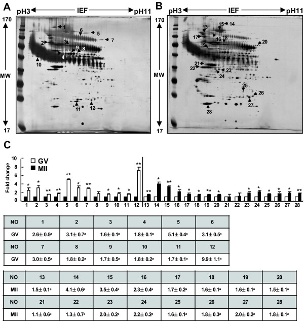
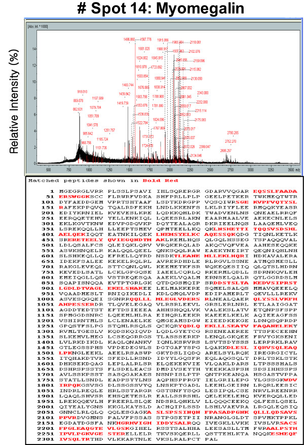
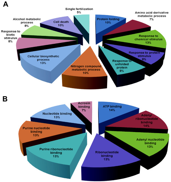
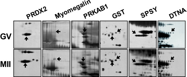
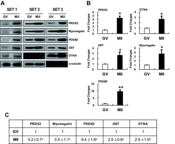
Result: A total of 16 up-regulated and 12 down-regulated proteins were identified. To investigate the IVM process, we specifically focused on the proteins that were up-regulated during the MII stage when compared with the GV stage, which included PRDX 2, GST, SPSY, myomegalin, PED4D, PRKAB 1, and DTNA. These up-regulated proteins were functionally involved in redox regulation and the cAMP-dependent pathway, which are essential for the intracellular signaling involved in oocyte maturation. Interestingly, the PDE4D and its partner, myomegalin, during the MII stage was consistently confirmed up-regulation by western blot analyses.

Conclusion: These results could be used to better understand some aspects of the molecular mechanisms underlying porcine oocyte maturation. This study identified some regulatory proteins that may have important roles in the molecular events involved in porcine oocyte maturation, particularly with respect to the regulation of oocyte meiotic resumption, MII arrest and oocyte activation. In addition, this study may have beneficial applications not only to basic science with respect to the improvement of oocyte culture conditions but also to mammalian reproductive biotechnology with potential implications.


Figures

Figure 1
Figure 1
Morphological characterization and orcein staining of porcine oocytes during IVM. (A) Oocyte released from a follicle containing an intact germinal vesicle (GV). (B) The M II stage and extrusion of the first polar body (I PB). Morphological characterization of the porcine oocytes in each stage (a, b, d, and e), and morphological evaluation of each of these stages using orcein staining (c, f). (C) Changes in the nuclear status of oocytes cultured in TCM-199 medium throughout the process of IVM. The oocytes were examined at four different stages of maturation: germinal vesicle (GV), chromosome condense (CC), metaphase I (M I) and metaphase II (M II).
Figure 2
Figure 2
Two-dimensional protein gels of GV- and M II-stage porcine oocytes. The proteins were separated using 2-DE gel electrophoresis, with the first dimension involving an 18-cm pH 3-11 NL IPG and the second dimension involving 10% gels (A: GV; B: MII), and visualized by silver staining. (C) Comparison of spot intensity in the 2-DE gels for the GV and MII-stage oocytes using quantitative analysis. The results, which were analyzed by the Phoretix Expression software, are presented in a bar graph *Value significantly differs from the control (*P < 0.05 and **P < 0.01). The numbered spots were identified by mass spectrometry, and the numbers correspond to their respective proteins in Tables 1 and 2.
Figure 3
Figure 3
MALDI-TOF MS peptide mass fingerprint spectrum of spot No. 14 (gi|114558492). The protein corresponding to spot No. 14 in Figure 2 was extracted from the 2-D gel and analyzed by MALDI-TOF MS to determine its identity.
Figure 4
Figure 4
Classification of differentially regulated proteins. An ontological classification of differentially regulated proteins in terms of (A) biological process and (B) molecular function using the Gene Ontology http://www.geneontology.org and UniProt http://www.expasy.uniprot.org websites. The compositions of the identified proteins are presented as percentages of all individually identified proteins.
Figure 5
Figure 5
Enlarged images of protein spots differentially expressed between the GV and M II stages. (A) The PRDX2, myomegalin, PRKAB1, GST, SPSY and DTNA proteins were up-regulated in the M II stage when compared with the GV stage. (B) Quantitative analysis of the expression levels of these proteins. The numbered spots were identified by mass spectrometry, and the numbers correspond to their respective proteins in Tables 1 and 2.
Figure 6
Figure 6
Western-blot analyses confirming the upregulation of particular proteins during the M II stage. (A) Western blot analyses of total protein extracts from GV and M II oocytes (lane 1, GV oocytes; lane 2, M II oocytes) using specific antibodies against proteins (PRDX2, Myomegalin, PDF4D, GST and DTNA) identified by 2-DE. Alpha-tubulin was used as a loading control. (B) Quantification of PRDX2, Myomegalin, PDF4D, GST and DTNA expression in GV and MII embryos. PRDX2, Myomegalin, PDF4D, GST and DTNA protein expression increased following MII embryos. *Value significantly differs from the control (*P <0.05 and **P <0.01). The results, which were analyzed by the TINA program, are presented in a bar graph.

References

    1. Kikuchi K, Somfai T, Nakai M, Nagai T. Appearance, fate and utilization of abnormal porcine embryos produced by in vitro maturation and fertilization. Soc Reprod Fertil Suppl. 2009;66:135–147. - PubMed
    1. Moor R, Dai Y. Maturation of pig oocytes in vivo and in vitro. Reprod Suppl. 2001;58:91–104. - PubMed
    1. Novak SPF, Savard C, Tremblay K, Sirard MA. Identification of porcine oocyte proteins that are associated with somatic cell nuclei after co-incubation. Biol Reprod. 2004;71:1279–1289. doi: 10.1095/biolreprod.103.027037. - DOI - PubMed
    1. Ellederova Z, Halada P, Man P, Kubelka M, Motlik J, Kovarova H. Protein patterns of pig oocytes during in vitro maturation. Biol Reprod. 2004;71:1533–1539. doi: 10.1095/biolreprod.104.030304. - DOI - PubMed
    1. Fair T, Carter F, Park S, Evans AC, Lonergan P. Global gene expression analysis during bovine oocyte in vitro maturation. Theriogenology. 2007;68(Suppl 1):S91–97. - PubMed