. 2011 Aug 15;356(2):398-410.
doi: 10.1016/j.ydbio.2011.05.666. Epub 2011 May 30.

The role of the Suppressor of Hairy-wing insulator protein in Drosophila oogenesis

Affiliations

The role of the Suppressor of Hairy-wing insulator protein in Drosophila oogenesis

Ryan M Baxley et al. Dev Biol. .

Abstract

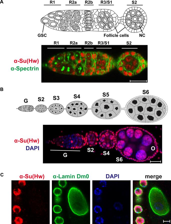
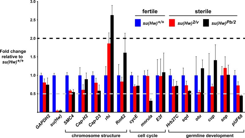
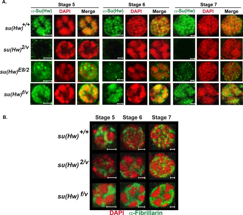
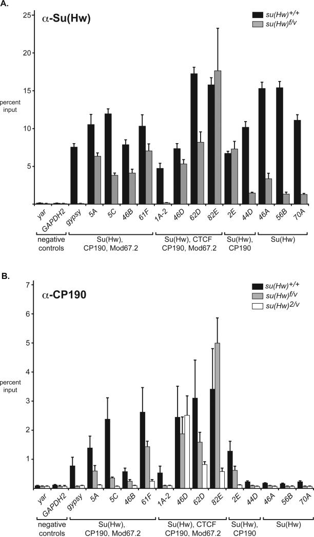
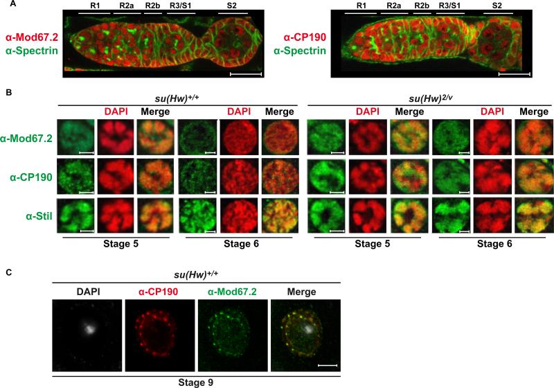
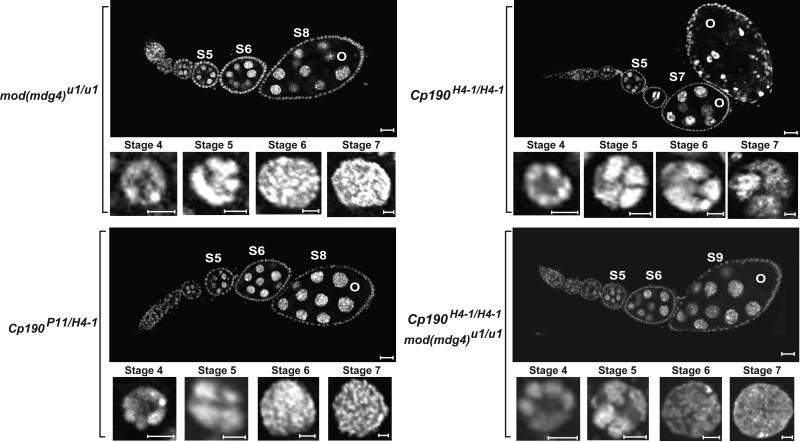
The Drosophila Suppressor of Hairy wing [Su(Hw)] insulator protein has an essential role in the development of the female germline. Here we investigate the function of Su(Hw) in the ovary. We show that Su(Hw) is universally expressed in somatic cells, while germ cell expression is dynamic. Robust levels accumulate in post-mitotic germ cells, where Su(Hw) localization is limited to chromosomes within nurse cells, the specialized cells that support oocyte growth. Although loss of Su(Hw) causes global defects in nurse cell chromosome structure, we demonstrate that these architectural changes are not responsible for the block in oogenesis. Connections between the fertility and insulator functions of Su(Hw) were investigated through studies of the two gypsy insulator proteins, Modifier of (mdg4)67.2 (Mod67.2) and Centrosomal Protein of 190kDa (CP190). Accumulation of these proteins is distinct from Su(Hw), with Mod67.2 and CP190 showing uniform expression in all cells during early stages of oogenesis that diminishes in later stages. Although Mod67.2 and CP190 extensively co-localize with Su(Hw) on nurse cell chromosomes, neither protein is required for nurse cell chromosome development or oocyte production. These data indicate that while the gypsy insulator function requires both Mod67.2 and CP190, these proteins are not essential for oogenesis. These studies represent the first molecular investigations of Su(Hw) function in the germline, which uncover distinct requirements for Su(Hw) insulator and ovary functions.


Figures

Figure 1
Figure 1. Molecular properties of su(Hw) alleles
A) Diagram of the su(Hw) genomic region, including the su(Hw) and RpII15 genes. The su(Hw) coding region is mapped onto the gene structure and includes two acidic domains (AD), the Mod67.2 interaction domain (MID) and the locations of the twelve zinc-fingers (black boxes). The su(Hw)v allele carries a deletion of the su(Hw) and RpII15 promoters. The positions of the insertions in su(Hw)2 (jockey) and su(Hw)Pb(Piggy-bac) are shown by inverted triangles, the su(Hw)E8 and su(Hw)f alleles contain point mutations in coding regions for zinc finger 7 and zinc finger 10, respectively. B) Shown is a western blot of protein extracts obtained from wild type (Canton S, CS) and su(Hw) mutant ovaries probed with Su(Hw) and alpha-Tubulin antibodies, with the latter used as a loading control.
Figure 2
Figure 2. Su(Hw) localizes to somatic and post-mitotic NC nuclei in the ovary
A) Top: Diagram of the germarium, marking regions 1 (R1), R2a, R2b, R3/egg chamber stage 1 (S1). The first egg chamber that emerges from the germarium is a stage 2 (S2) chamber. Germline stem cells (GSC) are located at the anterior end of the germarium. Egg chambers include germ cells and somatic follicle cells. NCs within the stage 2 egg chamber are labeled. Bottom: A representative image of the germarium from a su(Hw)+/+ (Canton S) female stained with anti-Spectrin (green) and anti-Su(Hw) (red) antibodies. Accumulation of Su(Hw) in R1/ R2a is limited to somatic cells. Cells labeled in R1/R2a are somatic cells. B) Top: Diagram of an ovariole that contains the germarium (G) and developing egg chambers up to stage 6 (S6) of development. Below: Shown is a representative image of an ovariole from su(Hw)+/+ (Canton S) stained with anti-Su(Hw) antibodies (red) and the DNA stain DAPI (blue). The germarium (G), egg chamber stages (S2 to S6) and oocyte (O) are indicated. Scale bars are 20 microns in length. C. Shown is a representative image of a wild type (Canton S) stage 9 oocyte nucleus (center) surrounded by follicle cells (right) that was stained with antibodies against Su(Hw) (red), laminDm0 (green) and DAPI (blue).
Figure 3
Figure 3. Mutations in the su(Hw) block oogenesis and produce apoptotic egg chambers
Representative images of DAPI stained ovarioles from wild type (su(Hw)+/+), su(Hw)2/v and su(Hw)f/v ovaries, oriented such that the germarium is at the left. Egg chamber stages (S4 to S8) and oocyte (O) are indicated. Magnified below each ovariole are representative NC nuclei from S4 to S7 egg chambers. Note that the late stage egg chamber in the su(Hw)2/v ovariole displays condensed DAPI foci, indicating apoptosis. Ovariole and nurse cell scale bars are 20 and 5 microns, respectively.
Figure 4
Figure 4. Loss of Su(Hw) does not alter expression of five-blob genes in the ovary
Levels of expression of genes required for NC chromosome development, referred to as five-blob genes, were determined using quantitative real time PCR. The fold change in expression of wild type to su(Hw)+/+ was set to 1 (blue). Two su(Hw) sterile backgrounds were tested, su(Hw)2/v (red) and su(Hw)Pb/2 (black). The functional class of the five-blob gene is noted. Error bars indicate standard deviation (n=3).
Figure 5
Figure 5. Mutations in su(Hw) alter nuclear architecture within NC nuclei
A) Magnified images of representative NC nuclei of stage 5 to 7 egg chambers from su(Hw)+/+ (Canton S), su(Hw)2/v, su(Hw)E8/2 and su(Hw)f/v egg chambers. Nuclei were stained with DAPI (red) and antibodies against Su(Hw) (green). B) Merged images of nurse cell nuclei representing stages 5 to 7 egg chambers from su(Hw)+/+ (Canton S), su(Hw)2/v and su(Hw)f/v ovaries stained with antibodies against the nucleolar marker Fibrillarin (green) and DAPI (red). Scale bars are 5 microns.
Figure 6
Figure 6. Properties of the ovary SBSs are similar to those in somatic cells
A) Su(Hw) binding in the ovary. Shown is ChIP data obtained using Su(Hw) antibodies on chromatin isolated from newly eclosed ovaries isolated from wild type (su(Hw)+/+) and su(Hw)f/v females. Noted below each SBS are the insulator protein partners present as determined in (Negre et al., 2010). Error bars indicate standard deviation (n=3). B) CP190 binding in the ovary. Shown ChIP data obtained using CP190 antibodies on chromatin isolated from newly eclosed ovaries isolated from su(Hw) wild type and mutant females. Axes are labeled as in A. Error bars indicate standard deviation (n=3).
Figure 7
Figure 7. Mod67.2 and CP190 display both Su(Hw)-dependent and -independent localization
A) Representative images of germaria from su(Hw)+/+ (Canton S) females stained with anti-Spectrin (green) and anti-Mod67.2 (red) or anti-CP190 (red) antibodies. Note Mod67.2 and CP190 staining in R1 and R2a, regions that lack Su(Hw). Scale bars are 20 microns. B) NC nuclei representing stages 5 to 6 egg chambers from su(Hw)+/+ (Canton S) and su(Hw)2/v ovaries. Nuclei were stained with antibodies against Mod67.2, CP190 or Stil (positive control) (green) and DAPI (red). Merged images are shown to display colocalization of each signal. Scale bars are 5 microns. C). A representative image of a wild type stage 8/9 oocyte nucleus (Canton S) is shown. This nucleus was stained with antibodies against CP190 (red), Mod67.2 (green) and DAPI (white), with the merged image of CP190 and Mod67.2 staining on the right.
Figure 8
Figure 8. Mutations in the mod(mdg4) and Cp190 genes have minor effects on oogenesis
Images of DAPI stained ovarioles isolated from mod(mdg4) mutant females and Cp190 mutant females carrying three different heteroallelic combinations. Genotypes of the females are indicated on the left. For each genotype, an ovariole and magnified NC nuclei are shown. Ovariole and nurse cell scale bars are 20 and 5 microns in length, respectively.

References

    1. Adryan B, Woerfel G, Birch-Machin I, Gao S, Quick M, Meadows L, Russell S, White R. Genomic mapping of Suppressor of Hairy-wing binding sites in Drosophila. Genome Biol. 2007;8:R167. - PMC - PubMed
    1. Bartkuhn M, Straub T, Herold M, Herrmann M, Rathke C, Saumweber H, Gilfillan GD, Becker PB, Renkawitz R. Active promoters and insulators are marked by the centrosomal protein 190. Embo J. 2009;28:877–888. - PMC - PubMed
    1. Burcin M, Arnold R, Lutz M, Kaiser B, Runge D, Lottspeich F, Filippova GN, Lobanenkov VV, Renkawitz R. Negative protein 1, which is required for function of the chicken lysozyme gene silencer in conjunction with hormone receptors, is identical to the multivalent zinc finger repressor CTCF. Mol Cell Biol. 1997;17:1281–1288. - PMC - PubMed
    1. Bushey AM, Dorman ER, Corces VG. Chromatin insulators: regulatory mechanisms and epigenetic inheritance. Mol Cell. 2008;32:1–9. - PMC - PubMed
    1. Bushey AM, Ramos E, Corces VG. Three subclasses of a Drosophila insulator show distinct and cell type-specific genomic distributions. Genes Dev. 2009;23:1338–1350. - PMC - PubMed

Publication types

LinkOut - more resources