Review
. 2011 Jul;138(13):2639-48.
doi: 10.1242/dev.048967.

Evolutionary crossroads in developmental biology: sea urchins

Affiliations
Review

Evolutionary crossroads in developmental biology: sea urchins

David R McClay. Development. 2011 Jul.

Abstract

Embryos of the echinoderms, especially those of sea urchins and sea stars, have been studied as model organisms for over 100 years. The simplicity of their early development, and the ease of experimentally perturbing this development, provides an excellent platform for mechanistic studies of cell specification and morphogenesis. As a result, echinoderms have contributed significantly to our understanding of many developmental mechanisms, including those that govern the structure and design of gene regulatory networks, those that direct cell lineage specification, and those that regulate the dynamic morphogenetic events that shape the early embryo.


Figures
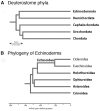
Fig. 1.
Fig. 1.
Phylogeny of the deuterostomes and the echinoderms. (A) A cladogram of deuterostome phyla. The timing of phylum separation is uncertain. (B) Phylogeny of echinoderms. Images courtesy of Greg Wray.
Fig. 2.
Fig. 2.
Commonly used sea urchin and star fish species. (A) Adult Strongylocentrotus purpuratus (photo courtesy of Spbase). (B) Adult Hemicentrotus pulcherrimus (courtesy of Takuya Minokawa) (C) Adult Lytechinus variegatus, showing three color variants. (D) The adult star fish Patiria miniata. (E) Two-week-old larva of S. purpuratus. (F) Pluteus larva of H. pulcherrimus. (G) Pluteus larva of L. variegatus visualized by polarized light to show the larval skeleton (courtesy of Rachel Fink). (H) Bipinnaria larva of P. miniata (courtesy of Veronica Hinman).
Fig. 3.
Fig. 3.
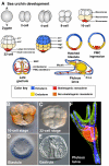
Early sea urchin development. (A) Sequence of sea urchin development from the zygote to the pluteus larva stage. At the 16-cell stage there are four micromeres (red) at the vegetal (V) pole, four central macromeres (light yellow) and eight mesomeres (grey) at the animal (A) pole. From the hatched blastula stage onwards, the embryo is shown as a mid-sagittal section. The colors indicate when the cells begin to be specified toward ectoderm (blue), mesoderm (red) and endomesoderm (yellow). Later, the ectoderm becomes subdivided (as indicated by different shades of blue), and the mesoderm (orange) separates from endoderm (dark yellow). (B-E) Selected stages of Paracentrotus lividus development: (B) 16-cell stage; (C) 32-cell stage; (D) blastula stage; and (E) mid-gastrula stage, showing the gut invaginating and the skeletogenic cells forming a ring of cells around the gut and beginning to synthesize the skeleton. (F) Pluteus larva stained to show the gut (red), the skeleton (blue) and the ectoderm (green). an, animal; veg, vegetal; PMC, primary mesenchyme cells (skeletogenic cells); SMC, secondary mesenchyme cells (non-skeletogenic mesoderm).
Fig. 4.
Fig. 4.
Sea urchin larval structures. (A) Larva of S. pururatus at 72 hours stained to show the hindgut (H) and midgut (Mi, red), skeleton (blue) and ectoderm (green). The mouth (Mo), foregut (F) and ciliary band (CB) are also shown. (B) Larva of H. pulcherrimus at 7 days showing the rudiment to the side of the gut (arrow). Image courtesy of Takuya Minokawa. (C) Larva of L. variegatus at 48 hours showing the sphincter muscles between the foregut and midgut (upper arrow), and between the midgut and hindgut (lower arrow). The ciliary band (green), the foregut (blue) and skeletogenic cells (red) are also shown. (D) Larva of L. variegatus at 72 hours showing the neural cells that include serotonergic neurons (yellow) and non-serotonergic neurons (red). The cell bodies and nerves of this system run within the ciliary band, with an additional circle of neurons surrounding the sphincter muscle between the foregut and midgut (compare C with D). Cell nuclei are shown in green.
Fig. 5.
Fig. 5.
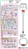
Gene regulatory network model for specification of the skeletogenic mesoderm lineage. A graphic model representing the GRN involved in skeletogenic micromere specification (see Box 3 for further details). At the top of the model, known maternal transcription factor inputs initiate specification in the micromeres at the 16-cell stage (shown as red cells in the embryo on the right). These cells activate Pmar1 [a transcription factor, (TF) and an obligate repressor], which then represses another repressor (HesC, TF), thereby activating Alx1 (TF), Ets1 (TF), Tbr (TF) and Delta [a ligand (L) for the Notch signal transduction pathway]. Over time, additional genes are activated as the embryo forms the mesenchyme blastula stage (depicted by the lower embryo on the right), at which point the skeletogenic cells (red) are fully specified and ingress into the blastocoel. Light gray indicates endoderm; dark gray indicates nonskeletogenic mesoderm. Modified, with permission, from E. Davidson (http://sugp.caltech.edu/endomes/). γ(2)α, cis regulatory sites of the Tbr transcription factor (TF); Blimp, TF; cβ, cytoplasmic β-catenin; CyP, a differentiation gene (DG); Dri, TF; ECNS, early cytoplasmic nuclearization system; Erg, TF; ES, early signal; Ets, TF; Fox, TF; GSK-3, enzyme in canonical Wnt signal transduction pathway; Hex, TF; Hnf6, TF; L1, transmembrane co-receptor of VEGFR; Msp, DG; nβ, TF; Nr1, neuralized, a ubiquitin ligase; Nucl., nucleus; Otx, TF; r11pm, cis-regulatory module of Delta signal; Sm, skeletal matrix; SoxC, TF; TCF, TF; Tel, TF; Tgif, TF; Ubiq, ubiquitous TF; VEGFR, VEGF signal transduction receptor; Wnt8, a signal in the Wnt signal transduction pathway.
None

References

    1. Angerer L. M., Oleksyn D. W., Logan C. Y., McClay D. R., Dale L., Angerer R. C. (2000). A BMP pathway regulates cell fate allocation along the sea urchin animal-vegetal embryonic axis. Development 127, 1105-1114 - PubMed
    1. Angerer L. M., Oleksyn D. W., Levine A. M., Li X., Klein W. H., Angerer R. C. (2001). Sea urchin goosecoid function links fate specification along the animal-vegetal and oral-aboral embryonic axes. Development 128, 4393-4404 - PubMed
    1. Arnone M. I., Dmochowski I. J., Gache C. (2004). Using reporter genes to study cis-regulatory elements. Methods Cell Biol. 74, 621-652 - PubMed
    1. Ben-Tabou de-Leon S., Davidson E. H. (2010). Information processing at the foxa node of the sea urchin endomesoderm specfication network. Proc. Natl. Acad. Sci. USA 107, 10103-10108 - PMC - PubMed
    1. Boveri T. (1902). Uber mehrpolige mitosen als mittel zur analyse des zellkerns. Verh. d. phys.-med. Ges. Wursburg N. F. 35, 67-90

Publication types