. 2011 Jul;138(13):2793-805.
doi: 10.1242/dev.063644.

A context-dependent combination of Wnt receptors controls axis elongation and leg development in a short germ insect

Affiliations

A context-dependent combination of Wnt receptors controls axis elongation and leg development in a short germ insect

Anke Beermann et al. Development. 2011 Jul.

Abstract

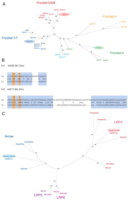
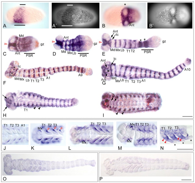
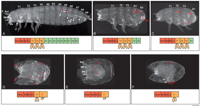
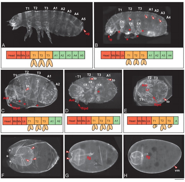
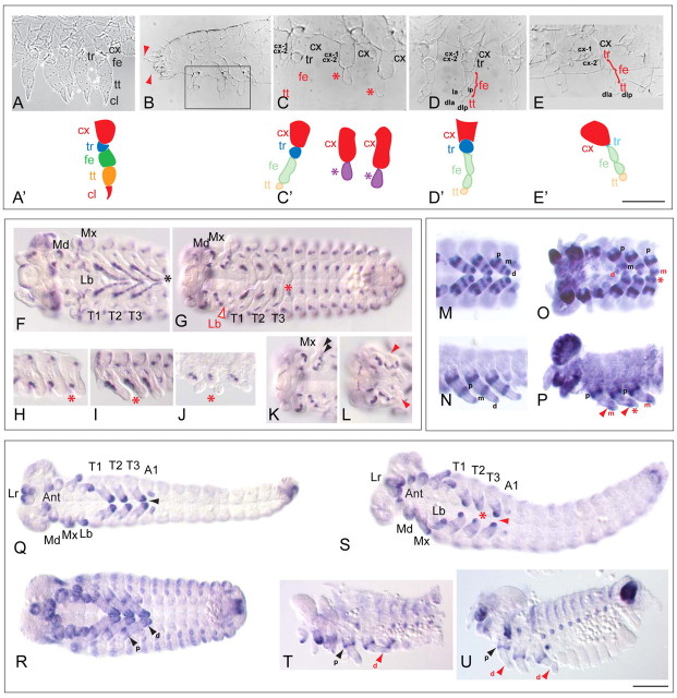
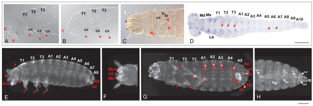
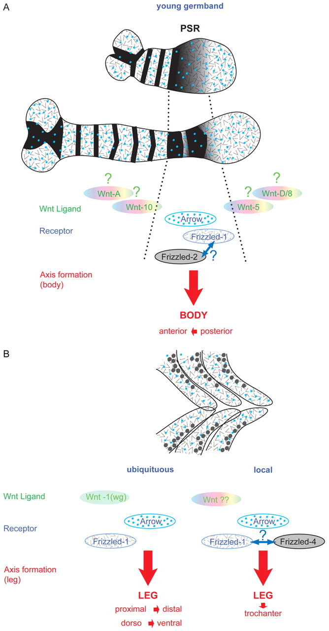
Short germ embryos elongate their primary body axis by consecutively adding segments from a posteriorly located growth zone. Wnt signalling is required for axis elongation in short germ arthropods, including Tribolium castaneum, but the precise functions of the different Wnt receptors involved in this process are unclear. We analysed the individual and combinatorial functions of the three Wnt receptors, Frizzled-1 (Tc-Fz1), Frizzled-2 (Tc-Fz2) and Frizzled-4 (Tc-Fz4), and their co-receptor Arrow (Tc-Arr) in the beetle Tribolium. Knockdown of gene function and expression analyses revealed that Frizzled-dependent Wnt signalling occurs anteriorly in the growth zone in the presegmental region (PSR). We show that simultaneous functional knockdown of the Wnt receptors Tc-fz1 and Tc-fz2 via RNAi resulted in collapse of the growth zone and impairment of embryonic axis elongation. Although posterior cells of the growth zone were not completely abolished, Wnt signalling within the PSR controls axial elongation at the level of pair-rule patterning, Wnt5 signalling and FGF signalling. These results identify the PSR in Tribolium as an integral tissue required for the axial elongation process, reminiscent of the presomitic mesoderm in vertebrates. Knockdown of Tc-fz1 alone interfered with the formation of the proximo-distal and the dorso-ventral axes during leg development, whereas no effect was observed with single Tc-fz2 or Tc-fz4 RNAi knockdowns. We identify Tc-Arr as an obligatory Wnt co-receptor for axis elongation, leg distalisation and segmentation. We discuss how Wnt signalling is regulated at the receptor and co-receptor levels in a dose-dependent fashion.


Figures

Fig. 1.
Fig. 1.
Three Frizzled and two Arrow/LRP proteins are encoded in the Tribolium genome. (A) Phylogenetic analysis places the Tc-Frizzled protein-sequences within three Frizzled classes: 1/7, 2/5/8 and 3/4/9/10. No Tribolium representative of the frizzled-3 gene family was found. (B) Sequence alignment of the C-terminus of the Drosophila and Tribolium Frizzled-1 and Frizzled-2 proteins. Both Tribolium Fz proteins share the specific binding sites for the protein Dishevelled (Wu and Mlodzik, 2008). Blue indicates conserved regions. Orange and asterisks indicate conserved binding sites. Dm, Drosophila melanogaster; Tc, Tribolium castaneum. (C) Phylogenetic analysis of the Tribolium Arrow and LRP proteins.
Fig. 2.
Fig. 2.
Tc-frizzled2 and Tc-frizzled4 show a distinct expression pattern throughout embryonic development whereas Tc-frizzled1 and Tc-arrow are ubiquitously expressed. Tc-fz2 (A-I), Tc-fz4 (J-N), Tc-fz1 (O), Tc-arrow (P) in situ hybridisation or Tc-fz2 and Tc-wg double in situ hybridisation (C,F,I) during Tribolium development. (A,A′) Blastoderm formation. Tc-fz2 is expressed in the prospective head region (bars). A′ shows DAPI staining. (B,B′) In the germ anlage, Tc-fz2 is expressed around the head lobes (asterisk). B′ shows DAPI staining. (C-E) Segmental stripes form, but in contrast to Tc-wg (red in C), the posterior growth zone (gz) stays free of Tc-fz2 expression (blue) at all stages. Tc-fz2 covers the presegmental region (PSR, bar), fading towards the posterior (C-E) (Schröder et al., 2008). Anteriorly, Tc-fz2 is seen in the prospective clypeolabrum and stomodeum (open arrowheads). The intercalary segment (Ic, E) appears as well as two broad stripes in the PSR (bar), possibly the future T3 and A1 segments (asterisks). (F) Within the segments, Tc-fz2 (blue) is expressed at the segmental border posterior to Tc-wg (red). The labrum is free of Tc-fz2 expression. Asterisks indicate antennal segments. (G) Segmental expression of Tc-fz2 in the fully elongated embryo. Arrowhead indicates the stomodeum. (H) Germband retraction. Segmental Tc-fz2 expression declines, Tc-fz2 expression appears in the visceral mesoderm, around the tracheal openings (arrowheads in H,I) and in the brain (arrows). (I) Fully retracted germband. Tc-fz2 expression (blue) in the foregut and the hindgut (open arrowheads), in the CNS (arrows) and laterally in the visceral mesoderm (black arrowheads). Tc-fz2 is at no time expressed in the legs, in contrast to Tc-wg (red). (J-N) Tc-fz4 expression in a proximo-dorsal position during appendage elongation (open red arrowheads in K). Late in embryogenesis (N) Tc-fz4 expression extends and marks the putative border of the trochanter (red arrowheads in N). No Tc-fz4 expression (black asterisks) is observed in the distal tip. (O,P) Tc-fz1 (O) and Tc-arrow (P) are ubiquitously expressed (representative stages shown). All views ventral, except A,A' (lateral) and C,D (dorsal). A1-A10, abdominal segments 1-10; Ant, antennae; gz, growth zone; Lb, labial segment; Ic, intercalary segment; Md, mandibular segment; Mx, maxillar segment; PSR, presegmental region; T1-T3, thoracic segments 1-3. Scale bars: 100 μm.
Fig. 3.
Fig. 3.
Tc-fz1/2-dependent Wnt signalling is required for posterior growth and segmentation. Autofluorescence cuticle preparations with schematics of the corresponding phenotype below. (A) Wild-type-like cuticle. Head with head appendages, thoracic segments 1-3 (T1-3) each with one pair of legs, and eight abdominal segments (A1-8) with urogomphi at abdominal segment 9 (A9). Autofluorescence of the hindgut cuticle can be observed. (B) Weak Tc-fz 1/2RNAi phenotype. Head and thorax with two abdominal segments marked by tracheal openings (arrowheads). A rudimentary hindgut (asterisk) is visible. (C) Intermediate Tc-fz 1/2RNAi phenotype. Head, thorax with malformed legs and one abdominal segment. Tracheal openings (arrowheads) indicate the identity of T2 and A1. (D) Stronger Tc-fz 1/2RNAi phenotype. Two thoracic segments with leg rudiments (asterisks). On the head only the antennal flagellae are missing. (E,F) Strongest Tc-fz 1/2RNAi phenotypes. Head with malformed gnathal appendages and T1 (gnarled legs, asterisks). A1-A10, abdominal segments 1-10; Ant, antennae; cl, pretarsal claw; cx, coxa; fe, femur; fl, flagella; hg, hindgut; Lb, labial segment; Md, mandibular segment; Mx, maxillar segment; to, tracheal opening; T1-T3, thoracic segments 1-3; tr, trochanter; tt, tibiotarsus; ug, urogomphi. Red asterisks indicate affected cuticle structures. Scale bar: 100 μm.
Fig. 4.
Fig. 4.
The growth zone collapses in Tc-fz1/2RNAi embryos. Tc-fz1/2RNAi embryos of increasing phenotypic strength analysed for the expression of marker genes. (A-J) Pair-rule gene product Eve (A-E) and odd-skipped (odd; F-J) expression in wild-type (A, F) and Tc-fz1/2RNAi (B-E,G-J) embryos. Even in the strongest phenotypes (C-E), a spot of Eve-positive cells is seen at the posterior, or a subterminal stripe of odd expression develops (red arrowhead in G,H; red asterisk in D,E,J). Both genes show only partial (weak phenotype in B) or no secondary pair-rule pattern. Pair-rule expression is initiated but not transformed into a segmental pattern anterior to the presegmental region (PSR; red asterisk), except for a weak stripe seen in B or I (open arrowhead). (K-O) Expression of the segment polarity gene product Engrailed (En) in wild-type (K) and Tc-fz1/2RNAi (L-O) embryos. Integrity of segmental borders seems normal anterior to the growth zone rudiment (red arrowhead). Red arrowheads indicate collapsed growth zone. (P-R) Expression of the Wnt ligand Tc-wnt5 in wild-type (P) and Tc-fz1/2RNAi (Q,R) embryos. Stripes in the abdomen and expression domains in the posterior growth zone are gone (red arrowhead) but head spots and antennal stripes are not affected in the Tc-fz1/2RNAi embryos. (S,T) Fgf ligand expression in wild-type (S) and Tc-fz1/2RNAi (T) embryos. Tc-fgf8 expression at the rims of the future mesoderm and within the segmented region is abolished but labral-specific domain (arrowhead) is unaffected in Tc-fz1/2RNAi (T) embryos. (U) Tc-caudal expression at a posterior position in a wild-type embryo. (V-X) Tc-caudal expression marks posterior cells in fz1/2RNAi embryos. (Y) Tc-brachyenteron (byn) marks the anlagen of the hindgut in the wild-type embryo. (Z-BB) Tc-byn expression is lost in strong fz1/2RNAi embryos (Z) but retained in weaker fz1/2RNAi embryos (AA,BB). A1, abdominal segment 1; T1, thoracic segment 1. Black asterisks indicate growth zone; red arrowheads indicate collapsed growth zone; red asterisks indicate affected structures. Scale bar: 100 μm.
Fig. 5.
Fig. 5.
Wnt co-receptor Arrow processes Wnt signals in axial growth, segmentation and leg formation. (A,B) Weak Tc-arrowRNAi phenotype. Few abdominal segments missing, leg defects in B. Arrowheads in B indicate tracheal openings. (C-E) Intermediate Tc-arrowRNAi phenotype. Few segments are developed, legs show abnormalities. (F-H) Strong Tc-arrowRNAi phenotype.. Cuticle spheres without signs of segmentation, with incomplete anterior head structures (white asterisks in F) and gut-like structure (fg) visible. A-C show a lateral view, D-F a ventral view. In A-E, schematics of the corresponding phenotypes are shown below. Red asterisks indicate affected cuticle structures. Open arrowheads in F,G indicate bristles. Ant, antennae; cx, coxa; fg, foregut; fl, flagella; Lr, labrum; pd, pedicellus; T1-T3, thoracic segments 1-3; to, tracheal opening; ug, urogomphi; vm, vitelline membrane; A1-A5 abdominal segments 1-5. Scale bar: 100 μm.
Fig. 6.
Fig. 6.
Tc-fz1 has an exclusive function in appendage differentiation. (A,A′) Wild-type larval leg showing coxa (cx), trochanter (tr), femur (fe), tibiotarsus (tt) and pretarsal claw (cl). Schematic of leg parts is shown in A′. (B-C′) Tc-fz1RNAi larva. Legs have defects of the proximal-distal axis. (B) Tc-fz1RNAi larva, anterior half. Antennae lack flagellae (red arrowheads). Boxed area in B is enlarged in C and illustrates a severe leg phenotype. Schematic is shown in C′. The coxa are unaffected according to coxa specific bristles cx-1 and cx-2 but femur and tibiotarsus are fused and twisted. There is no distinct identity of leg segments (asterisks and purple colour in C′), no pretarsal claw is present and the trochanter is fused to the coxa. (D-E′) Weaker Tc-fz1RNAi phenotypes. Schematics of leg phenotypes are shown in C′ and D′. (D,D′) Tc-fz1RNAi larva showing ‘non-pareille’ phenotype (Grossmann et al., 2009). Ectopic constriction in the femur, no pretarsal claw according to tibiotarsal bristle markers dla and dlp. Femur bristle markers la and lp are shown. (E,E′) Tc-fz1RNAi larva showing a stronger phenotype as in D. Trochanter is not clearly recognisable (blue in E′; ‘pearls on a chain’ phenotype) (Grossmann et al., 2009). (F) Tc-wg expression in wild type in all appendages, reaching in a ventral stripe to the tip of each leg (asterisk). (G) Tc-wg expression in a Tc-fz1RNAi embryo is restricted to the coxa (red asterisk), appendages are distally fused and twisted (red asterisk). There is normal Tc-wg expression in the segments. (H-J) Consecutively older Tc-fz1RNAi legs. Tc-wg expression remains proximal, restricted to coxa and never extends into the tips (asterisks). In J, the distal part of the legs shows the most severe defect (asterisks). (K) Tc-wg expression in wild-type head appendages. Maxillar lobes are present (arrowheads). (L) Tc-wg expression in Tc-fz1RNAi head appendages. Maxillar palp is missing (red arrowhead). (M,N) In wild-type legs (ventral and lateral view) Tc-Lim is expressed in three domains: the proximal ring (p, coxa), the median ring (m, femur/tibia) and the distal domain (d). (O,P) Tc-Lim expression in Tc-fz1RNAi legs, ventral and lateral view. Distal domain is missing or only partly present (red asterisks), the median ring (m) broadens (arrowheads). (Q,R) Tc-dachsous expression in wild-type appendages. In younger embryos, dachsous is expressed in a prominent distal domain in all appendages (Q), and later additionally in a proximal ring in the coxa (arrowheads in R). (S-U) Tc-dachsous expression in Tc-fz1RNAi embryos. Distal expression in the legs is incomplete or missing, only proximal expression remains. (S) Tc-fz1RNAi embryo with leg defects. The distal expression of Tc-dachsous is reduced (asterisk, arrowhead). (T,U) Older Tc-fz1RNAi legs. Only the proximal expression of Tc-dachsous together with a very faint distal domain (arrowheads). Leg segment bristle markers: cx-1, coxa-1; cx-2, coxa-2. A1, abdominal segment 1; Ant, antennae; cl, pretarsal claw; cx, coxa; d, distal ring; dla, distal dorsal anterior; dlp, distal dorsal posterior; fe, femur; la, dorsal anterior; Lb, labial segment; lp, dorsal posterior; Lr, labrum; m, median ring; Md, mandibular segment; Mx, maxillar segment; p, posterior ring; T1-T3, thoracic segments 1-3; tr, trochanter; tt, tibiotarsus. Red asterisks indicate affected structures. Scale bar: 100 μm.
Fig. 7.
Fig. 7.
TcFz4 and the enhancement of the TcFz1RNAi phenotype. (A-C) Tc-fz1/4RNAi leads to loss of distal leg structures. In weaker phenotypes, segments of unclear identity adhere to the proximal leg (A); only the coxa is left in strong Tc-fz1/4RNAi larvae (B). Tc-fz1/4RNAi larvae can hatch but die as L2 larvae (C). (D) Tc-wg expression is noticeably reduced in the legs and the abdominal segments (arrowheads). (E-G) Additional defects of Tc-fz1/4RNAi larvae. Slight head segmentation defects, missing urogomphi (E), maxillae and labium shortened (F). Hindgut is not elongated and divided (G). Subtle changes in the shape of the tracheal openings (open arrowheads, G). (H) Wild-type cuticularised hindgut (autofluorescence). Two loops (lp1,2), connected via the intersection (is) with two hooks (hk1, 2) to the outside. This division is absent in Tc-fz1/4RNAi larvae. A1-A10, abdominal segments 1-10; cx, coxa; fe, femur; fl, flagella; hg, hindgut; Lb, labial segment; Mx, maxillar segment; T1-T3, thoracic segments 1-3; to, tracheal opening; tr, trochanter; tt, tibiotarsus; ug, urogomphi. Red asterisks mark affected cuticle structures. Scale bar: 100 μm.
Fig. 8.
Fig. 8.
The ‘combinatorial code’ model. (A) Summary of expression patterns and possible receptor/co-receptor interactions in the presegmental region (PSR) during anterior-posterior axis formation. Tc-fz2 expression is shown in black in the segments and in the PSR. Within the PSR, Tc-fz2 shows a graded expression fading out towards the posterior. Tc-fz2 is always coexpressed with the ubiquitously expressed Tc-fz1 and Tc-arrow (chevrons and blue dots, respectively). In these regions, it is possible that various dimeric forms (homo- or heterodimers) could form. In the young germ anlage, Tc-wnt5 expression is close to the Tc-fz2 expression domain (Fig. 4P). Later in embryogenesis, Tc-WntA and Tc-Wnt10 can serve as additional/alternate ligands for Tc-Fz1 and Tc-Fz2 in the PSR. (B) Summary of expression patterns and possible receptor/co-receptor interactions in the leg during proximo-distal axis formation. Ubiquitously expressed Tc-Fz1 and Tc-Arrow are joined in a dorso-proximal position by Tc-Fz4 (black dots). Here, Tc-Fz4 supports Wnt signalling by an as yet unknown Wnt ligand. In more distal and ventral positions, Tc-Fz1 and Tc-Arrow exclusively transduce Wnt1 signalling.

References

    1. Angers S., Salahpour A., Bouvier M. (2002). Dimerization: an emerging concept for G protein-coupled receptor ontogeny and function. Annu. Rev. Pharmacol. Toxicol. 42, 409-435 - PubMed
    1. Arnold S. J., Stappert J., Bauer A., Kispert A., Herrmann B. G., Kemler R. (2000). Brachyury is a target gene of the Wnt/beta-catenin signaling pathway. Mech. Dev. 91, 249-258 - PubMed
    1. Aulehla A., Wiegraebe W., Baubet V., Wahl M. B., Deng C., Taketo M., Lewandoski M., Pourquié O. (2008). A beta-catenin gradient links the clock and wavefront systems in mouse embryo segmentation. Nat. Cell Biol. 10, 186-193 - PMC - PubMed
    1. Beermann A., Schröder R. (2008). Sites of Fgf signalling and perception during embryogenesis of the beetle Tribolium castaneum. Dev. Genes Evol. 218, 153-167 - PubMed
    1. Beermann A., Aranda M., Schröder R. (2004). The Sp8 zinc-finger transcription factor is involved in allometric growth of the limbs in the beetle Tribolium castaneum. Development 131, 733-742 - PubMed

Publication types

MeSH terms