. 2011 Aug 19;286(33):28772-28782.
doi: 10.1074/jbc.M110.205245. Epub 2011 Jun 7.

Protein kinase C (PKC) delta suppresses keratinocyte proliferation by increasing p21(Cip1) level by a KLF4 transcription factor-dependent mechanism

Affiliations

Protein kinase C (PKC) delta suppresses keratinocyte proliferation by increasing p21(Cip1) level by a KLF4 transcription factor-dependent mechanism

Yap Ching Chew et al. J Biol Chem. .

Retraction in

Abstract

PKCδ increases keratinocyte differentiation and suppresses keratinocyte proliferation and survival. However, the mechanism of proliferation suppression is not well understood. The present studies show that PKCδ overexpression increases p21(Cip1) mRNA and protein level and promoter activity and that treatment with dominant-negative PKCδ, PKCδ-siRNA, or rottlerin inhibits promoter activation. Analysis of the p21(Cip1) promoter upstream regulatory region reveals three DNA segments that mediate PKCδ-dependent promoter activation. The PKCδ response element most proximal to the transcription start site encodes six GC-rich DNA elements. Mutation of these sites results in a loss of PKCδ-dependent promoter activation. Gel mobility supershift and chromatin immunoprecipitation reveal that these DNA elements bind the Kruppel-like transcription factor KLF4. PKCδ increases KLF4 mRNA and protein level and KLF4 binding to the GC-rich elements in the p21(Cip1) proximal promoter. In addition, KLF4-siRNA inhibits PKCδ-dependent p21(Cip1) promoter activity. PKCδ increases KLF4 expression leading to enhanced KLF4 interaction with the GC-rich elements in the p21(Cip1) promoter to activate transcription.


Figures
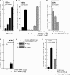
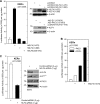
FIGURE 1.
FIGURE 1.
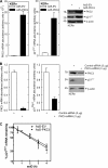
PKCδ activates p21Cip1 gene expression. A, human keratinocytes were transfected with the 0.5 μg of p21-2326 luciferase reporter plasmid and the indicated level of PKCδ expression vector. After 24 h, the cells were harvested, and lysates were assayed for luciferase activity. B, PKCδ activation of p21Cip1 promoter activity is reduced by PKCδ inhibitor. Keratinocytes were transfected with 0.5 μg of p21-2326 and 0.5 μg of PKCδ expression vector and after 24 h were treated with rottlerin or Go6976. After an additional 24 h, extracts were prepared for assay of luciferase activity. C, keratinocytes were transfected with 0.5 μg of p21-2326 and the indicated concentration of empty vector (EV) or dominant-negative (dn) PKCδ-encoding plasmid, and after 24 h, extracts were prepared for assay of luciferase activity. D, keratinocytes were electroporated with 3 μg of control or PKCδ-siRNA/1 × 106 cells. At 48 h, the cells were electroporated with 2 μg of endotoxin-free p21-2326, and after an additional 24 h, extracts were prepared and assayed for luciferase activity. E, keratinocytes were treated with control or PKCδ-siRNA as indicated above, and after 48 h, extracts were prepared for detection of PKCδ and β-actin. A similar reduction was observed at 72 h (not shown). F, keratinocytes were transfected with 0.5 μg of p21-2326 and 0.5 μg of plasmid encoding PKCδ-wt or PKCδ-Y311F, and after 24 h, extracts were prepared for luciferase activity assay. PKCδ-wt and PKCδ-Y311F are expressed at identical levels (not shown) (31). All of the values are the means ± S.D., and similar results were observed in a minimum of three experiments.
FIGURE 2.
FIGURE 2.
PKCδ controls p21Cip1 mRNA and protein level. A, keratinocytes were infected with 15 MOI of Ad5-EV or Ad5-PKCδ, and after 24 h, extracts were prepared for detection of PKCδ and p21Cip1 mRNA and protein. The mRNA abundance values are the means ± S.D. B, PKCδ knockdown reduces p21Cip1 mRNA and protein level. Keratinocytes were electroporated with 3 μg of the indicated siRNA, and after 48 h, the PKCδ and p21Cip1 mRNA and protein levels were monitored. Similar results were observed in three separate experiments. C, PKCδ does not alter p21Cip1 mRNA half-life. Keratinocytes were infected with 15 MOI of Ad5-EV or Ad5-PKCδ for 24 h. RNA was harvested for measurement of p21Cip1 mRNA level at 0–4 h after the addition of 5 μg/ml actinomycin D (actD). The log linear plot was used to determine first order decay constants and half-life as described under “Experimental Procedures.”
FIGURE 3.
FIGURE 3.
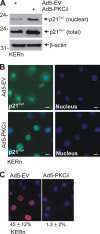
PKCδ affects p21Cip1 nuclear level. Keratinocytes were infected with 15 MOI of Ad5-EV or Ad5-PKCδ adenovirus. A, at 24 h after Ad5-PKCδ infection, total and nuclear extracts were prepared to detect p21Cip1. We electrophoresed four times more total than nuclear extract (based on cell equivalents). B, at 24 h post-infection, the cells were fixed and stained with anti-p21Cip1 (green) and Hoechst (nucleus, blue). The p21Cip1 panels are merged p21Cip1/Hoechst images. Cytoplasmic p21Cip1 staining was observed in 96 ± 3% of Ad5-EV and 1 ± 0.5% of Ad5-PKCδ-infected cells (mean ± S.D., n = 300). C, At 18 h post-infection, the cells were treated with 10 μm BrdUrd for 6 h. The cells were then fixed and stained with anti-BrdU, and we counted the percentage of BrdU-positive nuclei. The values are percentages of BrdU-positive nuclei presented as the means ± S.D. (n = 300).
FIGURE 4.
FIGURE 4.
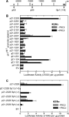
PKCδ activation of p21Cip1 promoter involves multiple elements. A, schematic of p21Cip1 promoter upstream regulatory region showing the p53 and GC-rich (Sp1) (nucleotides −120/−50) response elements (25). The distances are in nucleotides relative to the transcription start site. B, keratinocytes were transfected with 0.5 μg of each p21Cip1 promoter luciferase reporter plasmid in the presence of 0.5 μg of PKCδ or empty (−PKCδ) expression vector. After 24 h, the cells were harvested, and extracts were assayed for luciferase activity. C, keratinocytes were transfected, incubated, and assayed for luciferase activity as above. The respective plasmid names identify the mutated Sp1 sites (e.g. Δ1, Δ2, Δ1–6, etc.). We observed similar results in each of four experiments.
FIGURE 5.
FIGURE 5.
PKCδ expression increases KLF4 expression. A, keratinocytes were infected with 15 MOI of Ad5-EV or Ad5-PKCδ, and after 24 h, extracts were prepared for detection of the indicated transcription factors/co-factors and KLF4 mRNA. B, the KLF4 mRNA level was assessed by quantitative RT-PCR. The values are the means ± S.D. We observed similar results in three separate experiments.
FIGURE 6.
FIGURE 6.
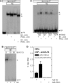
KLF4 interacts with GC-rich elements in the p21Cip1 proximal promoter. Keratinocytes were infected with 15 MOI Ad5-EV or Ad5-PKCδ. A, after 24 or 48 h, nuclear extracts were prepared, incubated with Sp1(1/2)-32P, and electrophoresed on a 4% acrylamide nondenaturing gel. The short arrow indicates the major band, and the long arrow indicates the fast mobility band observed in cells expressing elevated PKCδ levels. B, control extracts was prepared as above and incubated with Sp1(1/2)-32P in the presence of the indicated molar excess of radioinert probe. The arrow indicates band migration. C, extracts prepared as above were incubated with IgG, Sp1, Sp3, and KLF4 specific antibodies for 3 h at 4 °C, for 40 min with Sp1(1/2)-32P and then electrophoresed. D, ChIP assay was performed as described under “Experimental Procedures” using p21Cip1 promoter-derived PCR primers encoding the indicated range of nucleotides. NE, nuclear extract; EV, empty vector; FP, free probe.
FIGURE 7.
FIGURE 7.
KLF4 increases p21Cip1 promoter activity. A, keratinocytes were transfected with 0.5 μg of indicated luciferase reporter plasmid in the presence of 0.5 μg of hKLF4 expression vector or empty pcDNA3 vector (open box). At 24 h, the cells were harvested, and extracts were assayed for luciferase activity. To monitor KLF4 levels, extracts were prepared at 24 h post-transfection, and hKLF4 was detected by immunoblot. To compare the level of KLF4 in KLF4- and PKCδ-overexpressing cells, keratinocytes were infected with 15 MOI of the indicated virus or transfected with 0.5 μg of hKLF4 expression plasmid, and after 24 h, extracts were prepared for KLF4 immunoblot. B, keratinocytes were transfected with 0.5 μg of luciferase reporter, 0.5 μg of hKLF4(1–470) expression plasmid, and 0.5 μg of PKCδ expression vector. At 24 h, cell extracts were prepared for luciferase assay. C, keratinocytes were electroporated with 3 μg of the indicated siRNA and at 48 h transfected with 0.5 μg of luciferase reporter plasmid. After an additional 24 h, extracts were prepared for luciferase activity assay and immunoblot.
FIGURE 8.
FIGURE 8.
A, schematic of p21Cip1promoter upstream regulatory region. B, KLF4 regulates the p21Cip1 promoter via the proximal GC-rich elements. Keratinocytes were transfected with 0.5 μg of the indicated luciferase reporter plasmid in the presence or absence of 0.5 μg of hKLF4 expression plasmid, and at 24 h post-transfection, extracts were prepared for luciferase assay. The values are the means ± S.D. Similar results were observed in three experiments. EV, empty vector.
FIGURE 9.
FIGURE 9.
KLF4 is required for PKCδ-dependent responses. A, keratinocytes were electroporated with 3 μg of control or hKLF4-specific siRNA and after 48 h were infected with 15 MOI of Ad5-PKCδ. After an additional 24 h, extracts were prepared for detection of PKCδ and hKLF4. B, keratinocytes were electroporated with siRNA for 48 h and infected with virus for 24 h as above. At 72 h, the cells were harvested and counted. The open bar indicates cells counted on day 0 at the beginning of treatment. C, keratinocytes were electroporated with the indicated siRNA and after 48 h were infected with virus for 24 h. The cells were then harvested for assay of p21Cip1 mRNA level. The bars indicate the means ± S.D. Similar results were observed in each of two experiments.
FIGURE 10.
FIGURE 10.
PKCδ regulation of KLF4 and p21Cip1 protein level. Keratinocytes were electroporated with 3 μg of control or PKCδ-specific siRNA and after 48 h were infected with 15 MOI of tAd5-EV or tAd5-hKLF4 in the presence of 2.5 MOI of Ad5-TA. After an additional 24 h, extracts were prepared for detection of PKCδ, KLF4, and p21Cip1. Similar results were observed in two separate experiments.
FIGURE 11.
FIGURE 11.
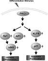
PKCδ coordinate regulation of keratinocytes differentiation and proliferation. PKCδ initiates a cascade the coordinately increases keratinocyte differentiation and suppresses keratinocyte proliferation. Active PKCδ increases AP1 and Sp1 transcription factor level and nuclear accumulation. These factors bind to the distal regulatory region of the hINV promoter to drive transcription and increase involucrin expression (i.e. differentiation) (29, 66). Active PKCδ also increases KLF4 level and binding to GC-rich elements in the p21Cip1 proximal promoter where it acts to increase p21Cip1 expression to suppress proliferation.

Comment in

  • Findings of Research Misconduct.
    [No authors listed] [No authors listed] Fed Regist. 2024 Aug 15;89(158):66420-66422. Fed Regist. 2024. PMID: 39161428 Free PMC article. No abstract available.

References

    1. Newton A. C. (1997) Curr. Opin. Cell Biol. 9, 161–167 - PubMed
    1. Nishizuka Y. (1992) Science 258, 607–614 - PubMed
    1. Rosse C., Linch M., Kermorgant S., Cameron A. J., Boeckeler K., Parker P. J. (2010) Nat. Rev. Mol. Cell Biol. 11, 103–112 - PubMed
    1. Gherzi R., Sparatore B., Patrone M., Sciutto A., Briata P. (1992) Biochem. Biophys. Res. Commun. 184, 283–291 - PubMed
    1. Matsui M. S., Chew S. L., DeLeo V. A. (1992) J. Invest. Dermatol. 99, 565–571 - PubMed

Publication types

MeSH terms

Substances

LinkOut - more resources