. 2011 Jun;8(3):223-72.
doi: 10.1513/pats.201012-071DW.

Stem cells and cell therapies in lung biology and lung diseases

Affiliations

Stem cells and cell therapies in lung biology and lung diseases

Daniel J Weiss et al. Proc Am Thorac Soc. 2011 Jun.

Abstract

The University of Vermont College of Medicine and the Vermont Lung Center, with support of the National Heart, Lung, and Blood Institute (NHLBI), the Alpha-1 Foundation, the American Thoracic Society, the Emory Center for Respiratory Health,the Lymphangioleiomyomatosis (LAM) Treatment Alliance,and the Pulmonary Fibrosis Foundation, convened a workshop,‘‘Stem Cells and Cell Therapies in Lung Biology and Lung Diseases,’’ held July 26-29, 2009 at the University of Vermont,to review the current understanding of the role of stem and progenitor cells in lung repair after injury and to review the current status of cell therapy approaches for lung diseases. These are rapidly expanding areas of study that provide further insight into and challenge traditional views of the mechanisms of lung repair after injury and pathogenesis of several lung diseases. The goals of the conference were to summarize the current state of the field, discuss and debate current controversies, and identify future research directions and opportunities for both basic and translational research in cell-based therapies for lung diseases.


Figures

Figure 1.
Figure 1.
A graphic representation of putative stem cell niches in the airway epithelium. (1) Basal cells in the gland duct; (2) surface basal cells typically present in the intercartilaginous zone; (3) variant Clara cells associated with pulmonary neuroendocrine cell (PNEC) bodies; (4) variant Clara cells present at the bronchiolar–alveolar duct junction. See text for further explanation. Reproduced with permission from Reference 49.
Figure 2.
Figure 2.
Endogenous bone marrow Ccsp+ cells can repopulate the airway epithelium. (A–K) Representative double immunofluorescence staining for GFP (green) and Ccsp (red) of lung sections from bone marrow transplant recipient mice that received Ccsp+Sca-1+ cells from GFP+ donors. Asterisks indicate a GFP+ cell that was Ccsp–. White arrows point to donor-derived Ccsp+ cells. Red arrows point to Ccsp+ cells that were not donor derived. (A) GFP+ (positive control) lung. (B) Isotype staining of lung from bone marrow transplant recipient. (C–E) Bone marrow transplant recipients had a wild-type background. (C) Low-power image. (D and E) High-power images. (F–H) Bone marrow transplant recipients were Ccsp–/– 60 days following bone marrow transplant without naphthalene. (F) Low-power image. (G and H) High-power image. (I and K) Thirty days after bone marrow transplant with naphthalene injury. (I) Low-power image. (J and K) High-power image. Scale bars: 20 μm. Original magnification: ×60 (A, B, C, F, and I); ×90 (D, H, and J); ×120 (E, G, K). Reprinted with permission from Reference 201.
Figure 3.
Figure 3.
Schematics depicting current thinking in isolation and identification of endothelial progenitor cells. Reprinted with permission from Reference 19.
Figure 4.
Figure 4.
Schematic illustrating the range of in vitro immune-modulating effects described for mesenchymal stem cells (MSCs). DC = dendritic cell; HGF = hepatocyte growth factor; IDO = indoleamine 2,3-dioxygenase; IFN-γ = interferon γ; Ig = Immunoglobulin; IL = Interleukin; IL-1RA = Interleukin-1 receptor antagonist; Mac = Macrophage; NK = natural killer; PGE2 = prostaglandin E-2; SDF-1 = stem-cell derived factor 1; TNF-α = tumor necrosis factor-α; TGF-β1 = transforming growth factor- β1; TLR = Toll-like receptor; VEGF = vascular endothelial growth factor. Reprinted with permission from Reference 412.
Figure 5.
Figure 5.
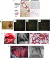
(A) Massome's trichrome and pentachrome staining of extracellular matrix proteins collagen and elastin in native and de-cellularized mouse lung. Reprinted with permission from Reference 497. (B, A) Gross image of AC rat lung (left) next to mESC-recellularized lung (right) after culture for 14 days showing contraction of the ECM. (B–D) Two-photon imaging: three-dimensional reconstructions of (B) normal fresh rat lung tissue, (C) AC lung, and (D) recellularized rat lung tissue. Green color corresponds to SHG showing collagen and red to autofluorescence of cells, elastin, and other ECM. (D) Recellularized lung tissue imaged at a depth of 22 mm. Reprinted with permission from Reference 498. (C, A) Tissue-engineered left lung was implanted into adult Fischer 344 rat recipient and photographed approximately 30 minutes later. (B) X-ray image of rat showing the implanted engineered left lung (white arrow) and the right native lung. (C) Hematoxylin and eosin stain of explanted lung. Red blood cells perfusing septa are evident, and some red blood cells are present in airspaces. Scale bar = 50 μm. Reprinted with permission from Reference 499. (D, a) Photograph of left rat chest after left anterior thoracotomy, left pneumonectomy, and orthotopic transplantation of a regenerated left lung construct. Recipient's left pulmonary artery (A), left main bronchus (B), and left pulmonary vein (V) are connected to regenerated left lung pulmonary artery (a), bronchus (b), and pulmonary vein (v). White arrowheads, the recipient's right lung (infracardiac and right lower lobe); black arrowheads, the regenerated left lung construct. (D, b) Radiograph of rat chest after left pneumonectomy and orthotopic transplantation of a regenerated left lung construct. White arrowheads, recipient's right lung; black arrowheads, regenerated left lung construct. Reprinted with permission from Reference 500.
Figure 6.
Figure 6.
Schematic of protocol used to differentiate esc into cells with phenotypic characteristics of type 2 alveolar epithelial cells (537). Embryonic stem cells (ESCs) can be effectively manipulated in vitro to differentiate into type 2 alveolar epithelial cells using lung development cell signaling pathway to guide ESCs differentiation. FGF-2 = Fibroblast growth factors-2, Pro Spc = Pro-surfactant protein C. Adapted with permission from Reference 537.
Figure 7.
Figure 7.
Derivation of transgene-free iPS cell line from tail tip of a CFTR knockout (KO) mouse. (A) The human floxed STEMCCA reprogramming vector. (B–D) generation of iPS cells by reprogramming human dermal fibroblasts, and their characterization by RT-PCR and immunostaining. (E) Southern blot of iPS cell clones generated from dermal fibroblasts from CF (DF508 CFTR) and α1-antitrypsin deficient (PiZZ) volunteers. The Southern blot shows high frequency of single copy STEMCCA integrants in the resulting clones prior to Cre-mediated excision of this vector to generate human transgene-free iPS cells. Adapted with permission from References and .
Figure 8.
Figure 8.
Schematic of challenges and issue for lung bioengineering. Slide courtesy of Dame Julia Polak DBE, MD, DSc.

References

    1. Weiss DJ, Berberich MA, Borok Z, Gail DB, Kolls JK, Penland C, Prockop DJ. Adult stem cells, lung biology, and lung disease. NHLBI/Cystic Fibrosis Foundation Workshop. Proc Am Thorac Soc 2006;3:193–207. - PubMed
    1. Weiss DJ, Kolls JK, Ortiz LA, Panoskaltis-Mortari A, Prockop DJ. Stem Cells and Cell Therapy Approaches for Lung Diseases. Conference Report. Proc Am Thorac Soc 2008;5:637–667. - PMC - PubMed
    1. Brown JK, Hogan BLM, Randell SH, Stripp B., Weiss DJ. Human embryonic stem cell research: An Official ATS Research Policy Statement. Am J Respir Crit Care Med 2006;173:1–3.
    1. Bellini A, Mattoli S. The role of the fibrocyte, a bone marrow-derived mesenchymal progenitor, in reactive and reparative fibroses. Lab Invest 2007;87:858–870. - PubMed
    1. Flotte TR, Ng P, Dylla DE, McCray PB Jr, Wang G, Kolls JK, Hu J. Viral vector-mediated and cell-based therapies for treatment of cystic fibrosis. Mol Ther 2007;15:229–241. - PubMed

Publication types

LinkOut - more resources