. 2011 Jul;134(Pt 7):1914-24.
doi: 10.1093/brain/awr128. Epub 2011 Jun 7.

Oxidative damage in multiple sclerosis lesions

Affiliations

Oxidative damage in multiple sclerosis lesions

Lukas Haider et al. Brain. 2011 Jul.

Abstract

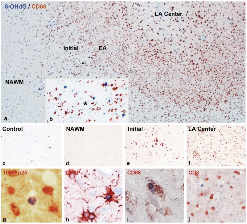
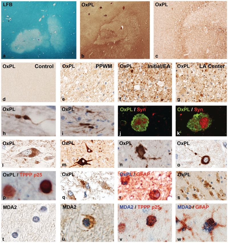
Multiple sclerosis is a chronic inflammatory disease of the central nervous system, associated with demyelination and neurodegeneration. The mechanisms of tissue injury are currently poorly understood, but recent data suggest that mitochondrial injury may play an important role in this process. Since mitochondrial injury can be triggered by reactive oxygen and nitric oxide species, we analysed by immunocytochemistry the presence and cellular location of oxidized lipids and oxidized DNA in lesions and in normal-appearing white matter of 30 patients with multiple sclerosis and 24 control patients without neurological disease or brain lesions. As reported before in biochemical studies, oxidized lipids and DNA were highly enriched in active multiple sclerosis plaques, predominantly in areas that are defined as initial or 'prephagocytic' lesions. Oxidized DNA was mainly seen in oligodendrocyte nuclei, which in part showed signs of apoptosis. In addition, a small number of reactive astrocytes revealed nuclear expression of 8-hydroxy-d-guanosine. Similarly, lipid peroxidation-derived structures (malondialdehyde and oxidized phospholipid epitopes) were seen in the cytoplasm of oligodendrocytes and some astrocytes. In addition, oxidized phospholipids were massively accumulated in a fraction of axonal spheroids with disturbed fast axonal transport as well as in neurons within grey matter lesions. Neurons stained for oxidized phospholipids frequently revealed signs of degeneration with fragmentation of their dendritic processes. The extent of lipid and DNA oxidation correlated significantly with inflammation, determined by the number of CD3 positive T cells and human leucocyte antigen-D expressing macrophages and microglia in the lesions. Our data suggest profound oxidative injury of oligodendrocytes and neurons to be associated with active demyelination and axonal or neuronal injury in multiple sclerosis.


Figures

Figure 1
Figure 1
Oxidized DNA in multiple sclerosis lesions, visualized by immunocytochemistry for 8-hydroxy-d-guanosine (8-OHdG). (a) Actively demyelinating lesion in a patient with primary progressive multiple sclerosis; the figure shows areas of the normal-appearing white matter (NAWM) (left), the zone of ongoing demyelinating activity with profound microglia activation and macrophage infiltration (brown immunoreactivity for the macrophage marker CD68) and the inactive portion of the lesion with low macrophage infiltration. Oxidized DNA (8-OHdG) reactivity is shown as blue nuclei, which are mainly present in the active lesion edge (×60). EA = early active demyelination with macrophages containing myelin-reactive degradation products; initial = initial stage of demyelination with profound microglia activation and oligodendrocyte apoptosis; LA centre = late active stage of demyelination in lesions centre; macrophages with neutral lipid degradation products. (b) Higher magnification of the ‘initial’ stage of the active plaque in a, shows profound microglia activation (brown cells; CD68 positive) and many blue nuclei, representing cells with DNA oxidation; the asterisks in a and b indicate the position of b within the lesion shown in a (×200). (c) Normal white matter of a control patient without neurological disease and brain lesions; only very few microglia cells show cytoplasmic CD68 reactivity (brown); there are no blue nuclei, suggesting DNA oxidation (×100). (d) Normal-appearing white matter (NAWM) of a patient with acute multiple sclerosis (Marburg’s type); similar pattern as in the control white matter, shown in c (×100). (e) Very early active lesion area in a patient with acute multiple sclerosis (Marburg’s type); profound microglia activation (brown cells; CD68) adjacent to cell nuclei, positive for 8-hydroxy-d-guanosine (blue) (×100). (f) Late active lesion centre in a patient with Marburg’s type of acute multiple sclerosis; numerous CD68-positive macrophages (brown), but no nuclei with blue 8-hydroxy-d-guanosine reactivity (×100). (g) Double staining for oxidized DNA (8-hydroxy-d-guanosine; blue) and the oligodendrocyte marker TPPP-p25 (red) at the lesion edge of an active lesion of acute multiple sclerosis. One of the six oligodendrocytes shows blue 8-hydroxy-d-guanosine reactivity in a nucleus with nuclear condensation and fragmentation (apoptotic cell) (×900). (h) Double staining for oxidized DNA (8-hydroxy-D-guanosine, blue) and glial fibrillary acidic protein (GFAP, red) at the edge of an active lesion of acute multiple sclerosis. One astrocyte contains a blue nucleus, indicating oxidized DNA (×600). (i) Double staining for oxidized DNA (8-hydroxy-d-guanosine, blue) and the macrophage marker CD68 (red) in an active lesion of acute multiple sclerosis. The macrophage contains a nucleus with oxidized DNA, possibly representing phagocytosis of an apoptotic cell (×600). (j) Double staining for oxidized DNA (8-hydroxy-d-guanosine, blue) and the T-cell marker CD3 (red) in an active lesion of acute multiple sclerosis. No DNA oxidation is seen in the CD3-positive T-cell population (×200).
Figure 2
Figure 2
Oxidized lipids in multiple sclerosis lesions. (a) Myelin loss in an active white matter lesion in acute multiple sclerosis, Marburg’s type (×8). (b) Adjacent serial section from a, stained with the marker E06 for oxidized phospholipids (OxPL) with profound immunoreactivity at the lesion edge and the adjacent peri-plaque white matter (×8). (c) Higher magnification of the sections shown in b, showing increased immunoreactivity for oxidized phospholipids at the lesion edge (×45). (d) Normal white matter of a control patient without neurological disease or CNS lesions, without immunoreactivity for oxidized phospholipids (×45). (e) Peri-plaque white matter (PPWM) from the area shown in b, with increased immunoreactivity, in comparison with the control white matter, but much lower reactivity as at the active lesion edge. Immunoreactivity is mostly associated with myelin (×150). (f) High immunoreactivity for oxidized phospholipids in myelin sheaths and some globular structures, representing axonal spheroids, are present at the edge of the active lesions shown in b (×150). (g) Inactive lesion centre of the same lesion shown in b, with low immunoreactivity for oxidized phospholipids; intensive immunoreactivity, however, is seen in some axonal spheroids (×150). (h–k) Oxidized phospholipids are selectively accumulated within dystrophic axons and axonal spheroids (h, i); the nature of dystrophic axons is documented by double staining confocal microscopy for oxidized phospholipids and synaptophysin (Syn: j and k) (h, i: ×900; j, k: ×2500). (l–o) Accumulation of oxidized phospholipids in neurons in patients with multiple sclerosis with active lesions: (l) lipofuscin reactivity in cortical neuron; (m) ballooned cortical neuron adjacent to destructive subcortical lesion with intense immunoreactivity for oxidized phospholipids; (n, o) neurons with intense cytoplasmic reactivity for oxidized phospholipids with fragmented cell processes (dendrites) in active lesions in the basal ganglia (h) and cortex (o) (×600). (p) Active multiple sclerosis lesions; double staining for the oligodendrocyte marker TPPP-p25 (red) and (black/brown), showing one of the two oligodendrocytes with intense cytoplasmic E06 immunoreactivity (×900). (q, r) In astrocytes, oxidized phospholipids are sequestered in the cytoplasm in the form of larger granules, possibly representing autophagic vacuoles (q: ×450; r: ×750). (s) Macrophages with granular cytoplasmic reactivity for oxidized phospholipids in the centre of an active multiple sclerosis plaque (×150). (t) Normal white matter of a control patient without neurological disease or brain lesions; no MDA2 immunoreactivity (×900). (u, v) MDA2 immunoreactivity in oligodendrocytes and myelin in active multiple sclerosis lesions; the nature of the cells as oligodendrocytes is shown by double staining in v, using TPPP-p25 as a marker (×900). (w) Granular cytoplasmic MDA2 immunoreactivity (blue) in the cytoplasm of a glial fibrillary acidic protein (GFAP)-positive astrocyte (red) in an active multiple sclerosis lesion (×800). LFB = Luxol fast blue.

References

    1. Aboul-Enein F, Rauschka H, Kornek B, Stadelmann C, Stefferl A, Brück W, et al. Preferential loss of myelin associated glycoprotein reflects hypoxia-like white matter damage in stroke and inflammatory brain diseases. J Neuropath Exp Neurol. 2003;62:25–33. - PubMed
    1. Bagasra O, Michaels FH, Zheng YM, Bobroski LE, Spitsin SV, Fu ZF, et al. Activation of the inducible form of nitric oxide synthase in the brains of patients with multiple sclerosis. Proc Natl Acad Sci USA. 1995;92:12041–5. - PMC - PubMed
    1. Barnett MH, Prineas JW. Relapsing and remitting multiple sclerosis: pathology of the newly forming lesion. Ann Neurol. 2004;55:458–68. - PubMed
    1. Bauer J, Elger CE, Hans VH, Schramm J, Urbach H, Lassmann H, et al. Astrocytes are a specific immunological target in Rasmussen's encephalitis. Ann Neurol. 2007;62:67–80. - PubMed
    1. Bizzozero OA, Dejesus G, Callaha K, Pastuszyn A. Elevated protein carbonylation in the brain white matter and grey matter of patients with multiple sclerosis. J Neurosci Res. 2005;81:687–95. - PubMed

Publication types

MeSH terms