. 2011 Nov;301(5):F1131-8.
doi: 10.1152/ajprenal.00589.2010. Epub 2011 Jun 8.

Establishment of conditionally immortalized human glomerular mesangial cells in culture, with unique migratory properties

Affiliations

Establishment of conditionally immortalized human glomerular mesangial cells in culture, with unique migratory properties

Ramadan M Sarrab et al. Am J Physiol Renal Physiol. 2011 Nov.

Abstract

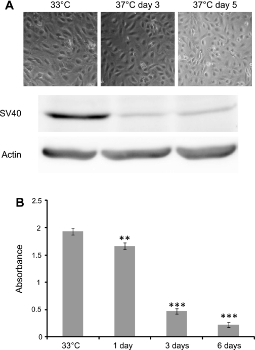
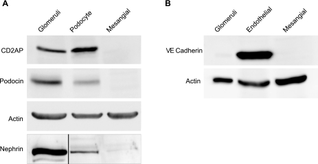
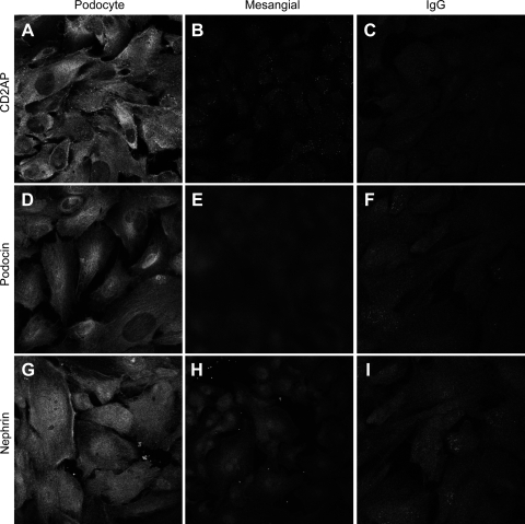
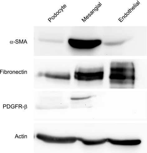
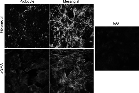
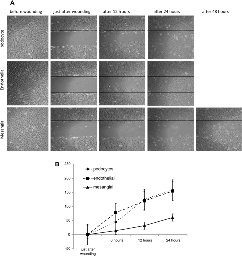
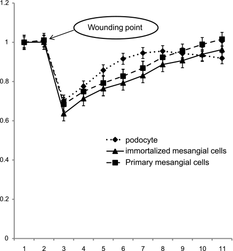
The aim of this study was to establish an immortalized human mesangial cell line similar to mesangial cells in vivo for use as a tool for understanding glomerular cell function. Mesangial cells were isolated from glomerular outgrowths from a normal human kidney, then retrovirally transfected with a temperature-sensitive SV40T antigen+human telomerase (hTERT). Mesangial cells exhibited features of compact cells with small bodies in a confluent monolayer at 33°C, but the cell shape changed to flat and stellate after 5 days in growth-restrictive conditions (37°C). Western blot and immunofluorescence analysis showed that podocyte markers (nephrin, CD2AP, podocin, Wilms' tumor-1) and an endothelial-specific molecule (VE-cadherin) were not detectable in this cell line, whereas markers characteristic of mesangial cells (α-SMA, fibronectin, and PDGFβ-R) were strongly expressed. In migration assays, a significant reduction in wound surface was observed in podocyte and endothelial cells as soon as 12 h (75 and 62%, respectively) and complete wound closure after 24 h. In contrast, no significant change was observed in mesangial cells after 12 h, and even after 48 h the wounds were not completely closed. Until now, conditionally immortalized podocyte and endothelial cell lines derived from mice and humans have been described, and this has greatly boosted research on glomerular physiology and pathology. We have established the first conditionally immortalized human glomerular mesangial cell line, which will be an important adjunct in studies of representative glomerular cells, as well as in coculture studies. Unexpectedly, mesangial cells' ability to migrate seems to be slower than for other glomerular cells, suggesting this line will demonstrate functional properties distinct from previously available mesangial cell cultures. This conditionally immortalized human mesangial cell line represents a new tool for the study of human mesangial cell biology in vitro.


Figures

Fig. 1.
Fig. 1.
A: demonstration of expression of SV40 large T antigen in protein lysate extracted from putative mesangial cells (MCs) at permissive temperature (33°C) and days 3 and 5 at 37°C. Actin was used as a loading control. SV40 large T antigen protein was strongly expressed in mesangial cells at 33°C. In contrast, at 37°C either after 3 or 5 days the expression of this protein was very faint and barely detected. This indicates that the transgene was active at the permissive temperature and thermoswitching the cells to the temperature of 37°C almost completely inactivates the transgene. B: measurement of cell proliferation upon thermoswitching using a bromodeoxyuridine (BrdU) assay. This shows, as predicted, a steadily declining proliferation rate postthermoswitching to detectable but very low level proliferation at day 6. **,*** P < 0.05.
Fig. 2.
Fig. 2.
Western blot analysis of podocyte- and endothelial glomerular cell-specific proteins in cultured mesangial cells. A: expression of podocyte-specific proteins including podocin, CD2AP, and nephrin in podocytes and mesangial cells. Actin loading control is shown for podocin and CD2AP. Nephrin blotting was performed using nephrin immunopreciptation (black line indicates where irrelevant lanes have been removed). B: expression of VE-cadherin, an endothelial marker, in mesangial and endothelial cells. Actin was used as a loading control in A and B, and whole glomeruli as a positive control for all proteins except for CD2AP, where we used HEK cells as a positive control. Podocytes and not mesangial cells expressed CD2AP, podocin, and nephrin. VE-cadherin is expressed only in endothelial cells and not in mesangial cells. These results validate the staining results and provide further evidence of the establishment of a mesangial-specific cell line in culture.
Fig. 3.
Fig. 3.
Specific indirect immunofluorescence characterizes and distinguishes mesangial cells from podocytes. AI: putative mesangial cells and previously characterized podocytes were grown separately and stained with the same antibodies. CD2AP, podocin, and nephrin staining was abundant in podocytes along the cell border and in the cytoplasm (A, D, and G); these were absent in mesangial cells (B, E, and H). Normal mouse/rabbit IgG were used to rule out nonspecific staining (C, F, and I). These results demonstrate that the mesangial cells grown in culture are distinct from podocytes. Magnification: ×40 in both cell lines.
Fig. 4.
Fig. 4.
Expression of mesenchymal markers α-smooth muscle actin (SMA) and fibronectin and platelet-derived growth factor receptor (PDGFβ-R). The figure shows α-SMA and fibronectin were abundantly expressed in mesangial cells, whereas α-SMA expression in podocytes was low and was entirely absent in glomerular endothelial cells. Fibronectin was expressed in the glomerular endothelial cell as well and was weakly expressed in podocytes. Actin was used as a loading control. PDGFβ-R was expressed exclusively in mesangial cells.
Fig. 5.
Fig. 5.
Indirect immunofluorescence shows expression of the mesenchymal markers fibronectin and α-SMA in mesangial cells and podocytes. Both fibronectin and α-SMA were highly expressed in the mesangial cells whereas in podocytes were barely detectable. Mouse IgG was used to rule out nonspecific staining. Magnifications: ×40 in both cell lines.
Fig. 6.
Fig. 6.
A: wound healing assay. Podocytes, glomerular endothelial, and mesangial cells were seeded at confluence into 6-well culture plates and allowed to adhere overnight. Then, the medium was altered to 0.2% FCS to reduce cell proliferation. Two days afterward, wounds were made. Images from 2 marked fields/well were taken under phase-contrast examination, just before wounding, immediately after wounding (0), and after 12, 24, and 48 h postwounding. Wounds in podocytes and endothelial cells wells were completely closed after 24 h, and unexpectedly in mesangial cells the wound was not completely closed even after 48 h. B: proportion of cells invading the scratch-made wound at different time points (after 6, 12, and 24 h); unexpectedly, mesangial cells migrate more slowly than podocytes and endothelial cells.
Fig. 7.
Fig. 7.
Measurement of cell monolayer resistance after wounding using cell impedance analyzer (electric cell substrate impedance sensing; ECIS). The curve shows the resistance measurements of primary and cloned mesangial cells and podocyte monolayers postwounding using ECIS technology. Cells were grown in ECIS 8W1E+ plates, submitted to an elevated voltage pulse of 40-kHz frequency and 3.5-V amplitude for a 30-sec duration. This led to detachment and loss of cells located on the small active electrode, leading to a wound normally healed by cells around the small active electrode that have not been subjected to the elevated voltage pulse. Wound healing was then followed by continuous resistance measurements for 12 h. The curves show a sharp drop in resistance following the high-voltage pulse. Resistance in podocytes increased more quickly than in mesangial cells (primary and cloned) to approach the prewounding level at ∼6 and 12 h, respectively, postwounding.

References

    1. Alpers CE, Hudkins KL, Gown AM, Johnson RJ. Enhanced expression of “muscle-specific” actin in glomerulonephritis. Kidney Int 41: 1134–1142, 1992 - PubMed
    1. Alpers CE, Seifert RA, Hudkins KL, Johnson RJ, Bowen-Pope DF. Developmental patterns of PDGF B-chain, PDGF-receptor, and alpha-actin expression in human glomerulogenesis. Kidney Int 42: 390–399, 1992 - PubMed
    1. Alpers CE, Seifert RA, Hudkins KL, Johnson RJ, Bowen-Pope DF. PDGF-receptor localizes to mesangial, parietal epithelial, and interstitial cells in human and primate kidneys. Kidney Int 43: 286–294, 1993 - PubMed
    1. Dubus I, L'Azou B, Gordien M, Delmas Y, Labouyrie JP, Bonnet J, Combe C. Cytoskeletal reorganization by mycophenolic acid alters mesangial cell migration and contractility. Hypertension 42: 956–961, 2003 - PubMed
    1. Ekblom P. Formation of basement membranes in the embryonic kidney: an immunohistological study. J Cell Biol 91: 1–10, 1981 - PMC - PubMed

Publication types

MeSH terms