Comparative Study
. 2011 Aug 1;71(15):5090-100.
doi: 10.1158/0008-5472.CAN-11-0568. Epub 2011 Jun 8.

Tumor microenvironment-derived proteins dominate the plasma proteome response during breast cancer induction and progression

Affiliations
Comparative Study

Tumor microenvironment-derived proteins dominate the plasma proteome response during breast cancer induction and progression

Sharon J Pitteri et al. Cancer Res. .

Abstract

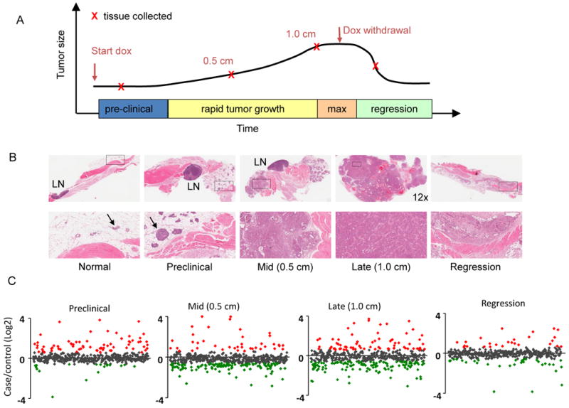
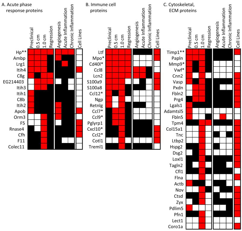
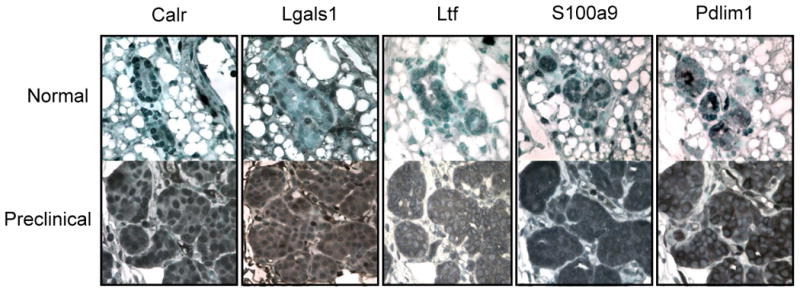
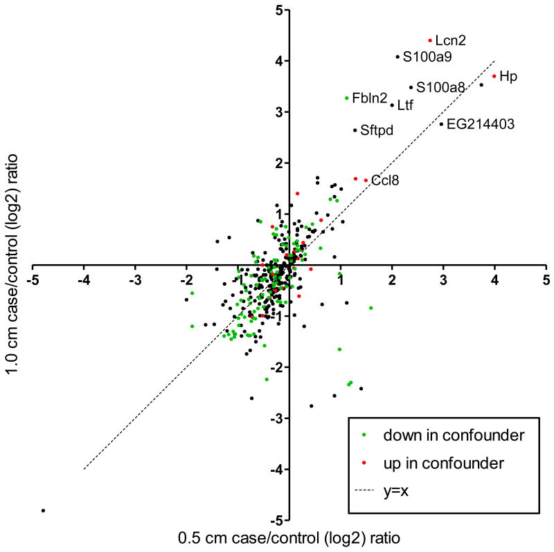
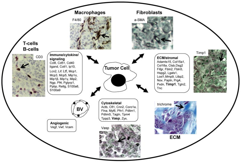
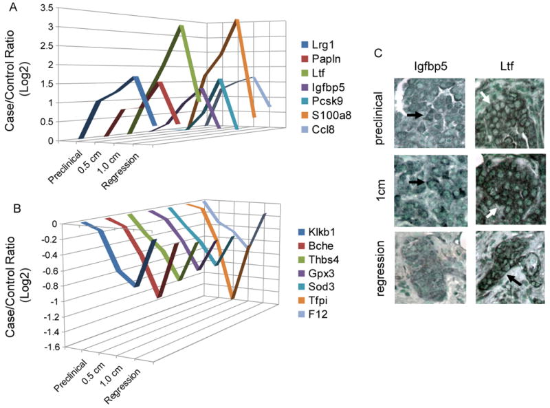
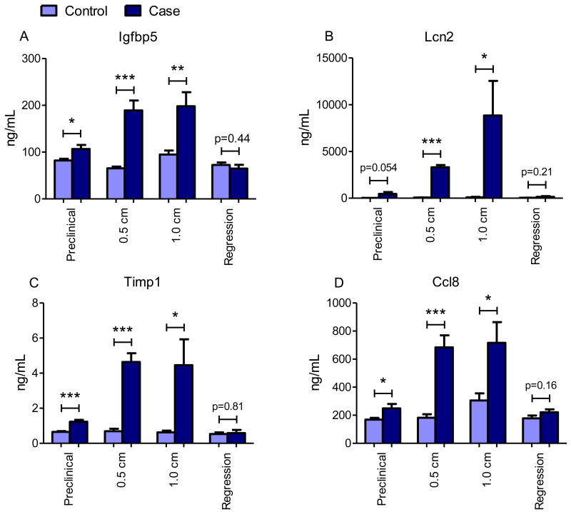
Tumor development relies upon essential contributions from the tumor microenvironment and host immune alterations. These contributions may inform the plasma proteome in a manner that could be exploited for cancer diagnosis and prognosis. In this study, we employed a systems biology approach to characterize the plasma proteome response in the inducible HER2/neu mouse model of breast cancer during tumor induction, progression, and regression. Mass spectrometry data derived from approximately 1.6 million spectra identified protein networks involved in wound healing, microenvironment, and metabolism that coordinately changed during tumor development. The observed alterations developed prior to cancer detection, increased progressively with tumor growth and reverted toward baseline with tumor regression. Gene expression and immunohistochemical analyses suggested that the cancer-associated plasma proteome was derived from transcriptional responses in the noncancerous host tissues as well as the developing tumor. The proteomic signature was distinct from a nonspecific response to inflammation. Overall, the developing tumor simultaneously engaged a number of innate physiologic processes, including wound repair, immune response, coagulation and complement cascades, tissue remodeling, and metabolic homeostasis that were all detectable in plasma. Our findings offer an integrated view of tumor development relevant to plasma-based strategies to detect and diagnose cancer.


Figures

Figure 1
Figure 1. Histologic and plasma proteomic analysis of HER2/neu mammary tumor progression
(A) Samples were collected from litter-matched case and control mice at indicated stages. (B) H&E staining of mammary tissue from bitransgenic case mice at the preclinical, 0.5 cm diameter, 1.0 cm diameter tumor stages and during tumor regression. Top row: 26× magnification unless otherwise indicated. Bottom row: 130× magnification of boxed sections from the top row. LN=lymph node. Arrows indicate breast epithelial cells within the mammary fat pads of normal and preclinical sections. (C) Case/control ratios of all quantified plasma proteins at all four tumor stages. Red indicates proteins increased >1.5 fold and green indicates proteins decreased >1.5 fold in plasma from case vs. control mice (p<0.05).
Figure 2
Figure 2. Wound healing response components: acute phase, immune cell, and extracellular matrix proteins
Plasma proteins that increased in tumor-bearing mice fell into major functional categories. (A) acute phase response proteins, mainly liver-derived, (B) proteins from tumor infiltrating immune cells (macrophages and neutrophils), and (C) proteins involved in cell movement and tumor microenvironment, e.g. cytoskeletal, extracellular matrix, and tumor stroma proteins. Red indicates case/control ratios >1.5 (p<0.05), black indicates no change between case and control plasma, and white indicates the proteins were not measured. For the breast cancer cell lines, red indicates the protein was identified by proteomic profiling and white indicates the protein was not identified. Unless otherwise indicated, levels were measured by LC-MS. *measured by Luminex/Rules Based Medicine. **measured by both LC-MS and Luminex/Rules Based Medicine.
Figure 3
Figure 3. IHC staining of selected proteins identified in plasma in normal breast and preclinical breast lesions
Calreticulin (Calr) shows some nuclear staining in normal breast epithelial and strong nuclear and cytoplasmic staining in preclinical tumor cells. Galectin-1 (Lgals1) shows some nuclear staining in normal and strong nuclear and cytoplasmic staining in preclinical tumor cells. Lactotransferrin (Ltf) shows some cytoplasmic staining in normal and strong cytoplasmic staining in tumor cells. Calgranulin B (S100a9) shows some nuclear staining in normal and strong nuclear and cytoplasmic staining in tumor cells. Pdz and lim domain protein 1 (Pdlim1) shows some cytoplasmic staining in normal and strong cytoplasmic staining in the tumor cells. 60× magnification.
Figure 4
Figure 4. Correlation of plasma protein abundance between tumor stages
Shown are case/control ratios for plasma proteins that were quantified in both 0.5 and 1.0 cm tumor stages. Green and red data points indicate proteins identified as increased or decreased in the plasma from the confounder models, respectively.
Figure 5
Figure 5. HER2/neu mammary tumor microenvironment schematic
In addition to the tumor cells, Her2/neu tumors contain cancer associated macrophages (F4/80), cancer associated fibroblasts (α-smooth muscle actin (aSMA)), lymphocytes (CD3), blood vessels (not shown), and extensive collagen deposition (trichrome). Also shown are IHC results of HER2/neu tumors for Timp1 and Vasp which were elevated in plasma from tumor-bearing mice. Additional proteins elevated in plasma from tumor-bearing mice are listed according to functional categories (boxes).
Figure 6
Figure 6. Dynamics of protein changes with tumor establishment, progression, and regression
(A) Proteins increased with tumor development and decreased after regression. (B) Proteins decreased with tumor development and increased after regression. (C) IHC revealed Ltf and Igfbp5 staining in tumor cells.
Figure 7
Figure 7. ELISA protein measurements in an independent mouse cohort
Plasma levels of (A) Insulin-like growth factor binding protein 5 (Igfbp5), (B) Lipocalin-2 (Lcn2), (C) Metallopeptidase inhibitor-1 (Timp1), and (D) C-C motif chemokine 8 (Ccl8) as determined by ELISA in a set of mouse plasmas independent of those used for LC-MS/MS experiments. For each time point n=6 controls and n=6 cases. Statistical significance was determined by two-tailed Student's t-test *p<0.05, **p<0.01, and ***p<0.0001.

References

    1. Polyak K, Kalluri R. The role of the microenvironment in mammary gland development and cancer. Cold Spring Harb Perspect Biol. 2010;2:a003244. - PMC - PubMed
    1. Kalluri R, Zeisberg M. Fibroblasts in cancer. Nat Rev Cancer. 2006;6:392–401. - PubMed
    1. Mueller MM, Fusenig NE. Friends or foes - bipolar effects of the tumour stroma in cancer. Nat Rev Cancer. 2004;4:839–49. - PubMed
    1. Grivennikov SI, Greten FR, Karin M. Immunity, inflammation, and cancer. Cell. 2010;140:883–99. - PMC - PubMed
    1. de Visser KE, Eichten A, Coussens LM. Paradoxical roles of the immune system during cancer development. Nat Rev Cancer. 2006;6:24–37. - PubMed

Publication types

MeSH terms