. 2011 Aug 12;286(32):28138-49.
doi: 10.1074/jbc.M110.204099. Epub 2011 Jun 8.

G Protein binding sites on Calnuc (nucleobindin 1) and NUCB2 (nucleobindin 2) define a new class of G(alpha)i-regulatory motifs

Affiliations

G Protein binding sites on Calnuc (nucleobindin 1) and NUCB2 (nucleobindin 2) define a new class of G(alpha)i-regulatory motifs

Mikel Garcia-Marcos et al. J Biol Chem. .

Abstract

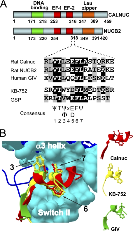
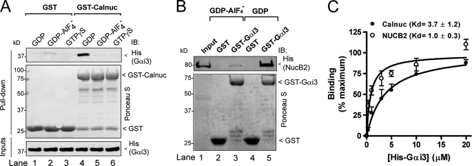
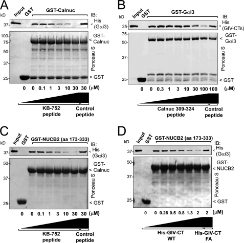
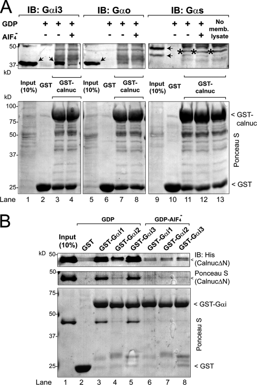
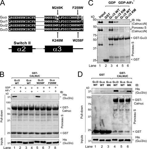
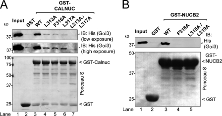
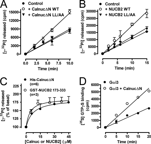
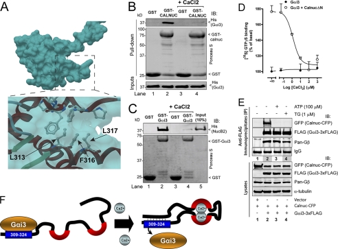
Heterotrimeric G proteins are molecular switches modulated by families of structurally and functionally related regulators. GIV (Gα-interacting vesicle-associated protein) is the first non-receptor guanine nucleotide exchange factor (GEF) that activates Gα(i) subunits via a defined, evolutionarily conserved motif. Here we found that Calnuc and NUCB2, two highly homologous calcium-binding proteins, share a common motif with GIV for Gα(i) binding and activation. Bioinformatics searches and structural analysis revealed that Calnuc and NUCB2 possess an evolutionarily conserved motif with sequence and structural similarity to the GEF sequence of GIV. Using in vitro pulldown and competition assays, we demonstrate that this motif binds preferentially to the inactive conformation of Gα(i1) and Gα(i3) over other Gα subunits and, like GIV, docks onto the α3/switch II cleft. Calnuc binding was impaired when Lys-248 in the α3 helix of Gα(i3) was replaced with M, the corresponding residue in Gα(o), which does not bind to Calnuc. Moreover, mutation of hydrophobic residues in the conserved motif predicted to dock on the α3/switch II cleft of Gα(i3) impaired the ability of Calnuc and NUCB2 to bind and activate Gα(i3) in vitro. We also provide evidence that calcium binding to Calnuc and NUCB2 abolishes their interaction with Gα(i3) in vitro and in cells, probably by inducing a conformational change that renders the Gα(i)-binding residues inaccessible. Taken together, our results identify a new type of Gα(i)-regulatory motif named the GBA motif (for Gα-binding and -activating motif), which is conserved across different proteins throughout evolution. These findings provide the structural basis for the properties of Calnuc and NUCB2 binding to Gα subunits and its regulation by calcium ions.


Figures

FIGURE 1.
FIGURE 1.
Identification of a putative Gα-binding motif in Calnuc and NUCB2. A, a sequence at the end of the Calnuc (aa 309–324) and NUCB2 (aa 311–326) second EF-hand shows a similarity to the GEF motif of GIV (aa 1678–1693) and the synthetic KB-752 and GSP peptides. Rat Calnuc, rat NUCB2, and human GIV sequences were obtained from the NCBI and aligned with the KB-752 (15) and GSP (33) sequences using ClustalW. Conserved identical residues are shaded in black and similar residues in gray. A consensus sequence of 7 aa based on this alignment and the phylogenetic analysis of Calnuc (Fig. S1) and GIV (14) is indicated. B, the Calnuc putative Gαi-binding motif has a structural similarity to the KB-752 peptide and the GEF motif of GIV. The coordinates of the Calnuc putative Gα-binding sequence (aa 309–320 (red)) were extracted from the Protein Data Bank (ID code: 1SNL) and the coordinates for the GEF motif of GIV (aa 1678–1689 (green)) from a previously described homology model (14). Both were threaded over the structure of KB-752 (yellow) in complex with Gαi1 (blue) (Protein Data Bank ID code: 1Y3A) using ICM-Browser-Pro. The Calnuc putative Gαi-binding motif, the GEF motif of GIV, and KB-752 form a helix in which the side chains of hydrophobic residues corresponding to positions 3, 6, and 7 from the consensus sequence depicted in A dock onto the α3/SwII hydrophobic cleft of the Gαi subunits (cyan surface).
FIGURE 2.
FIGURE 2.
Calnuc and NUCB2 bind inactive but not active Gαi3. A, His-Gαi3·GDP (lane 4) but not His-Gαi3·GDP·AlF4 (lane 5) or His-Gαi3·GTPγS (lane 6) binds to GST-Calnuc. No binding of Gαi3 to GST was detected under any of the conditions tested (lanes 1–3). 6 μg of His-Gαi3 preloaded with GDP (lanes 1 and 4), GDP and AlF4 (lanes 2 and 5), or GTPγS (30 μm, lanes 3 and 6) were incubated with ∼20 μg of purified GST (lanes 1–3) or GST-Calnuc (lanes 4–6) immobilized on glutathione beads. After extensive washing, bound proteins were separated by SDS-PAGE and analyzed by immunoblotting (IB) for His. Equal loading of GST proteins was confirmed by Ponceau S staining (middle panel), and equal loading of His-Gαi3 by His immunoblotting (lower panel). B, GST-Gαi3·GDP (lane 5) but not GST-Gαi3·GDP·AlF4 (lane 3) or GST (lanes 2 and 4) binds His-NUCB2. 10 μg of His-NUCB2 was incubated with ∼15 μg of purified GST (lanes 2 and 4) or GST-Gαi3 (lanes 3 and 5) preloaded with GDP (lanes 4 and 5) or GDP·AlF4 (lanes 2 and 3) immobilized on glutathione beads and analyzed as described in A. Input (lane 1), 1 μg of His-NUCB2. C, His-Gαi3·GDP binds to GST-Calnuc and GST-NUCB2-(173–333) with a Kd of 3.7 ± 1.2 μm (n = 4) and 1.0 ± 0.3 μm (n = 3), respectively. Increasing concentrations of His-Gαi3·GDP (0.3, 0.5, 1, 1.5, 3, 5, 10, and 20 μm) were incubated with 20 μg of GST-Calnuc (closed circles) or GST-NUCB2 (open circles) and analyzed as in A. His-Gαi3 binding was determined by quantitative immunoblotting using an Odyssey infrared imaging system, and data were fitted to a nonlinear, one-site binding hyperbola (solid lines) using Prism 4.0.
FIGURE 3.
FIGURE 3.
Calnuc and NUCB2 compete with KB-752 and GIV for binding to inactive Gαi3. A, the KB-752 peptide but not a control peptide (see “Experimental Procedures”) competes with GST-Calnuc for binding to His-Gαi3·GDP. Increasing concentrations of the KB-752 peptide (0, 0.1, 1, 3, 10, and 30 μm) or control peptide (30 μm) were incubated with 20 μg (∼0.9 μm) of GST-Calnuc in the presence of 6 μg (∼0.5 μm) of His-Gαi3 preloaded with GDP and analyzed as described in the legend for Fig. 2A. No binding of His-Gαi3·GDP GST was detected. Input, 0.2 μg of His-Gαi3·GDP. IB, immunoblot. B, a peptide corresponding to the Gα-binding motif of Calnuc 309–324 peptide, but not a control peptide, competes with His-GIV-CTs (aa 1660–1870, containing the GIV GEF motif) for binding to GST-Gαi3·GDP. Increasing concentrations of Calnuc 309–324 peptide (0, 0.3, 1, 3, 10, 30, and 100 μm) or a control peptide (100 μm) were incubated with 3 μg (∼250 nm) of GST-Gαi3 preloaded with GDP immobilized on glutathione beads in the presence of 2 μg (∼250 nm) of His-GIV-CTs and analyzed as described in the legend for Fig. 2A. No binding of His-GIV-CTs to GST is detected. Input, 0.1 μg of His-GIV-CTs. C, the KB-752 peptide but not a control peptide (see “Experimental Procedures”) competes with GST-NUCB2 for binding to His-Gαi3·GDP. Increasing concentrations of the KB-752 peptide (0, 0.1, 1, 3, 10, and 30 μm) or the control peptide (30 μm) were incubated with 20 μg (∼1.7 μm) of GST-NUCB2 in the presence of 6 μg (∼0.5 μm) of His-Gαi3 preloaded with GDP and analyzed as described in the legend for Fig. 2A. No binding of His-Gαi3·GDP to GST is detected. Input, 0.2 μg of His-Gαi3·GDP. D, His-GIV-CTs WT but not the Gαi binding-deficient His-GIV-CTs F1685A (FA) (14) competes with GST-NUCB2 for binding to His-Gαi3·GDP. Increasing concentrations of His-GIV-CTs WT (∼0, 0.26, 0.5, 0.8, 1.3, and 2 μm) or His-GIV-CTs F1685A (2 μm) were incubated with 20 μg (∼1.7 μm) of GST-NUCB2 in the presence of 5 μg (∼0.4 μm) of His-Gαi3 preloaded with GDP and analyzed as described in the legend for Fig. 2A. No binding of His-Gαi3·GDP to GST was detected. Input, 0.15 μg of His-Gαi3·GDP.
FIGURE 4.
FIGURE 4.
Calnuc binds to different Gαi subunits but not to Gαo or Gαs. A, Upper panel, GST-Calnuc binds Gαi3 (lane 3) but not Gαo (lane 7) or Gαs (lane 11) from rat brain membrane lysates in the presence of GDP but not GDP·AlF4 (lanes 4, 8, and 12). Solubilized proteins from 750 μg of rat brain membranes were incubated with ∼20 μg of purified GST (lanes 2, 6, and 10) or GST-Calnuc (lanes 3, 4, 7, 8, 11, 12, and 13) immobilized on glutathione beads in the presence of GDP (30 μm; lanes 2, 3, 6, 7, 10, and 11) or GDP and AlF4 (AlCl3, 30 μm; NaF, 10 mm; lanes 4, 8, and 12). An additional control without rat brain membrane lysate was performed to validate Gαs antibody specificity (lane 13). Input (lanes 1, 5, and 9), 10% of the membrane lysate. No binding of Gαi3, Gαo, or Gαs to the negative control GST was detected (lanes 2, 6, and 10). The arrows (lanes 1, 3, 5, and 9) denote the specific bands corresponding to the different Gα subunits (including the long and short splice forms of Gαs, lane 9), and the star (lanes 11, 12, and 13) denotes a nonspecific band recognized by the Gαs antibody. IB, immunoblot. Lower panel, equal loading of GST proteins was confirmed by Ponceau S staining. B, His-CalnucΔN binds to GST-Gαi1·GDP (lane 3) and GST-Gαi3·GDP (lane 5) to a greater extent (∼20-fold) than to GST-Gαi2·GDP (lane 4) and binds only marginally to either of the Gαi subunits preloaded with GDP·AlF4 (lanes 6, 7, and 8) or GST (lane 2). 10 μg of His-CalnucΔN was incubated with 15 μg of GST (lane 2), GST-Gαi1 (lanes 3 and 6), GST-Gαi2 (lanes 4 and 7), or GST-Gαi3 (lanes 5 and 8) preloaded with GDP (lanes 2–5) or GDP·AlF4 (lanes 6–8), immobilized on glutathione beads, and analyzed as described for Fig. 2A. Input (lane 1), 1 μg of His-CalnucΔN.
FIGURE 5.
FIGURE 5.
Lys-248 but not Trp-258 is responsible for preferential binding of Calnuc to Gαi3versuso. A, sequence alignment of Gαo, Gαi1, Gαi2, and Gαi3 indicating the Gαi3 and Gαo mutants studied. Rat Gαo, Gαi1, Gαi2, and Gαi3 sequences corresponding to the SwII and the α3 helix were obtained from the NCBI database and aligned using ClustalW. Conserved identical residues are shaded in black and similar residues in gray. The secondary structure elements (α = α-helix, β = β-sheet) indicated below the alignment are named according to their crystal structures. Residues within this region that are conserved among Gαi1, Gαi2, and Gαi3 but are different in Gαo were mutated in Gαi3 to the corresponding residues in Gαo (indicated below with arrows) or in Gαo to the corresponding residues in Gαi3 (indicated above with arrows). B, wild-type His-Gαi3·GDP (lane 1) and His-Gαi3·GDP W258F (lane 5) but not wild-type His-Gαo·GDP (lane 3) or His-Gαo·GDP F259W (lane 7) bind to GST-Calnuc. No binding of any of the Gα subunits loaded with GDP·AlF4 to GST-Calnuc was detected (lanes 2, 4, 6, and 8). 6 μg of His-Gαi3 (lanes 1 and 2), His-Gαo (lanes 3 and 4), His-Gαi3 W258F (lanes 5 and 6), or His-Gαo F259W (lanes 7 and 8) preloaded with GDP (lanes 1, 3, 5, and 7) or GDP and AlF4 (lanes 2, 4, 6, and 8) was incubated with ∼20 μg of purified GST-Calnuc immobilized on glutathione beads and analyzed as described in the legend for Fig. 2A. C, His-CalnucΔN binding to GST-Gαi3 K248M·GDP (Gαi3 Km (lane 4)) is reduced ∼80% compared with wild-type GST-Gαi3·GDP (Gαi3 WT, lane 3). No binding of His-CalnucΔN to GST (lane 2) or GDP·AlF4-loaded Gαi3 (lanes 5 and 6) is detected. 10 μg of His-CalnucΔN was incubated with purified GST (lane 2), wild-type GST-Gαi3 (lanes 3 and 5), or GST-Gαi3 K248M (lanes 4 and 6) preloaded with GDP (lanes 2–4) or GDP·AlF4 (lanes 5 and 6) immobilized on glutathione beads and analyzed as described in the legend for Fig. 2A. Input (lane 1), 1 μg of His-CalnucΔN. IB, immunoblot. D, wild-type His-Gαi3·GDP (Gαi3 WT (lane 4)) and His-Gαo M249K·GDP (Gαo MK (lane 6)) but not wild-type His-Gαo (Gαo WT (lane 5)) bind to GST-Calnuc. No binding of any of the Gα subunits to GST is detected (lanes 1–3). 6 μg of each His-Gα subunit preloaded with GDP was incubated with ∼20 μg of purified GST (lanes 1–3) or GST-Calnuc (lanes 4–6) immobilized on glutathione beads and analyzed as in the legend for Fig. 2A.
FIGURE 6.
FIGURE 6.
Identification of critical residues in Calnuc required for binding Gαi3. A, His-Gαi3·GDP binds to wild-type GST-Calnuc (lane 3) but not to GST-Calnuc mutants L313A (lane 4), F316A (lane 5), L317A (lane 6), or L313A/L317A (lane 7) or GST alone (lane 2). 6 μg of His-Gαi3 preloaded with GDP (30 μm) was incubated with ∼20 μg of purified GST (lane 2), wild-type GST-Calnuc (lane 3), or GST-Calnuc mutants L313A (lane 4), F316A (lane 5), L317A (lane 6), or L313A/L317A (lane 7) immobilized on glutathione beads and analyzed as described in the legend for Fig. 2A. Input (lane 1), 0.1 μg of His-Gαi3. IB, immunoblot. B, His-Gαi3·GDP binds to wild-type GST-NUCB2-(173–333) (lane 3) but not to GST-NUCB2 mutants F318A (lane 4) or L315A/L319A (lane 5) or to GST alone (lane 2). 6 μg of His-Gαi3 preloaded with GDP (30 μm) was incubated with ∼20 μg of purified GST (lane 2), wild-type GST-NUCB2 (lane 3), or GST-Calnuc mutants L313A (lane 4) or L315A/L319A (lane 5) immobilized on glutathione beads and analyzed as described in the legend for Fig. 2A. Input (lane 1), 0.1 μg of His-Gαi3.
FIGURE 7.
FIGURE 7.
Calnuc and NUCB2 activate Gαi3. A, His-CalnucΔN WT but not the L313A/L317A (LL/AA) mutant increases the steady-state GTPase activity of His-Gαi3. The steady-state GTPase activity of purified His-Gαi3 (50 nm) was determined in the absence (closed circles) or presence of 60 μm purified wild-type His-CalnucΔN (open circles) or His-CalnucΔN L315A/L319A mutant (inverted triangles) by quantifying the amount of [γ-32P]GTP (0.5 μm, ∼50 cpm/fmol) hydrolyzed at the indicated time points. Results are shown as mean ± S.D. of one representative experiment of three performed in duplicate. B, WT GST-NUCB2-(173–333) but not the L315A/L319A mutant increases the steady-state GTPase activity of His-Gαi3. The steady-state GTPase activity of purified His-Gαi3 (100 nm) was determined exactly as described in A except that 50 μm NUCB2 was used. C, dose-dependent activation of His-Gαi3 by His-CalnucΔN and GST-NUCB2. The steady-state GTPase activity of purified His-Gαi3 was determined in the presence of the indicated amounts of purified wild-type His-CalnucΔN (closed circles) or GST-NUCB2 (open circles) by quantification of the amount of [γ-32P]GTP (0.5 μm, ∼50 cpm/fmol) hydrolyzed in 10 min. Data expressed as percent of GTP hydrolyzed by the G protein alone (0 μm) were fitted to a nonlinear, one-site hyperbola (solid line) using Prism 4.0. Results are shown as mean ± S.E. of the indicated number of experiments (n). D, His-CalnucΔN increases the rate of GTPγS binding of His-Gαi3. Nucleotide exchange activity of purified His-Gαi3 (50 nm) was determined in the absence (closed circles) or presence of 25 μm purified wild-type His-CalnucΔN (open circles) by quantification of the amount of [35S]GTPγS (0.5 μm, ∼50 cpm/fmol) bound at the indicated time points. No significant binding of GTPγS binding was detected in the absence of His-Gαi3 or the presence of His-CalnucΔN alone (not shown). Results are shown as mean ± S.D. of one representative experiment of four performed in duplicate.
FIGURE 8.
FIGURE 8.
Effect of Ca2+ on Calnuc and NUCB2 binding to Gαi3. A, structural view of the Gαi-binding motif of Calnuc in the calcium-bound conformation. The coordinates of the NMR-resolved structure of Calnuc were extracted from the Protein Data Bank (ID code: 1SNL) and visualized using ICM-Browser-Pro. Residues of the Gαi-binding motif of Calnuc required for the interaction with Gαi3 (Fig. 6) are not solvent-exposed in the calcium-bound conformation of Calnuc because they were utilized to make an intramolecular contact. B, His-Gαi3 binding to GST-Calnuc is virtually abolished in the presence of CaCl2. 6 μg of His-Gαi3 preloaded with GDP (30 μm) was incubated with ∼20 μg of purified GST (lanes 1 and 3) or GST-Calnuc (lanes 2 and 4) immobilized on glutathione beads in the presence (lanes 3 and 4) or absence (lanes 1 and 2) of 8 mm CaCl2 (∼3 mm free Ca2+). Subsequent steps were performed as described in the legend for Fig. 2A. C, binding of GST-Gαi3 to His-NUCB2 is virtually abolished in the presence of CaCl2. 10 μg of His-NUCB2 was incubated with purified GST (lanes 1 and 3) or GST-Gαi3 (lanes 2 and 4) immobilized on glutathione beads in the presence (lanes 3 and 4) or absence (lanes 1 and 2) of 8 mm CaCl2 (∼3 mm free Ca2+). Subsequent steps were performed as described in the legend for Fig. 2B. Input (lane 5), 1 μg of His-NUCB2. D, activation of His-Gαi3 by His-CalnucΔN is inhibited by CaCl2 in a dose-dependent manner. Nucleotide exchange activity of purified His-Gαi3 (50 nm) at the indicated concentrations of CaCl2 was determined in the absence (closed circles) or presence of 25 μm purified wild-type His-CalnucΔN (open circles) by quantification of the amount of [35S] GTPγS (0.5 μm, ∼50 cpm/fmol) bound at 20 min. Results are shown as mean ± S.D. of one representative of three independent experiments performed in duplicate. CaCl2 does not affect the basal activity of Gαi3 alone (closed circles). E, stimulation of COS-7 cells with thapsigargin or ATP inhibits the interaction of Calnuc with Gαi3. Upper panels, co-immunoprecipitation of ΔSS-Calnuc-CFP but not Gβγ with Gαi3-FLAG is dramatically reduced after stimulation with thapsigargin (TG) (lane 3) or ATP (lane 4) compared with unstimulated cells (lane 2). No ΔSS-Calnuc-CFP or Gβγ was detected in FLAG immunoprecipitates of cells transfected with vector control (lane 1). COS-7 cells were transfected with empty vector (lane 1) or plasmids encoding Gαi3-FLAG and ΔSS-Calnuc-CFP (lanes 2, 3, and 4), stimulated with thapsigargin (lane 3) or ATP (lane 4) and immunoprecipitated (IP) as described under “Experimental Procedures.” Immunoprecipitation was followed by immunoblotting (IB) for FLAG (Gαi3), GFP (ΔSS-Calnuc-CFP), and Gβ. Equal IgG loading was confirmed by Ponceau S staining. Lower panels, aliquots of the lysates (10%) were analyzed by immunoblotting to confirm equal loading of Gαi3, Calnuc, Gβ, and α-tubulin. F, schematic illustration of how Ca2+ binding to Calnuc mediates a molecular rearrangement that prevents Gαi3 binding. Left, in the absence of Ca2+, the Calnuc calcium-binding domain (black line with red semicircles) is disordered (34) allowing the exposure of the Gαi-binding motif (aa 309–324 (blue box)). Right, in the presence of Ca2+, Calnuc undergoes a conformational change such that residues in the Gαi-binding motif (blue box) make an intramolecular interaction (dotted lines) and binding to Gαi3 is hindered because the Gαi-binding motif is not exposed. Presumably the same occurs for the NUCB2 Gαi-binding motif (aa 311–326) upon calcium binding.

References

    1. Sato M., Blumer J. B., Simon V., Lanier S. M. (2006) Annu. Rev. Pharmacol. Toxicol. 46, 151–187 - PubMed
    1. Blumer J. B., Smrcka A. V., Lanier S. M. (2007) Pharmacol. Ther. 113, 488–506 - PMC - PubMed
    1. Siderovski D. P., Willard F. S. (2005) Int. J. Biol. Sci. 1, 51–66 - PMC - PubMed
    1. De Vries L., Zheng B., Fischer T., Elenko E., Farquhar M. G. (2000) Annu. Rev. Pharmacol. Toxicol. 40, 235–271 - PubMed
    1. Ross E. M., Wilkie T. M. (2000) Annu. Rev. Biochem. 69, 795–827 - PubMed

Publication types

MeSH terms