Comparative Study
. 2011 Sep;106(3):1363-78.
doi: 10.1152/jn.00842.2010. Epub 2011 Jun 8.

Modules in the brain stem and spinal cord underlying motor behaviors

Affiliations
Comparative Study

Modules in the brain stem and spinal cord underlying motor behaviors

Jinsook Roh et al. J Neurophysiol. 2011 Sep.

Abstract

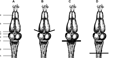
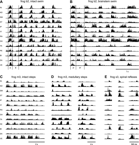
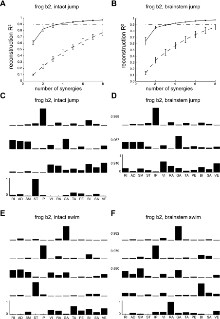
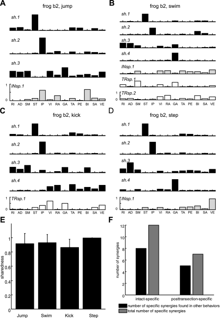
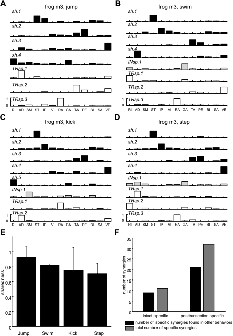
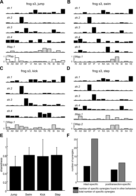
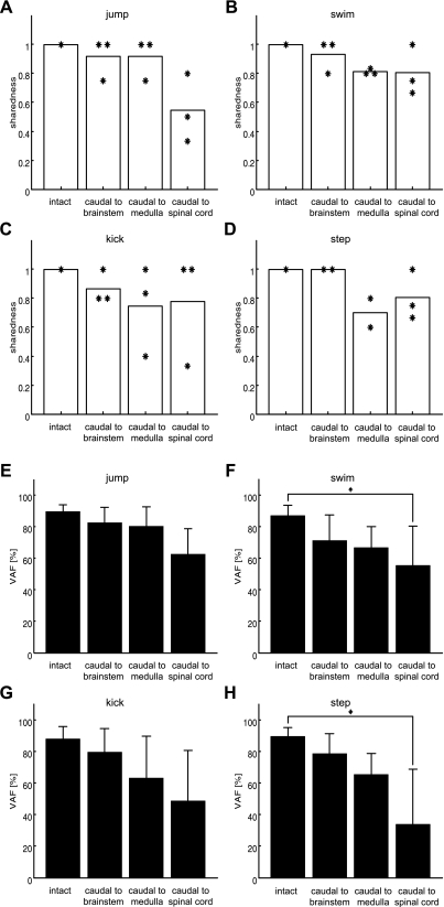
Previous studies using intact and spinalized animals have suggested that coordinated movements can be generated by appropriate combinations of muscle synergies controlled by the central nervous system (CNS). However, which CNS regions are responsible for expressing muscle synergies remains an open question. We address whether the brain stem and spinal cord are involved in expressing muscle synergies used for executing a range of natural movements. We analyzed the electromyographic (EMG) data recorded from frog leg muscles before and after transection at different levels of the neuraxis-rostral midbrain (brain stem preparations), rostral medulla (medullary preparations), and the spinal-medullary junction (spinal preparations). Brain stem frogs could jump, swim, kick, and step, while medullary frogs could perform only a partial repertoire of movements. In spinal frogs, cutaneous reflexes could be elicited. Systematic EMG analysis found two different synergy types: 1) synergies shared between pre- and posttransection states and 2) synergies specific to individual states. Almost all synergies found in natural movements persisted after transection at rostral midbrain or medulla but not at the spinal-medullary junction for swim and step. Some pretransection- and posttransection-specific synergies for a certain behavior appeared as shared synergies for other motor behaviors of the same animal. These results suggest that the medulla and spinal cord are sufficient for the expression of most muscle synergies in frog behaviors. Overall, this study provides further evidence supporting the idea that motor behaviors may be constructed by muscle synergies organized within the brain stem and spinal cord and activated by descending commands from supraspinal areas.


Figures

Fig. 1.
Fig. 1.
Schematic diagram of the bullfrog central nervous system (CNS) and transection at 3 different levels of the neuraxis. A: the bullfrog CNS. a, Olfactory lobes; b, cerebral hemispheres; c, pineal gland; d, thalamus; e, optic lobes; f, cerebellum; g, medulla; h, spinal cord. B: in the 1st set of experiments (intact vs. brain stem conditions), transection was performed at the level (marked by a solid line) of the caudal end of the 3rd ventricle to keep the entire brain stem connected to the musculoskeletal system. C: in the 2nd set of experiments (intact vs. medullary conditions), the level of transection was at the caudal end of the pons (by removing the deep cerebellar nuclei). D: in the 3rd set of experiments (intact vs. spinal conditions), spinalization was performed at the level of caudal end of the 4th ventricle. In this preparation, there was no descending command going to the spinal cord. For each set of comparisons, EMG data were recorded both before and after transection in the same animal.
Fig. 2.
Fig. 2.
Coactivation pattern across multiple muscles remained invariant after transection in each of these behavioral episodes. A and B: EMGs of swimming cycles recorded in the intact state (before transection) of frog b2 (A) and under the brain stem condition of frog b2 (B). A and B use the same timescale marked at the bottom of B. C and D: EMGs of stepping cycles recorded under the intact state of frog m3 (C) and under the medullary condition of frog m3 (D). The timescale is marked at the bottom of each panel. E: reflex episodes recorded in the spinal condition of frog s3. Note that the coactivations observed in A, such as those of semitendinosus (ST), iliopsoas (IP), and biceps femoris (BI), can still be seen even when the level of transection was lowered down from rostral midbrain in B with the reduced amplitude of ST activation, to rostral medulla in C, or to rostral spinal cord in D. The EMGs shown here were high-pass filtered and then rectified. In A, a and b indicate 2 phases in a cycle of swim; a′ and b′ in B refer to the 2 phases corresponding to phases a and b in A. RI, rectus internus; AD, adductor magnus; SM, semimembranosus; VI, vastus internus; RA, rectus anterior; GA, gastrocnemius; TA, tibialis anterior; PE, peroneus; SA, sartorius; VE, vastus externus.
Fig. 3.
Fig. 3.
Example of separate extraction of synergies in intact vs. brain stem conditions. To estimate the number of synergies underlying each behavior, we separately extracted synergies from EMGs recorded before and after transection. A and B: the fraction of total EMG data variation, for jump in intact and brain stem conditions, respectively, displayed as a function of the number of extracted synergies. In each plot, the curves show that the percentage of data variance accounted for by synergy combinations (R2; mean ± SD; n = 20) increased as the number of synergies extracted increased. The solid curves indicate how well a set of synergies extracted from the original EMG data set reconstructed the original data, while the dashed curves indicate how synergies extracted from the reshuffled EMGs reconstructed the original data. Note that the reconstruction R2 values for original EMG signals were significantly higher than the R2 values for reshuffled EMGs, suggesting that the extracted synergies capture structures in the original data set. C and D: sets of synergies separately extracted from EMGs collected during jump before (C) and after (D) transection in the same animal. The first 3 of 4 (C) synergies for natural jump were matched to 3 brain stem synergies that yielded maximal summation of scalar products between 2 synergy sets. E and F: sets of synergies separately extracted from EMGs collected during swims before (E) and after (F) transection in the same animal. The numbers between C and D and those between E and F are statistically significant scalar product values (P < 0.05).
Fig. 4.
Fig. 4.
Example of simultaneous extraction of shared and specific synergies in intact vs. brain stem conditions. Four sets of synergies for jump, swim, kick, and step, respectively, were extracted from the pooled EMG data sets recorded before and after transection in frog b2. In A–D, sh (black bars) refers to synergies shared between EMGs recorded in the intact and brain stem preparations, while INsp and TRsp (light gray and open bars, respectively) indicate individual intact-specific and posttransection-specific synergies. A: a set of 3 shared synergies and 1 intact EMG data set-specific synergy for jump. B: a set of 4 shared, 1 intact-specific, and 2 posttransection-specific synergies for swim. C: a set of 4 shared synergies and 1 posttransection-specific synergy for kick. D: a set of 4 shared synergies and 1 intact-specific synergy for step. Similarity between synergy sets was quantified by the sharedness measure. E: for each of 4 natural motor behaviors, sharedness (mean ± SD; n = 3) was defined as the average ratio of the number of shared synergies to the number of synergies that underlie the intact or brain stem EMGs, whichever number is smaller. High numbers of shared synergies resulted in high sharedness values (>0.8) in all 4 behaviors. F: across the 4 motor behaviors, in total, 8 of 12 and 5 of 7 intact- and posttransection-specific synergies appeared to be expressed by the brain stem and spinal circuitries and used to produce other movements. These findings support the idea that neural circuitries within the brain stem and spinal cord are sufficient to activate and express synergies for executing natural movements.
Fig. 5.
Fig. 5.
Example of simultaneous extraction of shared and specific synergies in intact vs. medullary conditions. A–D are analogous to A–D in Fig. 4. High sharedness values (E) found in all 4 behaviors (∼0.8) and many intact- and posttransection-specific synergies of a certain motor behavior identified as shared synergies for other behaviors (F) were observed, which suggests that the neural circuitries within and caudal to the medulla are sufficient for expressing the set of muscle synergies used for generating natural movements.
Fig. 6.
Fig. 6.
Example of simultaneous extraction of synergies in intact vs. spinal conditions. A–D here are analogous to A–D in Figs. 4 and 5. High sharedness values were found in kick, swim, and step (∼0.8) but not in jump (∼ 0.55) (E). In addition, only 36% (9 of 25) intact-specific synergies of a single motor behavior were observed in other movements as shared synergies (F). These results suggest that many of the synergies for natural behaviors are organized within the spinal cord, but the supraspinal circuits may contribute to the expression of synergies utilized for natural behaviors.
Fig. 7.
Fig. 7.
Summary of similarity measures of muscle synergies between pre- and posttransection. A–D show the mean (each bar) and distribution of sharedness values, with each asterisk denoting the value from each animal in 4 intact behaviors [jump (A), swim (B), kick (C), and step (D)]. The sharedness values indicate the degree to which individual synergies were common in pre- and posttransection for each behavior. For instance, the bar labeled “caudal to brain stem” shown in A implies that, on average, 92% of synergies for jump were found in brain stem preparations across the 3 animals. A–D demonstrate that >70% of the synergies in intact animals were preserved after transection at the level of rostral medulla across 4 behaviors. E–H show the mean variance of EMG episodes in 4 intact behaviors [jump (E), swim (F), kick (G), and step (H)] accounted for by synergies shared between the pre- and posttransection data sets [variance accounted for (VAF); means ± SD]. For instance, the bar labeled “caudal to brain stem” in F refers to how well synergies shared between pre- and posttransection at rostral brain stem could explain the variance of swim EMGs in intact animals (see main text for full description). In the cases of jump and kick, even when the level of transection was lowered to rostral spinal cord the VAFs obtained (bars labeled “caudal to spinal cord” in E and G) did not significantly decrease even when compared against the intact VAFs (2-way ANOVA with repeated measures, adjusted P > 0.05). This finding implies that the spinal circuitries are sufficient for expressing muscle synergies for jump and kick. In contrast, in the case of swim and step (F and H), as the level of transection was lowered to rostral spinal cord the VAF values significantly decreased (2-way ANOVA with repeated measures, adjusted P < 0.05), suggesting that supraspinal circuits within the brain stem are involved in expressing at least some of the swim and step muscle synergies.

Similar articles

Cited by

References

    1. Barale F, Corvaja N, Pompeiano O. Vestibular influences on postural activity in frog. Arch Ital Biol 109: 27–36, 1971 - PubMed
    1. Bizzi E, Cheung VC, d'Avella A, Saltiel P, Tresch M. Combining modules for movement. Brain Res Rev 57: 125–133, 2008 - PMC - PubMed
    1. Bizzi E, Mussa-Ivaldi FA, Giszter SF. Computations underlying the execution of movement: a biological perspective. Science 253: 287–291, 1991 - PubMed
    1. Cajigas-González I. Linear Control Model of the Spinal Processing of Descending Neural Signals (Master's thesis) Cambridge, MA: Massachusetts Inst. of Technology, 2003
    1. Cappellini G, Ivanenko YP, Poppele RE, Lacquaniti F. Motor patterns in human walking and running. J Neurophysiol 95: 3426–3437, 2006 - PubMed

Publication types

LinkOut - more resources