. 2011 Aug 1;22(15):2716-28.
doi: 10.1091/mbc.E11-01-0064. Epub 2011 Jun 8.

A laminopathic mutation disrupting lamin filament assembly causes disease-like phenotypes in Caenorhabditis elegans

Affiliations

A laminopathic mutation disrupting lamin filament assembly causes disease-like phenotypes in Caenorhabditis elegans

Erin M Bank et al. Mol Biol Cell. .

Abstract

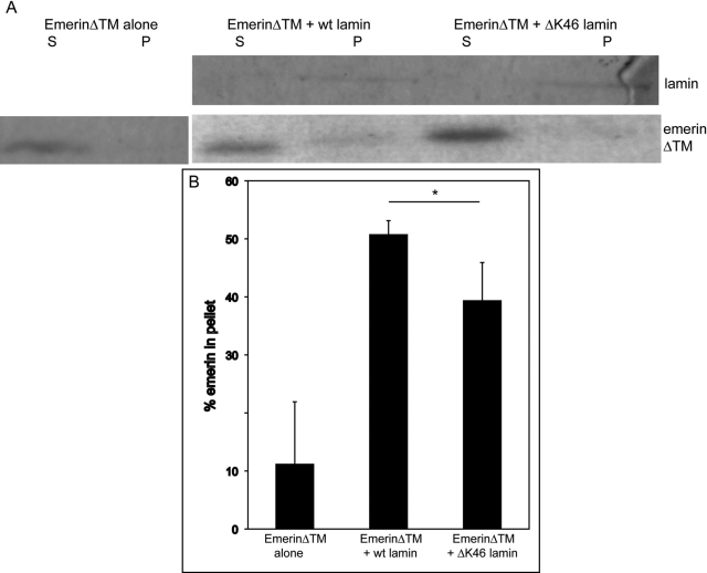
Mutations in the human LMNA gene underlie many laminopathic diseases, including Emery-Dreifuss muscular dystrophy (EDMD); however, a mechanistic link between the effect of mutations on lamin filament assembly and disease phenotypes has not been established. We studied the ΔK46 Caenorhabditis elegans lamin mutant, corresponding to EDMD-linked ΔK32 in human lamins A and C. Cryo-electron tomography of lamin ΔK46 filaments in vitro revealed alterations in the lateral assembly of dimeric head-to-tail polymers, which causes abnormal organization of tetrameric protofilaments. Green fluorescent protein (GFP):ΔK46 lamin expressed in C. elegans was found in nuclear aggregates in postembryonic stages along with LEM-2. GFP:ΔK46 also caused mislocalization of emerin away from the nuclear periphery, consistent with a decreased ability of purified emerin to associate with lamin ΔK46 filaments in vitro. GFP:ΔK46 animals had motility defects and muscle structure abnormalities. These results show that changes in lamin filament structure can translate into disease-like phenotypes via altering the localization of nuclear lamina proteins, and suggest a model for how the ΔK32 lamin mutation may cause EDMD in humans.


Figures

FIGURE 1:
FIGURE 1:
ΔK46 lamin forms abnormal filaments. (A) ClustalW (Larkin et al., 2007) alignment of coil 1A of the first rod domain of Ce-lamin (Ce_Lmn-1) with human lamins A and C (Hs_LMNA/C), human lamin B (Hs_LMNB), Xenopus lamin A (Xl_lam-A), and Drosophila lamin (Dm_lam). Conserved residues in all lamins are in bold; lysine 46 of Ce-lamin and homologous residues are shaded. (B) Electron micrographs of negative-stained wt and ΔK46 lamin filaments reveal thicker filaments of ΔK46 lamin (arrows). Scale bar = 100 nm. (C) Thick (30 nm) sections of tomograms of ΔK46 filaments show the altered structure of the IF-like filaments. The periodic arrangement of globular tail domains along protofilaments can be clearly seen as pairs (arrowheads) separated by an average distance of 14 nm. Scale bar = 50 nm. (D) Sections (10 nm) through tomograms of Ce-lamin filaments reveal a 7-nm shift of the globular tail domains in the ΔK46 filaments (left panel) compared with wt filaments (right panel). The magnification is similar in both sections. (E) 1-wt (left) and ΔK46 (right) lamin dimers assemble into a polar head-to-tail polymer of dimers. 2-Two antiparallel head-to tail polymers form a protofilament. The distances measured by the tomographic analysis in (D) are superimposed on a schematic 2D view of the head-to-tail polymer comprising the protofilaments. Both wt and ΔK46 Ce-lamin dimers associate into two head-to-tail polymers with 55 nm between the N-terminal head (yellow) and C-terminal tail (pink) domains. 3-The lateral assembly of these polymers into tetrameric protofilaments is disrupted in ΔK46 filaments.
FIGURE 2:
FIGURE 2:
GFP:ΔK46 lamin shows altered localization. (A) Representative Western blot analysis of GFP:lamin (arrow), lamin, and emerin protein levels in worm extracts from GFP:wt and GFP:ΔK46 animals. (B–D) Deconvoluted images of GFP-expressing worms. (B) GFP:ΔK46 can be seen in all cells in embryos with relatively normal nuclear localization, similar to GFP:wt. (C) GFP:ΔK46 is expressed in all cell types in L1 larval stage animals, with obvious nuclear aggregations in many nuclei (white arrows and inset). (D) Both GFP:wt and GFP:ΔK46 expression is strongest in the head (left panels) and tail (right panels) of early adult animals and does not appear in 100% of cells. Examples of GFP:ΔK46 aggregates are indicated with white arrows. Representative nuclei are enlarged in the inset (red outline) and show uniform shape even in the presence of GFP:ΔK46 aggregates. All scale bars are 10 μm in main images and 1 μm in insets. (E) Quantification of GFP aggregation confirms the visual data in (C) and (D).
FIGURE 3:
FIGURE 3:
LEM-2 localizes to GFP aggregates in nuclei expressing GFP:ΔK46. (A) L4 larval stage wt (GFP:wt, left panels) and mutant (GFP:ΔK46, right panels) animals were costained with DAPI (blue), anti-GFP (green), and anti-LEM-2 (red) antibodies. A representative slice from 3D deconvoluted images reveals that LEM-2 associates with the GFP-tagged lamin (yellow) in both GFP:wt and GFP:ΔK46 animals (arrowheads) and is found in the GFP aggregates in GFP:ΔK46 nuclei (arrows). Scale bars = 10 μm for all panels. (B) Quantification of GFP and LEM-2 aggregation confirms the visual data in A.
FIGURE 4:
FIGURE 4:
Emerin is mislocalized in nuclei expressing GFP:ΔK46. (A) L4 larval stage wt (GFP:wt, left panels) and mutant (GFP:ΔK46, right panels) animals were costained with anti-GFP (green) and anti-Ce-emerin (red) antibodies. A representative slice from 3D deconvoluted images showing the region posterior to the pharynx reveals that the localization of GFP:wt and emerin do not always overlap. Emerin staining is seen in most GFP-positive cells in wt animals (arrowheads); however, emerin staining is defective or absent (arrows) in many GFP-positive cells in mutant animals. Scale bars = 10 μm for all panels. (B) Quantification of Ce-emerin localization in GFP-positive cells reveals a significant difference in the colocalization of GFP and emerin in GFP:ΔK46 (n = 136) and GFP:wt (n = 86) nuclei (****p < 0.0001; Student's two-tailed t test).
FIGURE 5:
FIGURE 5:
Decreased association of emerin with ΔK46 lamin filaments in vitro. Heterologously expressed emerinΔTM was incubated alone or with wt lamin filaments or with ΔK46 lamin filaments. The samples were sedimented, and supernatant and pellet fractions were separated. (A) Representative Coomassie blue–stained SDS–PAGE gel. (B) Quantification of three individual experiments shows a significant reduction in the amount of emerin in the pellet when it is incubated with DK46 lamin filaments from when it is incubated with wt filaments (*p < 0.05; Student's two-tailed t test). Error bars represent SD.
FIGURE 6:
FIGURE 6:
Reduced swimming motility of GFP:ΔK46 animals. (A) Crawling motility of L4 larval stage worms was measured as the number of head turns per minute after placement on an agar plate. GFP:ΔK46 and wt (N2) animals showed similar motility. (B) Swimming motility of L4 larval stage worms was measured as the number of head turns to one side per minute after 15 min in a drop of water. GFP:ΔK46 animals were significantly impaired in their ability to move (****p = 0.0001). (C) Swimming motility was measured after feeding with an RNAi construct targeting GFP:ΔK46 (GFP) or an empty vector (L4440). Depletion of GFP:ΔK46 significantly rescued the swimming defect of GFP:ΔK46 L4 larval stage animals (**p = 0.005; ***p = 0.001; Student's two-tailed t test). Error bars represent SEM.
FIGURE 7:
FIGURE 7:
DIC analysis of early adult animals reveals structural abnormalities in GFP:ΔK46 animals. wt (GFP:wt) and GFP:ΔK46 animals were immobilized and photographed 1 d after reaching the L4 larval stage. Arrowheads indicate points of lesions, where muscles are detached from the cuticle, seen exclusively in mutant strains, in all worms examined. Scale bar = 10 μm for all panels.
FIGURE 8:
FIGURE 8:
TEM reveals muscle abnormalities in GFP:ΔK46 animals. wt longitudinal (long) (A) and transverse (trans) (D) sections show normal fiber (black arrows), dense body/attachment (white arrows), and subcuticle organization (white arrowhead with bracket), with few membrane-bound vesicles (black arrowheads). GFP:ΔK46 expression disrupts muscle organization, as evident in both longitudinal (B and C) and transverse (E and F) sections: Fibers are less uniformly arranged (black arrows); dense bodies and attachments are absent or mislocalized, causing sarcomere disorganization (white arrows); more aberrant membrane-bound structures are seen within sarcomeres and at cell edges (black arrowheads); and gross abnormalities between the sarcomere and the cuticle edge occur (white arrowheads). Sizes of all scale bars are indicated in the respective panels.

References

    1. Aebi U, Cohn J, Buhle L, Gerace L. The nuclear lamina is a meshwork of intermediate-type filaments. Nature. 1986;323:560–564. - PubMed
    1. Ben-Harush K, Wiesel N, Frenkiel-Krispin D, Moeller D, Soreq E, Aebi U, Herrmann H, Gruenbaum Y, Medalia O. The supramolecular organization of the C. elegans nuclear lamin filament. J Mol Biol. 2009;386:1392–1402. - PubMed
    1. Bercher M, Wahl J, Vogel BE, Lu C, Hedgecock EM, Hall DH, Plenefisch JD. mua-3, a gene required for mechanical tissue integrity in Caenorhabditis elegans, encodes a novel transmembrane protein of epithelial attachment complexes. J Cell Biol. 2001;154:415–426. - PMC - PubMed
    1. Bione S, Maestrini E, Rivella S, Mancini M, Regis S, Romeo G, Toniolo D. Identification of a novel X-linked gene responsible for Emery-Dreifuss muscular dystrophy. Nat Genet. 1994;8:323–327. - PubMed
    1. Bonne G, et al. Mutations in the gene encoding lamin A/C cause autosomal dominant Emery-Dreifuss muscular dystrophy. Nat Genet. 1999;21:285–288. - PubMed

Publication types

MeSH terms