. 2011 Aug 1;22(15):2664-79.
doi: 10.1091/mbc.E11-04-0374. Epub 2011 Jun 8.

Interaction of the human prostacyclin receptor with the PDZ adapter protein PDZK1: role in endothelial cell migration and angiogenesis

Affiliations

Interaction of the human prostacyclin receptor with the PDZ adapter protein PDZK1: role in endothelial cell migration and angiogenesis

Elizebeth C Turner et al. Mol Biol Cell. .

Abstract

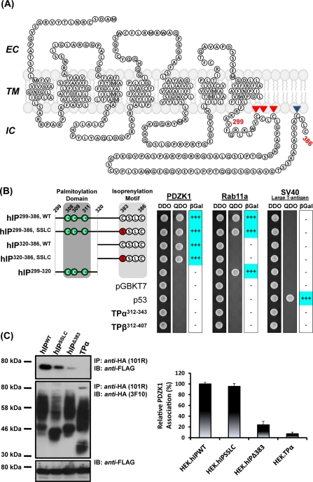
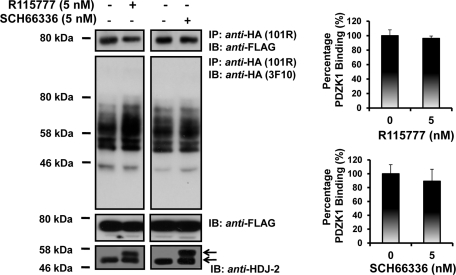
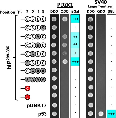
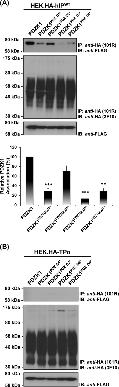
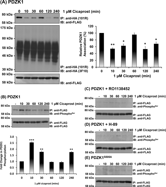
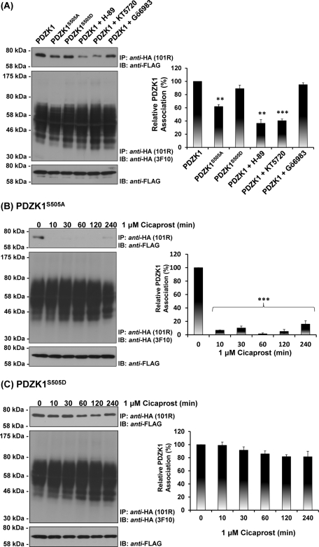
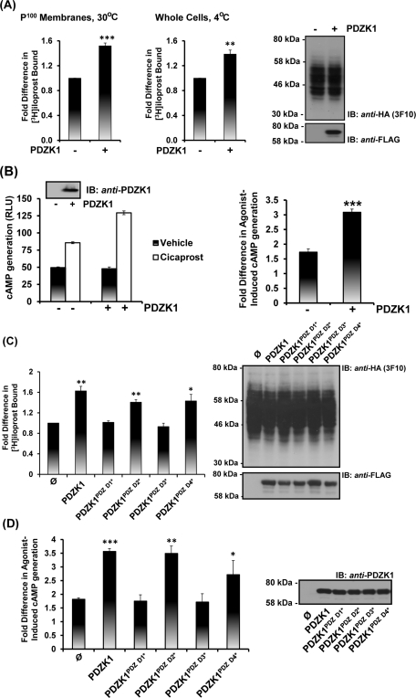
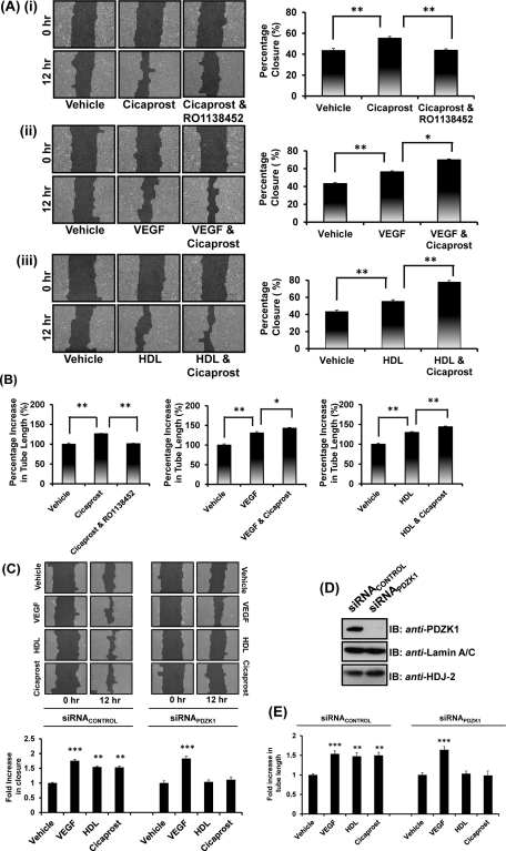
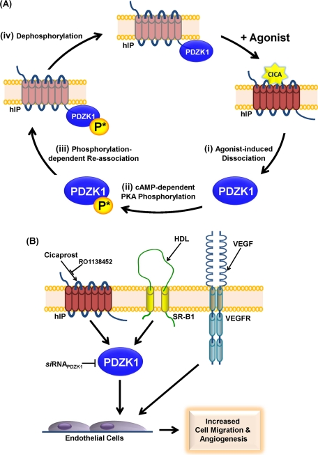
Prostacyclin is increasingly implicated in re-endothelialization and angiogenesis but through largely unknown mechanisms. Herein the high-density lipoprotein (HDL) scavenger receptor class B, type 1 (SR-B1) adapter protein PDZ domain-containing protein 1 (PDZK1) was identified as an interactant of the human prostacyclin receptor (hIP) involving a Class I PDZ ligand at its carboxyl terminus and PDZ domains 1, 3, and 4 of PDZK1. Although the interaction is constitutive, it may be dynamically regulated following cicaprost activation of the hIP through a mechanism involving cAMP-dependent protein kinase (PK)A-phosphorylation of PDZK1 at Ser-505. Although PDZK1 did not increase overall levels of the hIP, it increased its functional expression at the cell surface, enhancing ligand binding and cicaprost-induced cAMP generation. Consistent with its role in re-endothelialization and angiogenesis, cicaprost activation of the hIP increased endothelial cell migration and tube formation/in vitro angiogenesis, effects completely abrogated by the specific IP antagonist RO1138452. Furthermore, similar to HDL/SR-B1, small interfering RNA (siRNA)-targeted disruption of PDZK1 abolished cicaprost-mediated endothelial responses but did not affect VEGF responses. Considering the essential role played by prostacyclin throughout the cardiovascular system, identification of PDZK1 as a functional interactant of the hIP sheds significant mechanistic insights into the protective roles of these key players, and potentially HDL/SR-B1, within the vascular endothelium.


Figures

FIGURE 1.
FIGURE 1.
Interaction of PDZK1 with the human prostacyclin receptor. (A) Schematic of the hIP. The hIP undergoes palmitoylation at Cys308, Cys309, and Cys311 and isoprenylation/farnesylation at Cys383, which, together, are proposed to introduce fourth (IC4) and fifth (IC5) intracellular loops within the C-tail domain of the hIP. (B) S.c Y187 (pACT2:PDZK1) or, as controls, S.c Y187 (pACT2:Rab11a) and S.c Y187 (pTD1-1), encoding SV-40 large T-antigen, prey strains were mated with S.c AH109 bait strains transformed with recombinant pGBKT7 encoding the listed hIP subfragments and, as controls, pGBKT7.TPα, pGBKT7.TPβ and p53 or with the vector pGBKT7 alone. Diploids were selected on DDO medium, whereas interactants were selected on QDO medium and by their ability to express β-galactosidase (β-Gal). Data: n ≥ 3. (C) HEK.hIPWT, HEK.hIPSSLC, HEK.hIPΔ383 or, as controls, HEK.TPα cells, each transiently transfected with pCMVTag2C:PDZK1FL, were subject to immunoprecipitation with anti-HA 101R antibody. Immunoprecipitates (IP) were resolved by SDS–PAGE and immunoblotted (IB), as indicated. Uniform expression of Flag-tagged PDZK1 was verified by immunoblotting of whole cell lysates (50 μg/lane) with anti-FLAG antibody (bottom). The bar charts show mean relative levels of PDZK1-associated with the anti-HA 101R immunoprecipitates (relative protein, % ± SEM, n = 3) where levels associated with the anti-HA.hIP immunoprecipitates are expressed as 100%.
FIGURE 2.
FIGURE 2.
Effect of isoprenylation of the hIP on its interaction with PDZK1. HEK.hIP cells, transiently transfected with pCMVTag2C:PDZK1FL, were preincubated with vehicle, R115777 (5 nM) or SCH66336 (5 nM) for 24 h prior to immunoprecipitation with anti-HA 101R antibody. Immunoprecipitates (IP) were resolved by SDS–PAGE and immunoblotted (IB), as indicated. Uniform expression of Flag-tagged PDZK1 was verified by immunoblot analysis of whole cell lysates (50 μg/lane) with anti-FLAG antibody (middle panels). The efficacy of R115777 or SCH66336 to inhibit protein farnesylation was validated by immunoblot analysis of whole cell lysates (50 μg/lane) for the farnesylated (∼45–46 kDa) and nonfarnesylated (49 kDa) species of the molecular chaperone HDJ-2 (anti-HDJ-2; bottom panels). The bar charts show mean relative levels of PDZK1-associated with the anti-HA.hIP immunoprecipitates in the absence or of presence R115777 and SCH66336 (relative protein, % ± SEM, n = 3).
FIGURE 3.
FIGURE 3.
Characterization of the PDZ ligand within the C-tail domain of the hIP. S.c Y187 (pACT2:PDZK1) or, as a control, S.c Y187 (pTD1-1) prey strains were mated with S.c AH109 bait strains transformed with pGBKT7.hIP299-386 subfragment with its wild-type (-C383SLC386, corresponding to the positions (P)0, P−1, P−2, and P−3 of its PDZ ligand, respectively) carboxyl-terminal residues, or the listed mutated variants, and as controls, p53 or transformed with the vector pGBKT7 alone. Data: n ≥ 3.
FIGURE 4.
FIGURE 4.
Identification of the PDZ domains involved in the interaction of PDZK1 with the hIP. HEK.hIP (A) or, as controls, HEK.TPα (B) cells, each transiently transfected with pCMVTag2C encoding FLAG-tagged PDZK1FL, PDZK1PDZD1*, PDZK1PDZD2*, PDZK1PDZD3*, or PDZK1PDZD4*, were subject to immunoprecipitation with anti-HA 101R antibody. Immunoprecipitates (IP) were resolved by SDS–PAGE and immunoblotted (IB), as indicated. The bar charts show mean relative levels of the wild-type and mutated forms of PDZK1 associated with the anti-HA.hIP 101R immunoprecipitates (relative protein, % ± SEM, n = 3) where levels of the wild-type PDZK1 are expressed as 100%. The asterisks indicate where PDZK1 mutation resulted in significant reductions in complex associated PDZK1, where ** and *** indicate p < 0.01 and p < 0.001, respectively, for post hoc Dunnett's multiple comparison t-test analysis.
FIGURE 5.
FIGURE 5.
Effect of agonist activation of the hIP on the interaction and phosphorylation of PDZK1. (A) HEK.hIP cells, transiently transfected with pCMVTag2C:PDZK1, were stimulated with cicaprost (1 μM; 0–240 min). HA-tagged hIPs were immunoprecipitated with anti-HA 101R antibody; immunoprecipitates (IP) were resolved by SDS–PAGE and immunoblotted (IB), as indicated. The bar charts show mean relative levels of the PDZK1 associated with the anti-HA.hIP 101R immunoprecipitates as a function of cicaprost stimulation (relative protein, % ± SEM, n = 3) where levels in the absence of agonist are expressed as 100%. The asterisks indicate that cicaprost stimulation resulted in significant reductions in levels of PDZK1 associated with the hIP immune-complexes where * and ** indicate p < 0.05 and p < 0.01, respectively, for post hoc Dunnett's multiple comparison t-test analysis. (B–E) HEK.hIP cells, transiently transfected with either pCMVTag2C:PDZK1 (B–D) or pCMVTag2C:PDZKS505A (E), were preincubated with vehicle (B, E), RO1138452 (10 μM; 10 min) (C), or H-89 (10 μM; 10 min) (D) prior to stimulation with cicaprost (1 μM; 0–240 min). Cells were then subject to immunoprecipitation with anti-FLAG antibody to immunoprecipitate PDZK1. Immunoprecipitates (IP) were resolved by SDS–PAGE and immunoblotted (IB), as indicated.
FIGURE 6.
FIGURE 6.
Effect of agonist activation of the hIP on its interaction with PDZK1, PDZK1S505A, and PDZK1S505D. (A) HEK.hIP cells, transiently transfected with pCMVTag2C encoding PDZK1, PDZK1S505A, or PDZK1S505D, were incubated with vehicle (0.01% DMSO; 10 min), H-89 (10 μM; 10 min), KT5720 (5 μM; 10 min), or GÖ6983 (1 μM; 10 min), as indicated. Alternatively, HEK.hIP cells, transiently transfected with pCMVTag2C encoding PDZK1S505A (B) or PDZK1S505D (C), were stimulated with cicaprost (1 μM; 0–240 min). HA-tagged hIPs were immunoprecipitated with anti-HA 101R antibody; immunoprecipitates (IP) were resolved by SDS–PAGE and immunoblotted (IB), as indicated. The bar charts show mean relative levels of the PDZK1, PDZK1S505A, and PDZK1S505D -associated with the anti-HA.hIP 101R immunoprecipitates as a function of cicaprost stimulation (relative protein, % ± SEM, n = 3) where levels in the absence of agonist are expressed as 100%. The asterisks indicate that inhibiton of PKA (A) or cicaprost stimulation (B, C) resulted in significant reductions in PDZK1 levels in anti-HA immunoprecipitates, where ** and *** indicate p = 0.01 and p = 0.001, respectively, for post hoc Dunnett's multiple comparison t-test analysis.
FIGURE 7.
FIGURE 7.
Effect of PDZK1 on the expression and signaling of the hIP. (A, C) HEK.hIP cells were transiently transfected with pCMVTag2C encoding either PDZK1 (A) or PDZK1, PDZK1PDZ D1*, PDZK1PDZ D2*, PDZK1PDZ D3*, and PDZK1PDZ D4* or, as controls, pCMVTag2C vector (ø) alone (C). RLBA was performed 72 h posttransfection in the presence of 4 nM [3H]iloprost for 60 min using either crude membrane (P100) fractions (30°C; A, C) or whole cells (4°C; A). Data are presented as fold increases in [3H]iloprost bound as a function of PDZK1 expression where levels in the presence of wild-type PDZK1 are expressed as 1. (B, D) HEK 293 cells were transiently cotransfected with pHM6:hIP, pADVA, pCRE-LUC, and pRL-TK in the presence of pCMVTag2C encoding PDZK1FL (B) or PDZK1FL, PDZK1PDZ D1*, PDZK1PDZ D2*, PDZK1PDZ D3*, and PDZK1PDZ D4* or, as controls, pCMVTag2C vector (ø) alone (D). Cells were incubated with either vehicle or cicaprost (1 μM; 3 h) prior to determination of cAMP generation (RLU ± SEM; n = 3), where data are represented as levels of agonist-induced cAMP generation (B, left bar charts) and as fold inductions in agonist-induced cAMP accumulation (B, right bar charts; and D). Expression of the HA-tagged hIP and Flag-tagged PDZK1 proteins were verified by immunoblot analysis of the respective whole-cell lysates (50 μg/lane), as indicated. The asterisks indicate where ectopic expression of PDZK1 resulted in significant fold increases in [3H]iloprost bound (A, C) or agonist-induced cAMP accumulation (B, D) where *, **, and *** indicate p < 0.05, p < 0.01, and p < 0.001, respectively, for post hoc Dunnett's multiple comparison t-test analysis. Levels of [3H]iloprost binding in HEK.hIP cells were 1.1 ± 0.04 pmol/mg of cell protein (n = 4). Basal levels cAMP generation in HEK.hIP cells was 0.70 ± 0.04 pmol/mg of cell protein (n = 4) and was not affected by ectopic expression of PDZK1 or its mutated variants.
FIGURE 8.
FIGURE 8.
Effect of PDZK1 on hIP agonist-induced migration and endothelial tube formation in 1° HUVECs. (A, B) Migration after scratch wounds in 1° HUVEC monolayers (A) or tube formation of HUVECs seeded on Matrigel (B) in the presence of cicaprost (1 μM), VEGF (50 ng/ml), or HDL (50 μg/ml), alone or in combination and in the absence or presence of RO1138452 (10 μM; 10 min preincubation) were analyzed at 12 h. Bar charts represent mean percentage closure (% ± SEM; n = 3) at 12 h (A) or mean percentage of basal tube length at 12 h (% ± SEM; n = 3) (B). (C–E) 1° HUVECs were transfected with siRNAPDZK1 or siRNAcontrol, where immunoblot analysis confirmed specific disruption of PDZK1 expression (D). Migration (C) and tube formation (E) was analyzed in the presence of vehicle, VEGF, HDL or cicaprost, as indicated, at 0 h and 12 h. Bar charts represent mean fold increases (± SEM; n = 3) in either wound closure or tube length in the presence of vehicle, VEGF, HDL or cicaprost, as indicated, at 12 h. The asterisks indicate either significant agonist-induced increases in migration (A), significant agonist-induced increases in tube length (B), significant fold increases in agonist-induced migration in comparison to vehicle-treated cells (C), significant fold increases in agonist-induced tube length in comparison to vehicle-treated cells (E) where *, **, and *** indicate p < 0.05, p < 0.01, and p < 0.001, respectively, for post hoc Dunnett's multiple comparison t-test analysis.
FIGURE 9.
FIGURE 9.
Proposed model of the interaction of PDZK1 with the hIP and its implications for endothelial cell migration and in vitro angiogenesis. (A) In the absence of agonist, PDZK1 is constitutively associated in a complex with the hIP, where PDZK1 is either not phosphorylated or basally hypophosphorylated. On cicaprost stimulation, (i) the hIP undergoes an agonist-induced conformational activation leading to dissociation of PDZK1. (ii) Released PDZK1 is then subject to enhanced hIP induced cAMP-dependent PKA phosphorylation at Ser505, and (iii) this enhanced or nett hyperphosphorylated PDZK1 triggers its reassociation with the hIP. The reassociation of PDZK1 and hIP is coincident with regulated (iv) dephosphorylation of PDZK1 and its return to basal or hypophosphorylated levels. Consistent with this model, the phospho-defective PDZK1S505A is found in a constitutive complex with the hIP and undergoes agonist-induced dissociation but cannot undergo phosphorylation-induced reassociation in response to receptor activation. In contrast, the phospho-mimetic PDZK1S505D is hypothesized to mimic the hyperphosphorylated protein state (state iii), whereby any transient agonist-induced dissociation in the interaction of this mutant with the hIP is immediately recovered due to it mimicking the hyperphosphorylated state. (B) Agonist activation of the hIP, SR-B1, and VEGFR leads to enhanced endothelial cell migration and tube formation/in vitro angiogenesis. Consistent with a previous study (Zhu et al., 2008), HDL/SR-B1-, but not VEGF/VEGFR-, mediated endothelial cell migration is dependent on its interaction with PDZK1. Herein it was established that, similar to that of HDL/SR-B1, cicaprost activation of the RO1138452-sensitive hIP promotes endothelial cell migration and tube formation and that these effects are dependent on PDZK1. siRNA disruption of PDZK1 inhibits both cicaprost- and HDL-, but not VEGF-, induced endothelial cell responses.

References

    1. Arehart E, et al. Acceleration of cardiovascular disease by a dysfunctional prostacyclin receptor mutation: potential implications for cyclooxygenase-2 inhibition. Circ Res. 2008;102:986–993. - PMC - PubMed
    1. Biscetti F, et al. Selective activation of peroxisome proliferator-activated receptor (PPAR)alpha and PPAR gamma induces neoangiogenesis through a vascular endothelial growth factor-dependent mechanism. Diabetes. 2008;57:1394–1404. - PubMed
    1. Biscetti F, et al. Peroxisome proliferator-activated receptor alpha is crucial for iloprost-induced in vivo angiogenesis and vascular endothelial growth factor upregulation. J Vasc Res. 2009;46:103–108. - PubMed
    1. Biscetti F, Pola R. Endothelial progenitor cells and angiogenesis join the PPARty. Circ Res. 2008;103:7–9. - PubMed
    1. Clark RD, Jahangir A, Severance D, Salazar R, Chang T, Chang D, Jett MF, Smith S, Bley K. Discovery and SAR development of 2-(phenylamino) imidazolines as prostacyclin receptor antagonists [corrected] Bioorg Med Chem Lett. 2004;14:1053–1056. - PubMed

Publication types

MeSH terms