Randomized Controlled Trial
. 2011 Jul 15;187(2):1039-46.
doi: 10.4049/jimmunol.1001543. Epub 2011 Jun 8.

Increased frequencies of myelin oligodendrocyte glycoprotein/MHC class II-binding CD4 cells in patients with multiple sclerosis

Affiliations
Randomized Controlled Trial

Increased frequencies of myelin oligodendrocyte glycoprotein/MHC class II-binding CD4 cells in patients with multiple sclerosis

Khadir Raddassi et al. J Immunol. .

Abstract

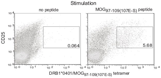
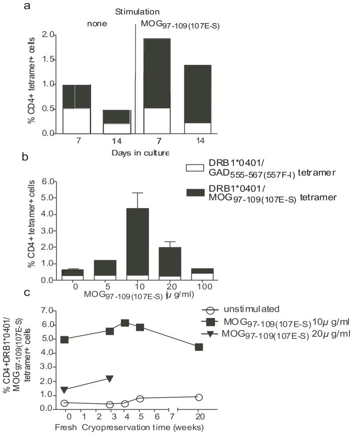
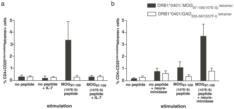
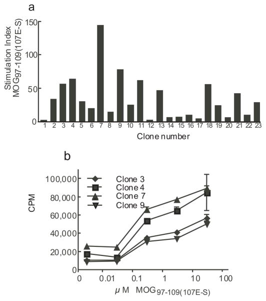
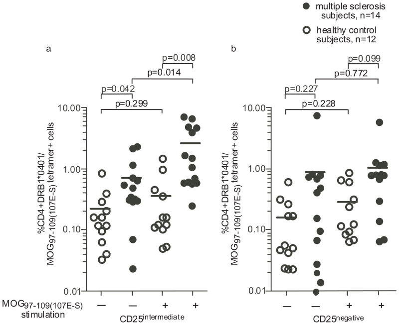
Multiple sclerosis (MS) is an autoimmune disease characterized by infiltration of pathogenic immune cells in the CNS resulting in destruction of the myelin sheath and surrounding axons. We and others have previously measured the frequency of human myelin-reactive T cells in peripheral blood. Using T cell cloning techniques, a modest increase in the frequency of myelin-reactive T cells in patients as compared with control subjects was observed. In this study, we investigated whether myelin oligodendrocyte glycoprotein (MOG)-specific T cells could be detected and their frequency was measured using DRB1*0401/MOG(97-109(107E-S)) tetramers in MS subjects and healthy controls expressing HLA class II DRB1*0401. We defined the optimal culture conditions for expansion of MOG-reactive T cells upon MOG peptide stimulation of PMBCs. MOG(97-109)-reactive CD4(+) T cells, isolated with DRB1*0401/MOG(97-109) tetramers, and after a short-term culture of PMBCs with MOG(97-109) peptides, were detected more frequently from patients with MS as compared with healthy controls. T cell clones from single cell cloning of DRB1*0401/MOG(97-109(107E-S)) tetramer(+) cells confirmed that these T cell clones were responsive to both the native and the substituted MOG peptide. These data indicate that autoantigen-specific T cells can be detected and enumerated from the blood of subjects using class II tetramers, and the frequency of MOG(97-109)-reactive T cells is greater in patients with MS as compared with healthy controls.


Figures

Figure 1
Figure 1. Detection of MOG97–109(107E-S) reactive T cells using DRB1*0401/MOG97–109(107E-S) tetramer in a MOG peptide expanded culture
Cryopreserved PMBC from a DRB1*0401 MS subject were thawed. CD4+ cells were isolated by negative selection and incubated with autologous adherent APC from the same donor loaded with or without 10μg/ml of MOG97–109(107E-S). The co-cultures were carried for 14 days in presence of recombinant human IL-2 (10 U/ml) on days 4, 7 and 10. The cells were stained with the DRB1*0401/MOG97–109(107E-S) tetramer, anti-CD4, anti-CD25 and 7AAD and analyzed using an LSR II flow cytometer. Live (7-AAD negative) and CD4+ cells were gated upon and used to enumerate the percentage of CD25intermediateDRB1*0401/MOG97–109(107E-S) tetramer+ cells. This is a representative sample from four subjects examined.
Figure 2
Figure 2. Optimum conditions for DRB1*0401/MOG97–109(107E-S) tetramer staining
The conditions for optimal tetramer staining was determined using PMBC from two DRB1*0401 MS patients with respect to in vitro culture duration, concentration of MOG97–109(107E-S) in a 14 day in vitro assay, and evaluation of fresh versus frozen PMBC for various periods of time. Triple color FACS staining was conducted to include DRB1*0401/MOG97–109(107E-S) tetramer-PE, CD4-APC, and 7-AAD. The percentage of tetramer positive cells was determined in the CD4 population by eliminating the 7-AAD-stained cells. The averages (+/− SD) from two samples are shown. In (a), the signal to noise ratio for cells stained with the specific tetramer (loaded with substituted MOG peptide) versus the irrelevant tetramer (loaded with the GAD peptide) was best at Day 14 of culture (ratio: 9.7) as compared to those at Day 7 of culture (ratio: 4.0). In 14 day cultures (b), the maximal number of DRB1*0401/MOG97–109(107E-S) tetramer+ cells was detected with an initial peptide stimulation concentration of 10 μg/ml. In (c), the number of DRB1*0401/MOG97–109(107E-S) tetramer+ cells was similar from fresh as compared to PBMC cryopreserved for various lengths of time in 14 day assays with the initial peptide stimulation of 10 μg/ml MOG97–109(107E-S).
Figure 3
Figure 3. Addition of IL-7 or treatment with neuraminidase decreases specific binding of the DRB1*0401 MOG97–109(107E-S) tetramer to MOG97–109(107E-S) stimulated PBMC
Cryopreserved PMBC from DRB1*0401 MS subjects were thawed. CD4+ cells were isolated by negative selection and incubated with autologous adherent APC from the same donor loaded with 10μg/ml of MOG97–109(107E-S). The co-cultures were carried for 14 days in presence of added IL-2 on day 4, 7 and 10. (a) IL-7 (5ng/ml) was added in the culture on the first day. (b) Cells were incubated with or without 0.5 U/ml of neuraminidase for 30 minutes at 37°C before staining with the tetramer and antibodies on Day 14. The cells were counterstained with anti-CD4, anti-CD25, and 7AAD. Data shown is the average (+/− standard error) of samples (n=4 for a, IL-7 addition, and n=5 for b, neuraminidase treatment).
Figure 4
Figure 4. Reactivity of T cell clones generated with the DRB1*0401/MOG97–109 tetramer
T cell clones were generated by single cell cloning of CD4+CD25intermediateDRB1*0401/MOG97–109(107E-S) tetramer+ cells as described in methods. Irradiated Priess (DRB1*0401 +/+) B cells were pulsed with MOG97–109(107E-S) peptide for two hours, washed and plated at 10,000/well. T cell clones were added at 25,000 cells/well and incubated for 48 hours. Wells were then pulsed with 1 μCi/well of tritiated thymidine and harvested 28 hours later. The stimulation index (antigen pulsed culture CPM/no antigen pulsed culture CPM) of representative 23 clones is shown in (a); concentration of MOG97–109(107E-S) peptide in this experiment was 10 μM. A clone was considered to be positive to the peptide SI>5 (horizontal line) and ΔCPM>10,000. The dose response of 4 representative clones to the native MOG97–109(107E-S) peptide is shown in (b).
Figure 5
Figure 5. MOG97–109(107E-S) reactive T cells are more frequent after culture with MOG97–109(107E-S) from multiple sclerosis subjects than from healthy subjects
Frequency of CD4+CD25intermediate DRB1*0401/MOG97–109(107E-S) reactive T cells (a) was determined by flow cytometry after 14 days of culture with MOG97–109(107E-S) as described in methods. CD4+CD25negative DRB1*0401/MOG97–109(107E-S) tetramer+ T cells were enumerated as a comparison (b). CD4+CD25intermediate DRB1*0401/MOG97–109(107E-S) reactive T cells were more frequently detected from the MS patients after 14 day culture with or without MOG97–109(107E-S) stimulation. P values comparing data between groups are shown in the figure.

References

    1. Agrawal SM, V, Yong W. Immunopathogenesis of multiple sclerosis. International Review of Neurobiology. 2007;79:99–126. - PubMed
    1. Minagar A, Carpenter A, Alexander JS. The destructive alliance: interactions of leukocytes, cerebral endothelial cells, and the immune cascade in pathogenesis of multiple sclerosis. International Review of Neurobiology. 2007;79:1–11. - PubMed
    1. Peterson JW, Trapp BD. Neuropathobiology of multiple sclerosis. Neurologic Clinics. 2005;23:107–129. - PubMed
    1. Hafler DA, Slavik JM, Anderson DE, O’Connor KC, De Jager P, Baecher-Allan C. Multiple sclerosis. Immunological Reviews. 2005;204:208–231. - PubMed
    1. Martin R, Howell MD, Jaraquemada D, Flerlage M, Richert J, Brostoff S, Long EO, McFarlin DE, McFarland HF. A myelin basic protein peptide is recognized by cytotoxic T cells in the context of four HLA-DR types associated with multiple sclerosis. J Exp Med. 1991;173:19–24. - PMC - PubMed

Publication types

MeSH terms