. 2011 Jun 8;31(23):8476-90.
doi: 10.1523/JNEUROSCI.1598-11.2011.

Mesocorticolimbic glutamatergic pathway

Affiliations

Mesocorticolimbic glutamatergic pathway

Tsuyoshi Yamaguchi et al. J Neurosci. .

Abstract

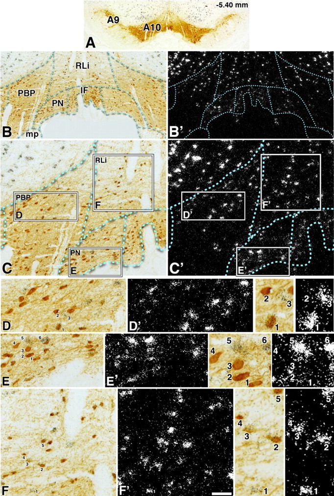
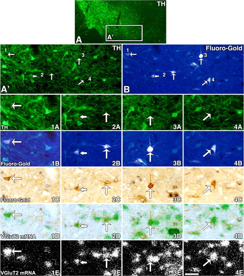
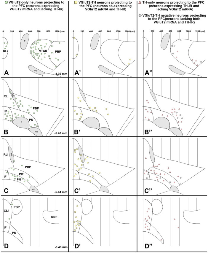
The mesocorticolimbic dopamine (DA) system plays important roles in reward, motivation, learning, memory, and movement. This system arises from the A10 region, comprising the ventral tegmental area and three adjacent midline nuclei (caudal linear nucleus, interfascicular nucleus, and rostral linear nucleus of the raphe). DAergic and GABAergic neurons are intermingled in this region with recently discovered glutamatergic neurons expressing the vesicular glutamate transporter 2 (VGluT2). Here, we show by in situ hybridization and immunohistochemistry that there are two subpopulations of neurons expressing VGluT2 mRNA in the A10 region: (1) a major subpopulation that expresses VGluT2 but lacks tyrosine hydroxylase (TH; VGluT2-only neurons), present in each nucleus of the A10 region, and (2) a smaller subpopulation that coexpresses VGluT2 and TH (VGluT2-TH neurons). By quantitative real-time PCR, we determined the mRNA copy numbers encoding VGluT2 or TH in samples of individual microdissected TH immunoreactive (IR) neurons. Data from both in situ hybridization and from mRNA quantification showed that VGluT2 mRNA is not present in every TH-IR neuron, but restricted to a subset of TH-IR neurons located in the medial portion of the A10 region. By integration of tract tracing, in situ hybridization, and immunohistochemistry, we found that VGluT2-only neurons and VGluT2-TH neurons each innervate both the prefrontal cortex and the nucleus accumbens. These findings establish that in addition to the well-recognized mesocorticolimbic DA-only and GABA-only pathways, there exist parallel mesocorticolimbic glutamate-only and glutamate-DA pathways.


Figures

Figure 1.
Figure 1.
Cellular expression of VGluT2 mRNA but lack of expression of either VGluT1 or VGluT3 mRNAs in the PBP, RLi, and the IF (radioactive in situ hybridization). A–H′, Coronal sections incubated with anti-TH antibodies and hybridized with either VGluT2 antisense (A–B′), VGluT2 sense (C–D′), VGluT1 antisense (E–F′), or VGluT3 antisense radioactive riboprobes (G–H′). A, C, Coronal sections at low magnification under bright-field microscopy showing TH immunoreactivity (dark brown label) in the A9 [substantia nigra compacta (SNC)] and the A10 regions (PBP, RLi, IF). Delimited areas in A and C are shown at higher magnification under bright-field microscopy for visualization of TH immunoreactivity (B, D) or under epiluminescence microscopy for visualization of cells expressing VGluT2 mRNA (silver white grains in B′). There are numerous cells expressing VGluT2 mRNA in the PBP, RLi, and IF at this rostral level (−5.04 mm from bregma). D′, Note lack of signal in the section hybridized with sense radioactive VGluT2 riboprobe. E, G, Coronal sections at low magnification under bright-field microscopy showing TH immunoreactivity (dark brown label) in the A9 and A10 regions. Delimited areas in E and G are shown at higher magnification under bright-field microscopy for visualization of TH immunoreactivity (F, H) or under epiluminescence microscopy for visualization of cells expressing VGluT1 mRNA (F′) or VGluT3 mRNA (H′). Note lack of detection of either VGluT1 mRNA (F′) or VGluT3 mRNA (H′) in the A9 or A10 region. High expression of VGluT1 mRNA is seen in the lateral–medial geniculata nucleus (E, arrow). fr, Fasciculus retroflexus. Scale bar: (in H′) A, C, E, G, 1100 μm; B, B′, D, D′, F, F′, H, H′, 165 μm. A, C, E, −5.04 mm from bregma; G, −5.28 mm from bregma.
Figure 2.
Figure 2.
Two distinct subpopulations of neurons expressing VGluT2 mRNA in the A10 region (−5.28 mm from bregma). A, Low magnification of a 5-μm-thick coronal section with TH immunoreactivity in the A9 and A10 regions. B–C′ correspond to the same section at two different magnifications showing TH immunoreactivity under bright-field microscopy (B, C) or VGluT2 mRNA under epiluminescence microscopy (B′, C′) in the different divisions of the A10 region. In C and C′, the boxes delimit areas shown at higher magnification of the lateral parabrachial pigmental area [PBP(l)] in D and D′, medial PBP [PBP(m)] in E and E′, the RLi in F and F′, and the IF in G and G′. D–G′, TH-IR neurons coexpressing VGluT2 mRNA (thin arrows), examples of neurons expressing VGluT2 mRNA and lacking TH immunoreactivity (thick arrows), and examples of TH-IR neurons with background levels of silver grains (arrowheads). fr, Fasciculus retroflexus. Scale bar: (in G′) A, 393 μm; B, B′, 238 μm; C, C′, 145 μm; D–G′, 50 μm.
Figure 3.
Figure 3.
Two distinct subpopulations of neurons expressing VGluT2 mRNA in the A10 region (−5.40 mm from bregma). A, Low magnification of a 5-μm-thick coronal section with TH immunoreactivity in the A9 and A10 regions. B–C′ correspond to the same section at two different magnifications showing TH immunoreactivity under bright-field microscopy (B, C) or VGluT2 mRNA under epiluminescence microscopy (B′, C′) in the different divisions of the A10 region. In C and C′, the boxes delimit areas shown at higher magnification of the PBP in D and D′, the PN in E and E′, and the RLi in F and F′. D–F′, The numbers indicate different cells shown at higher magnification in the adjacent panels. In D, D′, and the two adjacent panels, the numbers indicate three different classes of neurons within the PBP (cell 1 shows coexpression of VGluT2 mRNA and TH immunoreactivity, cell 2 shows TH immunoreactivity with background levels of silver grains, and cell 3 shows expression of VGluT2 mRNA without TH immunoreactivity). At this level of the PBP, there are only few cells coexpressing VGluT2 mRNA and TH immunoreactivity, contrasting to the more rostral level of the PBP (Fig. 2) that contains many double-labeled neurons. In E, E′, and the two adjacent panels, the numbers indicate two different classes of neurons within the PN (cells 1–4 show TH immunoreactivity with background levels of silver grains, and cells 5 and 6 shows expression of VGluT2 mRNA without TH immunoreactivity). In F, F′, and the two adjacent panels, the numbers indicate three different classes of neurons within the RLi (cell 1 shows expression of VGluT2 mRNA without TH immunoreactivity; cells 2, 3 and 5 show coexpression of VGluT2 mRNA and TH immunoreactivity; cell 4 shows TH immunoreactivity with background levels of silver grains). Note that cells in the RLi (F) are smaller than those in either the PBP (D) or the PN (E). Scale bar: (in F′) A, 705 μm; B, B′, 220 μm; C, C′, 100 μm; D–F′, 50 μm; frames showing high magnification of selected cells, 25 μm.
Figure 4.
Figure 4.
Summary diagram of the cellular heterogeneity within the A10 region. Differential distribution of VGluT2-only neurons, VGluT2-TH neurons, and TH-only neurons within each subdivision of the A10 region. A–D, Distribution of VGluT2-only neurons (neurons expressing VGluT2 mRNA but lacking TH immunoreactivity). These VGluT2-only neurons are present throughout the rostrocaudal levels of each subdivision of the A10 region with a lateromedial increasing gradient of concentration. VGluT2-only neurons are infrequent in the most lateral region of the PBP, a region with a high concentration of TH-only neurons (A″, B″, C″). A′–D′, Distribution of VGluT2-TH neurons (neurons coexpressing VGluT2 mRNA and TH immunoreactivity). VGluT2-TH neurons are restricted to the most rostromedial aspects of the PBP and PN; however, they are present at all rostrocaudal and mediolateral levels of the RLi, CLi, and IF. A″–D″, Distribution of TH-only neurons (neurons lacking expression of VGluT2 mRNA but containing TH immunoreactivity). The TH-only neurons are present throughout the rostrocaudal levels of each subdivision of the A10 region, including the lateral aspects of the PBP. Each panel represents the average number of labeled neurons found in three sections, each section from a different rat. PIF, Parainterfascicular nucleus; mtg, mammillotegmental tract; fr, fasciculus retroflexus; mp, mammillary peduncle; RRF, retrorubral field.
Figure 5.
Figure 5.
Quantification of the expression of TH or VGluT2 transcripts within individual TH-IR cells located in the A10 region (qRT-PCR analysis). A, A′, Low magnification of a 10-μm-thick coronal section incubated with anti-TH antibodies showing TH-IR cells before (A) or after (A′) microdissection. Rectangles in A and A′ indicate regions within the lateral PBP shown at higher magnification in B and B′, respectively. Arrows in B indicate the selected TH-IR neurons for microdissection, seen as black holes after microdissection (B′). Scale bar: (in B′) A, A′, 242 μm; B, B′, 50 μm. C, D, PCR amplification plots obtained for TH (C) or VGluT2 (D) detection from individual TH-IR neurons located in the lateral PBP. All single TH-IR cells in the lateral PBP (50 individual cells) showed detectable levels of TH mRNA (C). In contrast, most of these TH-IR cells lack VGluT2 mRNA amplification (D); VGluT2 mRNA was detected in only 3 of the 50 TH-IR tested cells. ΔRn, Changes in fluorescence.
Figure 6.
Figure 6.
FG injection sites within the PFC or the nAcc. A–O, Schematic representation of FG injected sites within the PFC (four rats; A–E) or the nAcc (four rats; F–O). A–E are arranged rostrocaudally from 3.72 to 2.52 mm from bregma. F–O are arranged rostrocaudally from 2.52 to 1.28 mm from bregma. aca, Anterior commissure; Cg1, cingulate cortex area 1; CPu, caudate–putamen; fmi, forceps minor of the corpus callosum; IL, infralimbic cortex; PrL, prelimbic cortex.
Figure 7.
Figure 7.
Two subpopulations of neurons expressing VGluT2 mRNA innervate the PFC. A, Low magnification of a coronal section (18 μm in thickness) from a rat injected with FG into the PFC. TH immunofluorescent is seen in cell bodies and fibers within the A10 region. A′ and B correspond to the same region delimited by a rectangle in A. The arrows in A′ (TH immunofluorescent) and B (fluorescent FG) indicate cells seen at higher magnification in 1A–4A (TH immunofluorescent), 1B–4B (fluorescent FG detection), 1C–4C (FG detection with an anti-FG antibody), 1D–4D (VGluT2 mRNA, aggregates of green grains), and 1E–4E (VGluT2 mRNA, aggregates of white grains). Long arrows in 1A–4E point to examples of four FG-labeled cells lacking TH immunoreactivity but containing VGluT2 mRNA. Note that in 2A the short arrow indicates a TH-IR neuron and the long arrow indicates a cell lacking TH; both cells contain FG (2B, 2C) and VGluT2 mRNA (2D, 2E). Scale bar: (in 4E) A, 275 μm; A′, B, 180 μm; 1A–4E, 40 μm.
Figure 8.
Figure 8.
Summary diagram of the rostrocaudal and mediolateral distribution of four different phenotypes of mesocortical neurons. A–D, Mesocortical VGluT2-only neurons (FG-labeled neurons expressing VGluT2 mRNA but lacking TH immunoreactivity) with a decreasing rostrocaudal gradient of distribution. A′–D′, Mesocortical VGluT2-TH neurons (FG-labeled neurons coexpressing VGluT2 mRNA and TH immunoreactivity) are concentrated in the caudal aspects of both the RLi and the IF. A″–D″, Mesocortical TH-only neurons (FG-labeled neurons lacking expression of VGluT2 mRNA but containing TH immunoreactivity) are intermingled with mesocortical VGluT2-TH-negative neurons (FG-labeled neurons lacking both VGluT2 mRNA and TH immunoreactivity). The symbols in each panel represent the pool of FG-labeled neurons found in four brain sections, with each section selected from a different rat. mtg, Mammillotegmental tract; fr, fasciculus retroflexus; mp, mammillary peduncle; VTAR, ventral tegmental area rostral; RRF, retrorubral field.
Figure 9.
Figure 9.
Two subpopulations of neurons expressing VGluT2 mRNA innervate the nAcc. A–D, Low magnification of coronal sections (18 μm thick) from a rat injected with FG into the nAcc. TH immunofluorescent in cell bodies and fibers (A, C) or FG-labeled cells (B, D). Boxed areas in A–D are shown in A′–D′, respectively, in which arrows indicate FG-labeled cells shown at higher magnification in 1A–6A (TH immunofluorescent), 1B–6B (FG fluorescent), 1C–6C (FG detection with an anti-FG antibody), 1D–6D (VGluT2 mRNA seen as aggregates of green grains), and 1E–6E (VGluT2 mRNA seen as aggregates of white grains). 1A–1E, A FG-labeled cell containing TH immunoreactivity and lacking VGluT2 mRNA (arrowheads). 2A–2E, A FG-labeled cell lacking TH immunoreactivity but containing VGluT2 mRNA (long arrows). 3A–3E, A FG-labeled cell lacking both TH and VGluT2 mRNA (double-headed arrows). 4A–4E, A FG-labeled cell with TH immunoreactivity and VGluT2 mRNA (short arrows). 5A–6E, FG-labeled cells lacking TH immunoreactivity but containing VGluT2 mRNA (long arrows). Scale bar: (in 6E) A–D, 258 μm; A′–D′, 80 μm; 1A–6E, 40 μm.
Figure 10.
Figure 10.
Summary diagram of the rostrocaudal and mediolateral distribution of four different phenotypes of mesoaccumbal neurons. A–D, Mesoaccumbal VGluT2-only neurons (FG-labeled neurons expressing VGluT2 mRNA but lacking TH immunoreactivity) are concentrated in the medial aspects of the A10 region. A′–D′, Mesoaccumbal VGluT2-TH neurons (FG-labeled neurons coexpressing VGluT2 mRNA and TH immunoreactivity) are clustered in the medial aspects of the A10 region, the RLi, CLi, and IF. A″–D″, Mesoaccumbal TH-only neurons (FG-labeled neurons lacking expression of VGluT2 mRNA but containing TH immunoreactivity) are intermingled with mesoaccumbal VGluT2-TH-negative neurons (FG-labeled neurons lacking both VGluT2 mRNA and TH immunoreactivity). The subpopulation of mesoaccumbal TH-only neurons has a more lateral distribution than the subpopulation of mesoaccumbal VGluT2-TH neurons. The symbols in each panel represent the average of cells found in brain sections of four different rats (8 sections for A, A″; 16 sections for B, B″; 22 sections for C, C″; 15 sections for D, D″). PIF, Parainterfascicular nucleus; mtg, mammillotegmental tract; fr, fasciculus retroflexus; mp, mammillary peduncle; VTAR, ventral tegmental area rostral; RRF, retrorubral field.
Figure 11.
Figure 11.
A10 region VGluT2 mRNA-expressing neurons: distribution, degree of colocalization with TH immunoreactivity, and projections. A, Incidence of VGluT2-only neurons, VGluT2-TH neurons, and TH-only neurons within each subdivision of the A10 region. VGluT2-only neurons (green) are prevalent in each subdivision of the A10 region. These neurons are more frequent than the TH-IR neurons in the medial PBP [PBP(m)], medial PN [PN(m)], lateral PN [PN(l)], and RLi. In contrast, the subpopulation of VGluT2-TH neurons (yellow) is a small fraction of the total population of neurons that expresses VGluT2 mRNA. These VGluT2-TH neurons are also a small fraction of the total population of TH-IR neurons. The TH-only neurons (pink) are prevalent in the lateromedial aspects of the PBP(t). For PBP(t) and PN(t), cells were counted from −5.28 to −5.64 mm from bregma. Total number of cells counted in the PBP [PBP(t)] or in the PN [PN(t)] from −5.28 to −5.64 mm from bregma. B, Four different phenotypes of neurons within the A10 region differentially innervate the PFC (mesocortical neurons) or the nAcc (mesoaccumbal neurons). The proportionality of neurons projecting to the PFC (left bar) or to the nAcc (right bar) is shown. The VGluT2-only neurons (green) are the largest subpopulation of neurons innervating the PFC, but the smallest innervating the nAcc. In contrast, most of the neurons projecting to the nAcc are TH-only neurons (pink). The subpopulation of VGluT2-TH-positive neurons (yellow), and the subpopulation of VGluT2-TH-negative neurons (gray) innervate with similar frequency the PFC and the nAcc (counted neurons are indicated in each bar).

References

    1. Arluison M, Dietl M, Thibault J. Ultrastructural morphology of dopaminergic nerve terminals and synapses in the striatum of the rat using tyrosine hydroxylase immunocytochemistry: a topographical study. Brain Res Bull. 1984;13:269–285. - PubMed
    1. Bérube-Carriére N, Riad M, Dal Bo G, Levesque D, Trudeau LE, Descarries L. The dual dopamine-glutamate phenotype of growing mesencephalic neurons regresses in mature rat brain. J Comp Neurol. 2009;517:873–891. - PubMed
    1. Birgner C, Nordenankar K, Lundblad M, Mendez JA, Smith C, le Greves M, Galter D, Olson L, Fredriksson A, Trudeau LE, Kullander K, Wallen-Mackenzie A. VGLUT2 in dopamine neurons is required for psychostimulant-induced behavioral activation. Proc Natl Acad Sci U S A. 2010;107:389–394. - PMC - PubMed
    1. Bourque MJ, Trudeau LE. GDNF enhances the synaptic efficacy of dopaminergic neurons in culture. Eur J Neurosci. 2000;12:3172–3180. - PubMed
    1. Bouyer JJ, Joh TH, Pickel VM. Ultrastructural localization of tyrosine hydroxylase in rat nucleus accumbens. J Comp Neurol. 1984;227:92–103. - PubMed

Publication types

MeSH terms