. 2011 Jun 8;31(23):8585-94.
doi: 10.1523/JNEUROSCI.0659-11.2011.

Functional dissection of the Oct6 Schwann cell enhancer reveals an essential role for dimeric Sox10 binding

Affiliations

Functional dissection of the Oct6 Schwann cell enhancer reveals an essential role for dimeric Sox10 binding

Noorjahan B Jagalur et al. J Neurosci. .

Abstract

The POU domain transcription factor Pou3f1 (Oct6/Scip/Tst1) initiates the transition from ensheathing, promyelinating Schwann cells to myelinating cells. Axonal and other extracellular signals regulate Oct6 expression through the Oct6 Schwann cell enhancer (SCE), which is both required and sufficient to drive all aspects of Oct6 expression in Schwann cells. Thus, the Oct6 SCE is pivotal in the gene regulatory network that governs the onset of myelin formation in Schwann cells and provides a link between myelin promoting signaling and activation of a myelin-related transcriptional network. In this study, we define the relevant cis-acting elements within the SCE and identify the transcription factors that mediate Oct6 regulation. On the basis of phylogenetic comparisons and functional in vivo assays, we identify a number of highly conserved core elements within the mouse SCE. We show that core element 1 is absolutely required for full enhancer function and that it contains closely spaced inverted binding sites for Sox proteins. For the first time in vivo, the dimeric Sox10 binding to this element is shown to be essential for enhancer activity, whereas monomeric Sox10 binding is nonfunctional. As Oct6 and Sox10 synergize to activate the expression of the major myelin-related transcription factor Krox20, we propose that Sox10-dependent activation of Oct6 defines a feedforward regulatory module that serves to time and amplify the onset of myelination in the peripheral nervous system.


Figures

Figure 1.
Figure 1.
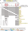
Comparative genomic and functional analysis of the mouse Oct6 locus. A, VISTA alignment of the mouse, rat, and human Oct6 locus spanning ∼50 kb. The position of the Oct6 transcription unit and the SCE are indicated above the plot. The Oct6 open reading frame is in red, with the POU domain in dark blue and the 3′ untranslated region in light blue. The gray arrowhead marks the position of the ultraconserved sequence, and numbered arrows mark the position of the previously mapped DNaseI hypersensitive sites (Mandemakers et al., 2000). Repeat sequences present in the mouse genome are indicated. SINE, Short interspersed elements; LINE, long interspersed elements; LTR, long terminal repeat. Note that the homology threshold has been adjusted to 90% for the rat sequence. B, The enlarged section of the SCE region shows the four regions of strong homology, HR1a, HR1b, HR2a, and HR2b, between mouse and human sequences. The approximate position of HSS6 and HSS7 as well as the short repeat sequences (in green) are indicated. The black lines represent the segments of the SCE that were cloned behind the Hsp68LacZ reporter construct. Individual constructs were tested for activity in transgenic mice, and the number of transgenic founders (nr of tg) and those that express the Hsp68LacZ in the peripheral nerves (nr LacZ+) are indicated. A dissected sciatic nerve of a P2 mouse transgenic for construct 9 is shown as a typical example. C, The FAIRE plot of the rat Oct6 SCE is shown at the same scale as the VISTA plot in B to allow a direct visual comparison of the two plots. The coordinates of the rat genome sequence (RN4) are indicated. The red bar indicates sequences not represented on the custom-made microarray used in this experiment.
Figure 2.
Figure 2.
Dissection of Oct6 SCE functional elements in differentiating Schwann cell cultures. A, SCE constructs were cloned behind the minimal SV40 promoter-driven luciferase cassette (pGL3) and transfected in rat Schwann cells. B, The SCE constructs tested are represented with a black line and aligned with the VISTA plot. Luciferase activity of each construct is expressed as a percentage of the activity of the full SCE in these experiments. All transfections were performed in triplicate. C, The sequence of HR1a3, which has strong enhancer activity in the luciferase assay, shows potential binding sites for transcription factors known to be involved in Schwann cell biology. These potential binding sequences were identified using the phylogenetic footprinting program ConSite. The sequence of the c2 element, containing the consensus Sox binding site, is underlined.
Figure 3.
Figure 3.
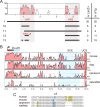
Functional analysis of SCE elements in transgenic mice and comparative genomics. A, Schematic depiction of the SCE constructs tested in transgenic mice. The SCE constructs are represented by black lines aligned with the VISTA plot of the mouse SCE to visualize the presence or absence of conserved sequences in the various constructs. The total number of transgenic animals (nr of tg) and the number of animals that express the transgene in neonatal peripheral nerves (nr LacZ+) is indicated. All animals were analyzed as founders. B, VISTA alignment of human SCE sequence with mouse, dog (Canis familiaris), opossum (Monodelphis domestica), and zebrafish (Danio rerio). Note that the lower homology threshold has been set to 30% for opossum and zebrafish to allow for the visualization of more diverged homologous regions. The position of core element c1 in the VISTA plot of opossum is indicated by an arrow. UCS, Ultra-conserved sequence. C, Alignment of core element c1 in the SCE of human, mouse, opossum, and wallaby (Macropus eugenii). Hundred percent homologous nucleotides are boxed in black, 75% homology in light blue, and 50% homology in ochre.
Figure 4.
Figure 4.
Developmental control of Schwann-cell-specific expression through SCE13. A, Southern blot of genomic DNA of the two transgenic lines analyzed here shows that they carry a comparable number of transgenes. The transgene is detected with a LacZ probe that hybridizes to a 3.5 kb BamHI restriction fragment derived from the transgenic construct. The Oct6 locus is detected with a probe that hybridizes with two fragments of 2.8 and 2.4 kb in length. The equal intensity of these endogenous band shows that equal amounts of DNA were loaded in both lanes. B, Transgenic animals carrying a LacZ reporter gene driven by either the Oct6 promoter or the Hsp68 promoter and controlled by SCE construct 13, Oct6LacZSCE13 and Hsp68LacZSCE13, respectively, were stained for β-galactosidase activity in Schwann cells of the sciatic nerve at different stages of prenatal and postnatal development. Developmental expression of β-galactosidase activity in nerves of the two transgenic lines was compared with the β-galactosidase activity in Oct6 β-galactosidase–neomycin (Oct6-βgeo) heterozygous knock-in animals (Jaegle et al., 1996; Mandemakers et al., 2000).
Figure 5.
Figure 5.
Biochemical and functional analysis of core element c1. A, EMSA with radiolabeled double-stranded oligonucleotides c1 and c2. The sequence of these probes is depicted in E. Oligonucleotide probes were incubated with nuclear extracts from RT4–D6P2T cells or from HEK293 cells overexpressing Sox10 (labeled Sox10), in the presence (+) or absence (−) of Sox10 antibodies, as indicated. The three major DNA–protein complexes formed on c1 with RT4–D6P2T nuclear proteins are marked with an asterisk. The Sox10 monomeric and dimeric complexes are indicated with arrows. B, EMSA with radiolabeled double-stranded oligonucleotide c1 and c1mut. Oligonucleotide probes were incubated with increasing amounts of nuclear extracts from HEK293 cells expressing Sox10 MIC. C, ChIP of Oct6 SCE sequences using Sox10 antibodies. Chromatin of RT4–D6P2T cells was precipitated with Sox10 or rabbit IgG antibodies. The nonspecific rabbit IgG antibodies served as a control. Quantitative PCR on immune-precipitated and purified DNA using primers for the c1 region (c1), a region 13 kb upstream of the Oct6 promoter (−13 kb), and a region associated with the IgG2a promoter (IgG2aP) was performed in triplicate and was used to determine the relative enrichment for these loci by Sox10 over IgG immunoprecipitation. The experiment was performed twice. Data from both experiments are presented (1 and 2). D, Functional analysis of Sox binding sites in element c1. The in vivo activity of the various SCE constructs was assessed in newborn founder animals. The number of transgenic animals (nr of tg) and the number of animals that express LacZ in the peripheral nerves (nr LacZ+) is indicated. Constructs 14, 15, and 16 were created from construct 13 by replacing the c1 sequence for that of c1mut, C/C', and C, respectively. E, Sequence of c1 and related elements and its various mutant derivatives. Red asterisks indicate the nucleotides mutated in c1mut. The position and orientation of the Sox10 binding sites is indicated by green boxes and arrows. The C/C' and C sequence is derived from the Mpz promoter (Peirano et al., 2000). Differences between C/C' and C are indicated with red asterisks. The consensus Sox DNA binding motif was derived by Harley et al. (1994).
Figure 6.
Figure 6.
The figure provides a schematic representation of the gene regulatory network that governs the transition from a premyelinating Schwann cell to a myelinating cell and incorporates findings described in this study. Different signaling pathways converge on the regulatory elements in this network and cooperate with Sox10 to control Oct6, Krox20, and myelin gene expression (Svaren and Meijer, 2008). The Sox10-dependent activation of Oct6 and subsequent synergistic activation of Krox20 defines a feedforward (FF) regulatory module. Sox10 then cooperates with Krox20 to control expression of myelin-related genes, through closely spaced Krox/Sox (K/S) binding elements, defining a second feedforward loop. These feedforward loops serve to time and amplify the onset of myelination in the PNS. For additional details, see Discussion.

References

    1. Arroyo EJ, Bermingham JR, Jr, Rosenfeld MG, Scherer SS. Promyelinating Schwann cells express Tst-1/SCIP/Oct-6. J Neurosci. 1998;18:7891–7902. - PMC - PubMed
    1. Arthur-Farraj P, Wanek K, Hantke J, Davis CM, Jayakar A, Parkinson DB, Mirsky R, Jessen KR. Mouse schwann cells need both NRG1 and cyclic AMP to myelinate. Glia. 2011;59:720–733. - PMC - PubMed
    1. Bansal R, Pfeiffer SE. Regulated galactolipid synthesis and cell surface expression in Schwann cell line D6P2T. J Neurochem. 1987;49:1902–1911. - PubMed
    1. Bejerano G, Pheasant M, Makunin I, Stephen S, Kent WJ, Mattick JS, Haussler D. Ultraconserved elements in the human genome. Science. 2004;304:1321–1325. - PubMed
    1. Bermingham JR, Jr, Scherer SS, O'Connell S, Arroyo E, Kalla KA, Powell FL, Rosenfeld MG. Tst-1/Oct-6/SCIP regulates a unique step in peripheral myelination and is required for normal respiration. Genes Dev. 1996;10:1751–1762. - PubMed

Publication types

Substances