. 2011 Sep;301(3):C695-704.
doi: 10.1152/ajpcell.00322.2010. Epub 2011 Jun 8.

Regulation of VEGF-induced endothelial cell migration by mitochondrial reactive oxygen species

Affiliations

Regulation of VEGF-induced endothelial cell migration by mitochondrial reactive oxygen species

Youxue Wang et al. Am J Physiol Cell Physiol. 2011 Sep.

Abstract

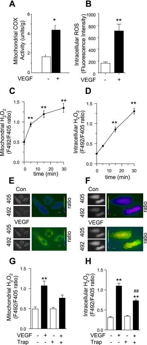
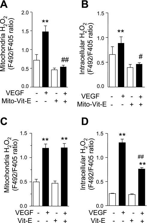
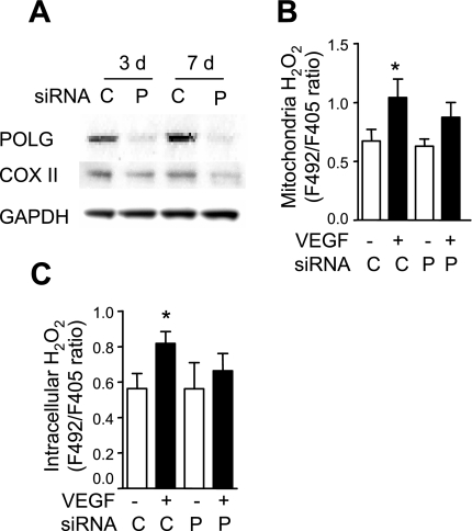
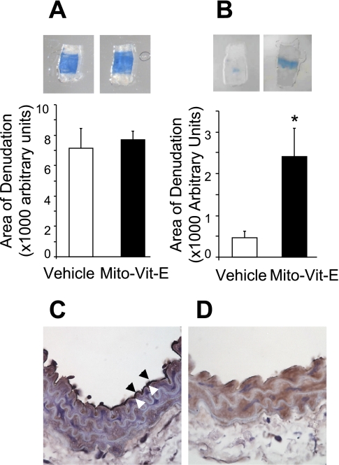
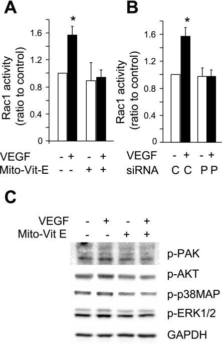
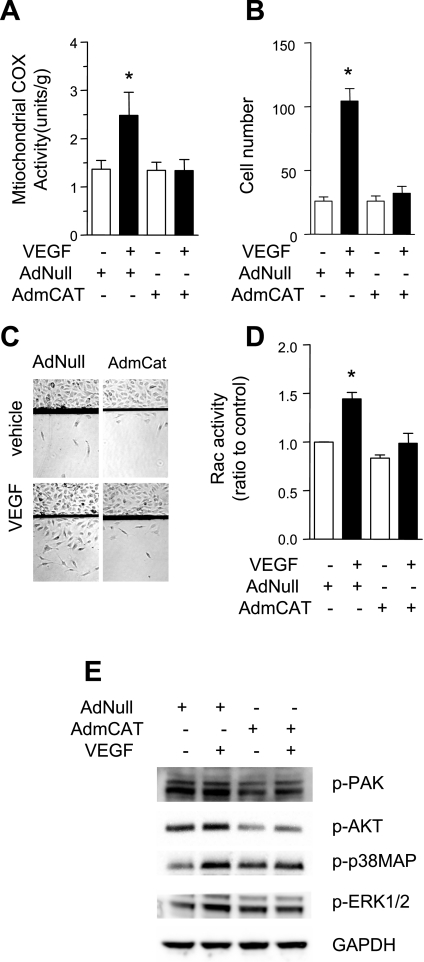
Endothelial migration is a crucial aspect of a variety of physiologic and pathologic conditions including atherosclerosis and vascular repair. Reactive oxygen species (ROS) function as second messengers during endothelial migration. Multiple intracellular sources of ROS are regulated by cellular context, external stimulus, and the microenvironment. However, the predominant source of ROS during endothelial cell (EC) migration and the mechanisms by which ROS regulate cell migration are incompletely understood. In this study, we tested the hypothesis that mitochondria-derived ROS (mtROS) regulate EC migration. In cultured human umbilical vein endothelial cells, VEGF increased mitochondrial metabolism, promoted mtROS production, and induced cell migration. Either the targeted mitochondrial delivery of the antioxidant, vitamin E (Mito-Vit-E), or the depletion of mitochondrial DNA abrogated VEGF-mediated mtROS production. Overexpression of mitochondrial catalase also inhibited VEGF-induced mitochondrial metabolism, Rac activation, and cell migration. Furthermore, these interventions suppressed VEGF-stimulated EC migration and blocked Rac1 activation in endothelial cells. Constitutively active Rac1 reversed Mito-Vit-E-induced inhibition of EC migration. Mito-Vit-E also attenuated carotid artery reendothelialization in vivo. These results provide strong evidence that mtROS regulate EC migration through Rac-1.


Figures

Fig. 1.
Fig. 1.
VEGF increased mitochondria cytochrome-c activity, as well as mitochondrial and intracellular H2O2 production. A: human umbilical vein endothelial cells (HUVEC) were stimulated with vehicle or VEGF (50 ng/ml) for 5 min, and mitochondrial fractions were isolated and cytochrome-c oxidase (COX) activity was determined. B: HUVEC were loaded with dichlorofluorescein (DCF), and fluorescence intensity was determined after cells were stimulated with vehicle or VEGF (50 ng/ml) for 15 min. ROS, reactive oxygen species. CH: HUVEC were transfected with pHyPer-dMito (C, E, and G) or pHyPer-cyto (D, F, and H), and mitochondrial (C, E, and G) and intracellular (D, F, and H) H2O2 levels were determined by fluorescence ratio (F492/F405 nm) imaging before and after VEGF (50 ng/ml) treatment. For the inhibition of VEGF effects, VEGF was preincubated with VEGF-Trap (300 ng/ml) for 30 min before VEGF was used to stimulate the cells (G and H). Data shown are means ± SE of at least 3 independent experiments (AD, G, and H) and representative images at 15 (E) or 30 (F) min after VEGF treatment. Bars, 20 μm. Con, control. *P < 0.05, **P < 0.01 vs. vehicle control or before VEGF stimulation; ##P < 0.01 vs. without VEGF-Trap preincubation.
Fig. 2.
Fig. 2.
Mitochondria-targeted vitamin E (Mito-Vit-E) inhibits VEGF-induced mitochondrial and intracellular H2O2. HUVEC were transfected with pHyPer-dMito (A and C) or pHyPer-cyto (B and D). Cells were pretreated with Mito-Vit-E (1 μM) (A and B), Vit-E (1 μM) (C and D), or vehicle for 6 h before mitochondrial and intracellular H2O2 was determined by fluorescence ratio imaging (F492/F405 nm) before and after VEGF (50 ng/ml) stimulation for 15 (A and C) or 30 (B and D) min. Data are expressed as means ± SE of at least 3 independent experiments. **P < 0.01 vs. before VEGF stimulation. #P < 0.05, ##P < 0.01 vs. VEGF-stimulated cells without Mito-Vit-E or Vit-E pretreatment.
Fig. 3.
Fig. 3.
DNA polymerase gamma (POLG) knockdown decreases VEGF-induced mitochondrial and intracellular H2O2 production. A: at 3 and 7 days (d) after HUVEC cells were transfected with scrambled control small interfering (si)RNA (C) or POLG siRNA (P), the expression of POLG and COX II was analyzed using Western blot. B and C: HUVEC cells were transfected with scrambled control or POLG siRNA on day 0, and they were transfected with pHyPer-dMito (B) or pHyPer-cyto (C) on day 5. On day 7, mitochondrial and intracellular H2O2 levels were determined by fluorescence ratio imaging before and after VEGF stimulation (50 ng/ml) for 15 (B) or 30 (C) min. Data are expressed as means ± SE of at least 3 independent experiments. *P < 0.05 vs. before VEGF stimulation.
Fig. 4.
Fig. 4.
Mitochondrial ROS are involved in VEGF-induced endothelial migration. A: HUVEC infected or uninfected with adenovirus expressing mutationally activated Rac1 [Rac(V12)] were pretreated with vehicle or Mito-Vit-E (1 μM) for 6 h before they were exposed to vehicle or VEGF (50 ng/ml, 10 min). Cell migration was measured for 6 h by determining the change of transmonolayer electrical resistance after a wounding. B: HUVEC with (P) or without (C) POLG gene knockdown with siRNA were stimulated with vehicle or VEGF (50 ng/ml, 10 min) before cell migration was measured for 6 h by transmonolayer electrical resistance after a wounding. C and E: HUVEC were pretreated with vehicle, Vit-E (1 μM), or Mito-Vit-E (1 μM) for 6 h before a defined area of the cells was removed with a razor blade. Cells were then treated with vehicle or VEGF (50 ng/ml) for 36 h, and the number of cells that migrated past the wound edge was counted. D and F: HUVEC with or without POLG knockdown were treated with vehicle or VEGF, and migration assay was done in a similar way as in C and E. G: HUVEC were pretreated with vehicle, Vit-E (1 μM), or Mito-Vit-E (1 μM) for 6 h before they were stimulated with vehicle or VEGF (50 ng/ml) for 48 h and the MTT assay was performed. Data are expressed as means ± SE of at least 3 independent experiments (A, B, E, F, and G) or representative images (C and D). *P < 0.05 vs. without VEGF stimulation.
Fig. 5.
Fig. 5.
Mito-Vit-E inhibited endothelial cell migration in vivo. C57BL/6 mice received Mito-Vit-E by oral gavage for 3 days before perivascular carotid electric injury (day 0), and the Mito-Vit-E treatment was continued until the end point of the experiment. Animals were euthanized at day 1 (A) or day 4 (B), and the area of denudation was quantified. C and D: immunostaining of von Willebrand Factor (vWF) was carried out on carotid arteries on day 1 after sham operation (C) or electric injury (D). Arrowheads indicate endothelium positively stained for vWF. Results from 6–8 mice/group were analyzed by Mann-Whitney's rank sum test. *P < 0.05.
Fig. 6.
Fig. 6.
Inhibition of mitochondrial ROS production prevents VEGF-induced Rac1 activation. A: HUVEC pretreated with vehicle or Mito-Vit-E (1 μM) for 6 h were stimulated with vehicle or VEGF (50 ng/ml) for 5 min, and Rac1 activity was determined. B: at 7 days after transfection with control (C) or POLG siRNA (P), HUVEC cells were stimulated with vehicle or VEGF (50 ng/ml) for 5 min and Rac1 activity was determined. C: HUVEC were pretreated with vehicle or Mito-Vit-E (1 μM) for 6 h and then stimulated with VEGF for 60 min, and whole cell lysates were collected for Western blot analysis of phosphorylated PAK, Akt, p38MAP, and ERK1/2. *P < 0.05 vs. vehicle.
Fig. 7.
Fig. 7.
Mitochondrially targeted catalase inhibited endothelial migration. HUVEC were infected with AdNull or AdmCat 48 h before experiments. A: HUVEC were stimulated with vehicle or VEGF (50 ng/ml) for 5 min, mitochondrial fractions were isolated, and cytochrome-c oxidase activity was determined. B and C: a defined area of the cells was removed. Cells were then treated with vehicle of VEGF (50 ng/ml) for 36 h and the number of cells that migrated past the wound edge was counted. D: HUVEC were stimulated with VEGF (50 ng/ml) for 5 min and Rac1 activity was determined. E: cells were stimulated with VEGF (50 ng/ml) for 60 min before whole cell lysates were collected for Western blot analysis of phosphorylated PAK, Akt, ERK1/2, and p38MAP. *P < 0.05 vs. vehicle.

References

    1. Abid MR, Spokes KC, Shih SC, Aird WC. NADPH oxidase activity selectively modulates vascular endothelial growth factor signaling pathways. J Biol Chem 282: 35373–35385, 2007 - PubMed
    1. Adlam VJ, Harrison JC, Porteous CM, James AM, Smith RA, Murphy MP, Sammut IA. Targeting an antioxidant to mitochondria decreases cardiac ischemia-reperfusion injury. FASEB J 19: 1088–1095, 2005 - PubMed
    1. Ahmad IM, Aykin-Burns N, Sim JE, Walsh SA, Higashikubo R, Buettner GR, Venkataraman S, Mackey MA, Flanagan SW, Oberley LW, Spitz DR. Mitochondrial O2*− and H2O2 mediate glucose deprivation-induced stress in human cancer cells. J Biol Chem 280: 4254–4263, 2005 - PubMed
    1. Bai J, Rodriguez AM, Melendez JA, Cederbaum AI. Overexpression of catalase in cytosolic or mitochondrial compartment protects HepG2 cells against oxidative injury. J Biol Chem 274: 26217–26224, 1999 - PubMed
    1. Burridge K, Wennerberg K. Rho and Rac take center stage. Cell 116: 167–179, 2004 - PubMed

Publication types

MeSH terms

Substances